. 2025 Apr 22;16(1):3446.
doi: 10.1038/s41467-025-57960-x.

Structure of a lasso peptide bound ETB receptor provides insights into the mechanism of GPCR inverse agonism

Affiliations

Structure of a lasso peptide bound ETB receptor provides insights into the mechanism of GPCR inverse agonism

Wataru Shihoya et al. Nat Commun. .

Abstract

Lasso peptides exhibit a unique lariat-like knotted structure imparting exceptional stability and thus show promise as therapeutic agents that target cell-surface receptors. One such receptor is the human endothelin type B receptor (ETB), which is implicated in challenging cancers with poor immunotherapy responsiveness. The Streptomyces-derived lasso peptide, RES-701-3, is a selective inhibitor for ETB and a compelling candidate for therapeutic development. However, meager production from a genetically recalcitrant host has limited further structure-activity relationship studies of this potent inhibitor. Here, we report cryo-electron microscopy structures of ETB receptor in both its apo form and complex with RES-701-3, facilitated by a calcineurin-fusion strategy. Hydrophobic interactions between RES-701-3 and the transmembrane region of the receptor, especially involving two tryptophan residues, play a crucial role in RES-701-3 binding. Furthermore, RES-701-3 prevents conformational changes associated with G-protein coupling, explaining its inverse agonist activity. A comparative analysis with other lasso peptides and their target proteins highlights the potential of lasso peptides as precise drug candidates for G-protein-coupled receptors. This structural insight into RES-701-3 binding to ETB receptor offers valuable information for the development of novel therapeutics targeting this receptor and provides a broader understanding of lasso peptide interactions with human cell-surface receptors.


Conflict of interest statement

Competing interests: O.N. is a co-founder and scientific advisor for Curreio. P.A.J., A.L., B.K.O., G.C.M.C., H.M., and M.J.B. are employed by Lassogen Inc. All other authors declare no competing interests.

Figures

Fig. 1
Fig. 1. Cryo-EM structure determination of ETB using a three-point fusion strategy.
a Concept design of the three-point fusion strategy. b Purification of the ETB-CN-FKBP12 complex. Protein purifications were successfully reproduced at least twice. c, d Representative 2D cryo-EM averages of the ETB–CN-FKBP12 complex in the apo state (c) and bound to RES-701-3 (d). e, f Cryo-EM density maps and 3D models of the ETB–CN-FKBP12 complexes in the apo state (e) and complex with RES-701-3 (f), viewed from the side and top.
Fig. 2
Fig. 2. Interactions between ETB and calcineurin.
a Structural comparison of the crystal structure of the calcineurin-FKBP12 complex (PDB 1TCO) and the current apo ETB-CN-FKBP12 complex. b, c Close-up views of the N-terminus and C-terminus of calcineurin. d Electrostatic surface potentials of ETB and calcineurin in the apo-ETB-CN-FKBP12 complex. e Charged residues at the interface of ETB and calcineurin. f–h Structural comparison of the apo-ETB-CN-FKBP12 complex and the AF-predicted ETB-CN structure.
Fig. 3
Fig. 3. Structural comparison of inactive ETB structures.
a–c Superimposition of the apo ETB-CN-FKBP12 complex and the apo-crystal structure of ETB-mT4L (PDB 5GLI), viewed from the extracellular side (a) and the membrane plane (b and c). The mT4L and CN-B fusions into ICL3 are shown in (c).
Fig. 4
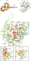
Fig. 4. RES-701-3 binding mode.
a Schematic illustration of the lasso peptide RES-701-3. b Cryo-EM map of RES-701-3. c Residues involved in RES-701-3 binding within 4.5 Å. Black dashed lines indicate hydrogen bonds. d, e Close-up views of W10 (d) and W3 (e). Bulky residues are shown as CPK models.
Fig. 5
Fig. 5. Structural comparison of the ETB receptors.
a–d Structural changes upon binding of ET-1 (a, PDB 8IY5), RES-701-3 (b), bosentan (c, PDB 5 × 93), and IRL2500 (d, PDB 6KIQ), focused on TM6. Apo state and compound-bound structures are colored gray and light-green, respectively. Black arrows indicate conformational changes upon drug binding.
Fig. 6
Fig. 6. Binding modes of cyclic peptides.
a Cavity for RES-701-3. The ETB receptor is shown as a molecular surface. b, c Cavities for MccJ25 in bacterial RNA polymerase (PBD 6N60) (b) and the siderophore receptor FhuA (PBD 4CU4) (c). d Crystal structures of the P. furiosus MATE transporter bound to the thioether-macrocyclic peptides MaD3S (PBD 3VVS) and MaL6 (PBD 3WBN).

References

    1. Maksimov, M. O., Pan, S. J. & James Link, A. Lasso peptides: structure, function, biosynthesis, and engineering. Nat. Prod. Rep.29, 996–1006 (2012). - PubMed
    1. Hegemann, J. D., Zimmermann, M., Xie, X. & Marahiel, M. A. Lasso peptides: an intriguing class of bacterial natural products. Acc. Chem. Res.48, 1909–1919 (2015). - PubMed
    1. Tietz, J. I. et al. A new genome-mining tool redefines the lasso peptide biosynthetic landscape. Nat. Chem. Biol.13, 470–478 (2017). - PMC - PubMed
    1. Cheng, C. & Hua, Z.-C. Lasso peptides: heterologous production and potential medical application. Front. Bioeng. Biotechnol.8, 571165 (2020). - PMC - PubMed
    1. Si, Y., Kretsch, A. M., Daigh, L. M., Burk, M. J. & Mitchell, D. A. Cell-free biosynthesis to evaluate lasso peptide formation and enzyme–substrate tolerance. J. Am. Chem. Soc.143, 5917–5927 (2021). - PMC - PubMed

LinkOut - more resources