. 2025 Apr 22;16(1):3755.
doi: 10.1038/s41467-025-59045-1.

Boosting hydrogel conductivity via water-dispersible conducting polymers for injectable bioelectronics

Affiliations

Boosting hydrogel conductivity via water-dispersible conducting polymers for injectable bioelectronics

Hossein Montazerian et al. Nat Commun. .

Erratum in

Abstract

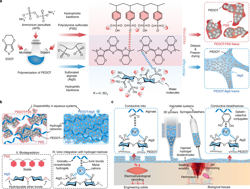
Bioelectronic devices hold transformative potential for healthcare diagnostics and therapeutics. Yet, traditional electronic implants often require invasive surgeries and are mechanically incompatible with biological tissues. Injectable hydrogel bioelectronics offer a minimally invasive alternative that interfaces with soft tissue seamlessly. A major challenge is the low conductivity of bioelectronic systems, stemming from poor dispersibility of conductive additives in hydrogel mixtures. We address this issue by engineering doping conditions with hydrophilic biomacromolecules, enhancing the dispersibility of conductive polymers in aqueous systems. This approach achieves a 5-fold increase in dispersibility and a 20-fold boost in conductivity compared to conventional methods. The resulting conductive polymers are molecularly and in vivo degradable, making them suitable for transient bioelectronics applications. These additives are compatible with various hydrogel systems, such as alginate, forming ionically cross-linkable conductive inks for 3D-printed wearable electronics toward high-performance physiological monitoring. Furthermore, integrating conductive fillers with gelatin-based bioadhesive hydrogels substantially enhances conductivity for injectable sealants, achieving 250% greater sensitivity in pH sensing for chronic wound monitoring. Our findings indicate that hydrophilic dopants effectively tailor conducting polymers for hydrogel fillers, enhancing their biodegradability and expanding applications in transient implantable biomonitoring.


Conflict of interest statement

Competing interests: The authors declare no competing interests.

Figures

Fig. 1
Fig. 1. Design and synthesis of water-dispersible poly(3,4-ethylenedioxythiophene) (PEDOT)-based conducting polymers using dopants with hydrophilic backbones.
a Doping of PEDOT via negatively charged macromolecules with hydrophilic and hydrophobic backbones, i.e., sulfonated alginate (AlgS), and poly(styrene sulfonate) (PSS), respectively. b Doping of PEDOT with AlgS as compared with PSS leads to enhanced dispersibility in aqueous solutions, improved molecular degradability, and high ionic integrability with ionically cross-linkable hydrogel matrices. c The PEDOT:AlgS polymers serve as high-concentration dispersible fillers in hydrogel pre-polymers for the development of injectable and 3D-printable hydrogel bioelectronics for wearable physiological recordings as well as wound closure and monitoring.
Fig. 2
Fig. 2. Characterization of freeze-dried sulfonated alginate-doped PEDOT polymers.
a Polymerization and doping of PEDOT. The AlgS samples are labeled as AlgSx where x represents the % w/v concentration of CSA used in sulfonation reaction. b UV-vis spectra of PEDOT:AlgS2 at different polymerization times. EDOT 3,4-ethylenedioxythiophene, APS ammonium persulfate. The inset shows the absorbance of PEDOT:AlgS reaction solutions at 800 nm before freeze-drying. Data in the inset is presented as mean ± standard deviation. c Effect of alginate sulfonation on the molecular weight distribution of freeze-dried PEDOT:AlgS2 tested via size-exclusion chromatography (SEC) (PEDOT polymerized for 1 d). d Freeze-dried PEDOT forming large aggregates when doped with PSS while doping with AlgS2 results in homogeneously distributed small nanoparticles. e Scanning electron microscopy (SEM) images of freeze-dried PEDOT:AlgS2 and PEDOT:PSS. Values represent the mean (n = 3 independent samples).
Fig. 3
Fig. 3. Aqueous re-dispersion properties of PEDOT:AlgS.
a Facilitated re-dispersion of PEDOT in aqueous solutions via hydrophilic AlgS dopants. b The number size distribution of PEDOT polymers obtained from dynamic light scattering (DLS) tests. The inset represents the average hydrodynamic sizes of PEDOT re-dispersions in water (n = 3 independent samples). c Results of zeta potential for aqueous PEDOT:AlgS (n = 3 independent samples). d Comparison of the colloidal stability of 2% w/v PEDOT:AlgS2 with PEDOT:PSS after 1 week at rest. e Dispersibility of freeze-dried PEDOT:AlgS2 in water at various sulfonation degrees and PEDOT:PSS. Tubes represent 10% w/v PEDOT:PSS in water forming self-associated gels and 20% w/v PEDOT:AlgS remaining in liquid phase. f Viscosity-shear rate profiles for 10% w/v solutions of PEDOT:AlgS2 and PEDOT:PSS in water. g Ionic crosslinking of PEDOT-based polymers with exposure to Fe3+ cations in terms of storage (Gʹ) and loss (Gʹʹ) modulus. h Hematoxylin and eosin (H&E) staining of intradermally injected solutions of PEDOT:AlgS2 and PEDOT:PSS (5% w/v) in vivo after 1-week implantation (n = 4 independent samples with similar results). i Hydrolysis-driven degradability assessment of PEDOT:PSS and PEDOT:AlgS2 tested via SEC. j Air dry coating of PEDOT doped with PSS and AlgS on I: glass substrate and their II: atomic force microscopy (AFM) phase plots obtained from PEDOT dispersions (5% w/v) (n = 3 independent samples with similar results).
Fig. 4
Fig. 4. 3D-printable conductive PEDOT:AlgS inks for wearable sensing.
a Formulation and crosslinking scheme of conductive inks comprised of alginate with PEDOT fillers doped with AlgS and PSS. b Dispersibility limit of PEDOT:AlgS and PEDOT:PSS in 3% w/v alginate solutions in water. c The relative increase in conductivity of alginate (7.1 × 10⁴ S m−1) as a result of introducing conductive PEDOT fillers at their dispersibility limit before crosslinking with Fe3+. The p-value is determined from a two-tailed Student’s t-test with unequal variances (n = 3 independent samples). d Effect of PEDOT dopants on the impedance spectroscopy characteristics of alginate inks. The inset shows Nyquist plots corresponding to the ink formulations. e Illustration of the multilayer patterns of PEDOT:AlgS in alginate after crosslinking in 25 mM FeCl3 solutions. f SEM images of the freeze-dried hydrogels based on the mixtures of PEDOT:PSS and PEDOT:AlgS (4 and 20% w/v, respectively) in alginate (n = 3 independent samples with similar results). g Temperature sensitivity of PEDOT-incorporated alginate hydrogels (n = 3 independent samples). h, i Results of electrocardiogram (ECG) and electromyography (EMG) recordings using conductive hydrogels as electrode interfaces, respectively. Values in (c) and (g) represent the mean and the standard deviation (n = 3 independent samples).
Fig. 5
Fig. 5. Injectable conductive bioadhesives for implantable bioelectronics applications.
a Formulation of bioadhesives involving gelatin-catechol (GelCA), synthesized via coupling caffeic acid to gelatin, as hydrogel bioadhesive matrices. PEDOT doped with PSS and AlgS are incorporated separately within GelCA at their dispersibility limits (4 and 20% w/v, respectively) and crosslinked ionically by Fe3+ for pH sensing in wound monitoring applications. b Hydrogel pre-polymer composites of GelCA (12% w/v) with PEDOT:PSS (4% w/v) and PEDOT:AlgS (20% w/v) after shaking on a vortex for 1 min. c Viscosity-shear rate of injectable bioadhesives. d Impedance spectroscopy of injectable bioadhesive pre-polymers. e Ex vivo porcine lung burst pressure adhesion testing of hydrogels (n = 3 independent samples). f Clotting time assays in terms of relative decrease in coagulation time for the assessment of hemostatic activity after hydrogel crosslinking. Clotting time for blank controls was 21.7 ± 0.6 min. Statistical analysis was performed via one-way ANOVA (n = 3 independent samples). g In vitro pH sensitivity of conductive bioadhesives obtained by chronoamperometric testing of hydrogels in various pH levels. The inset shows current variations with time at pH 7 (n = 3 independent samples). h In vivo monitoring of wound infection using conductive bioadhesive hydrogels. The data in (eh) represents the mean and the standard deviation (n = 3 independent samples).

Similar articles

References

    1. Shi, J. et al. Monolithic-to-focal evolving biointerfaces in tissue regeneration and bioelectronics. Nat. Chem. Eng.1, 73–86 (2024).
    1. Li, P., Shi, J. & Tian, B. An electronic pill for non-invasive gastric monitoring. Nat. Electron.7, 434–435 (2024).
    1. Xu, S., Xiao, X., Manshaii, F. & Chen, J. Injectable fluorescent neural interfaces for cell-specific stimulating and imaging. Nano Lett.24, 4703–4716 (2024). - PubMed
    1. Zhao, X. et al. Permanent fluidic magnets for liquid bioelectronics. Nat. Mater.23, 703–710 (2024). - PubMed
    1. Montgomery, M. et al. Flexible shape-memory scaffold for minimally invasive delivery of functional tissues. Nat. Mater.16, 1038–1046 (2017). - PubMed

LinkOut - more resources