. 2025 Apr 22;15(1):13868.
doi: 10.1038/s41598-025-88793-9.

Three-dimensional bioprinted in vitro glioma tumor constructs for synchrotron microbeam radiotherapy dosimetry and biological study using gelatin methacryloyl hydrogel

Affiliations

Three-dimensional bioprinted in vitro glioma tumor constructs for synchrotron microbeam radiotherapy dosimetry and biological study using gelatin methacryloyl hydrogel

John Paul O Bustillo et al. Sci Rep. .

Abstract

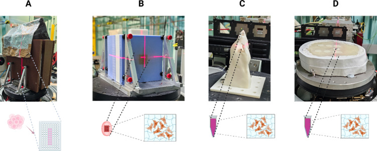
Synchrotron microbeam radiotherapy (MRT) is an innovative cancer treatment that uses micron-sized of ultra-high dose rate spatially fractionated X-rays to effectively control cancer growth while reducing the damage to surrounding healthy tissue. However, the current pre-clinical experiments are commonly limited with the use of conventional two-dimensional cell cultures which cannot accurately model in vivo tissue environment. This study aims to propose a three-dimensional (3D) bioprinting gelatin methacryloyl (GelMA) hydrogel protocol and to characterize 3D bioprinted glioma relative to cell monolayer and spheroid models for experimental MRT using 9L rat gliosarcoma and U87 human glioma. Synchrotron broad-beam (SBB) and MRT beams were delivered to all cell models using 5, 10, and 20 Gy. 3D bioprinting enables the creation of 3D cell models that mimic in vivo conditions using bioinks, biomaterials, and cells. Synchrotron dosimetry, Monte Carlo simulation, in vitro cell viability, and fluorescence microscopy were performed to understand the relationship of the radiation dosimetry with the radiobiological response of different cancer models. Encapsulated gliomas were placed inside 3D printed human and rat phantoms to mimic scattering conditions. Results showed that MRT kills more gliomas relative to SBB for all cell models. The 3D bioprinted culture detected the spatial clustering of dead cells due to MRT high peak doses as seen in fluorescence imaging. The result of this study progresses MRT research by integrating 3D bioprinting techniques in radiobiological experiments. The study's bioprinting protocol and results will help in reducing the use of animal experiments and possibly in clinical translation of MRT.

Keywords: 3D Printing; Biofabrication; Bioprinting; Brain Cancer; GelMA; Glioma; Microbeam Radiation Therapy; Spatial fractionation; Synchrotron Radiation.


Conflict of interest statement

Declarations. Competing interests: The authors declare no competing interests.

Figures

Fig. 1
Fig. 1
(A) Illustration of encapsulating glioma cells in GelMA hydrogel scaffolding and selected parameters that contribute to the printability of 3D bioprinted structure. Created in BioRender. Bustillo, J. (2025) https://BioRender.com/o77d581, (B-D) Pictures of the actual 3D bioprinted 8% w/v GelMA encapsulated glioma scaffolding wherein C shows its image under microscope (1 mm scale bar), (E) 3D REDI bioprinter with bioink setup inside a biosafety cabinet.
Fig. 2
Fig. 2
3D bioprinting protocol used in this study to create 3D in vitro models. (Created in BioRender. Bustillo, J. (2024) https://BioRender.com/b38r747)
Fig. 3
Fig. 3
Simplified illustration (not to scale) of the experimental setup at the Australian Synchrotron- Imaging and Medical Beamline with approximated distances relative to the SCMPW radiation source: (A) hutch 3B for the synchrotron computed tomography imaging in attenuation coefficient measurement, (B) hutch 2B in delivering synchrotron broad-beam (SBB) and microbeam radiation therapy (MRT), (C & D) Geant4 Monte Carlo simulated SBB and MRT beam relative dose profiles in arbitrary units (a.u.), respectively. Superconducting multipole wiggler (SCMPW), dual-crystal Laue monochromator (DCLM), beam defining aperture (BDA), removable multislit collimator (MSC), air ionization chamber (AIC).
Fig. 4
Fig. 4
(A) 3D printed head phantom in a CT simulation machine for CT number characterization, (B) 3D printed adult brain with cylindrical inserts, (C) Selected external layer of the 3D printed head phantom, (D) Actual MRI brain slice of a human head, (E) radiochromic film 2D SBB dose map inside the 3D printed head phantom showing the skull and brain, (F) CT image and profile of the 3D printed head phantom.
Fig. 5
Fig. 5
Different irradiation setup used to deliver synchrotron broad-beam and microbeam radiation beams to the in vitro samples: (A) Combination of RMI457 slabs and PLA + slab insert for 96-well plate (Lead sheet was used to localize the beam during column irradiation.), (B) 3D printed PLA + slab insert for cell culture dish together with Solid Water® HE, (C) 3D printed rat phantom, and (D) 3D printed adult anthropomorphic head phantom. Created in BioRender. Bustillo, J. (2025) https://BioRender.com/f96g579.
Fig. 6
Fig. 6
(A) Experimental monochromatic attenuation coefficient based on synchrotron CT imaging, and (B) CT number measurement of hydrogel samples using Siemens Inveon CT machine. Error bars represent the standard deviation. (n = 5 repeats).
Fig. 7
Fig. 7
(A) Geant4 Monte Carlo calculated microbeam dose profile at 20 mm depth, and (B) Two-dimensional percent depth dose in a Solid Water HE Phantom. Peak and valley doses were verified experimentally for all the phantoms used.
Fig. 8
Fig. 8
U87 (A, C, E) and 9L (B, D, F) cell line normalized cell viability for both synchrotron broad-beam (SBB) and microbeam radiation therapy (MRT) delivered to 2D monolayer cell model (blue) and 3D GelMA cell model (orange). Cell viability assay was done 72 h. after irradiation. Four replicates were done per treatment dose. Error bars represent standard error of the mean and asterisk * represents p < 0.05. Note that the percentages were normalized wherein 1 is equivalent to 100% cell viability.
Fig. 9
Fig. 9
Propidium iodide average fluorescence intensity ± standard deviation of the mean (in arbitrary units, a.u.) normalized to the control (0 Gy) for 2D monolayer, 3D spheroid, and 3D GelMA encapsulated cells measured three days after treatment delivery. (n = 5).
Fig. 10
Fig. 10
(A) Representative brightfield images of spheroids for different doses of synchrotron broad-beam (SBB) and microbeam radiation therapy (MRT) two days after treatment delivery, (B and C) Line graphs showing the average spheroid diameter ± standard deviation of the mean for different time periods (n = 6 for each experiment). Note that the down arrow shown in figures B & C refers to the time of radiation treatment beam delivery.
Fig. 11
Fig. 11
3D bioprinted lattice structure for MRT experiments: (A) Brightfield and PI fluorescence imaging of the U87 bioprinted lattice detecting damaged cells due to high peak doses of MRT beam, (B & C) Red channel of the PI image and imaging profile showing two gray value peaks due to MRT peak doses, and (D) Comparison of three cancer models used in this study. Created in BioRender. Bustillo, J. (2025) https://BioRender.com/l60k787.

References

    1. Yalamarty, S. S. K. et al. Mechanisms of Resistance and Current Treatment Options for Glioblastoma Multiforme (GBM), Apr. 01, MDPI. (2023). 10.3390/cancers15072116 - PMC - PubMed
    1. Lee, J. H. & Wee, C. W. Treatment of adult gliomas: a current update. Brain Neurorehabilitation. 15 (3). 10.12786/bn.2022.15.e24 (2022). - PMC - PubMed
    1. Hau, E. et al. The evolving roles and controversies of radiotherapy in the treatment of glioblastoma, Jun. 01, John Wiley and Sons Ltd. (2016). 10.1002/jmrs.149 - PMC - PubMed
    1. Grigorieva, E. V. Radiation effects on Brain Extracellular Matrix. Oct. 02 2020 Front. Media S A10.3389/fonc.2020.576701 - PMC - PubMed
    1. Tsai, P. F. et al. Hippocampal dosimetry correlates with the change in neurocognitive function after hippocampal sparing during whole brain radiotherapy: a prospective study. Radiat. Oncol.10 (1). 10.1186/s13014-015-0562-x (Dec. 2015). - PMC - PubMed

LinkOut - more resources