. 2025 Aug;32(8):1383-1395.
doi: 10.1038/s41594-025-01543-3. Epub 2025 Apr 22.

The mechanochemical cycle of reactive full-length human dynein 1

Affiliations

The mechanochemical cycle of reactive full-length human dynein 1

Pengxin Chai et al. Nat Struct Mol Biol. 2025 Aug.

Abstract

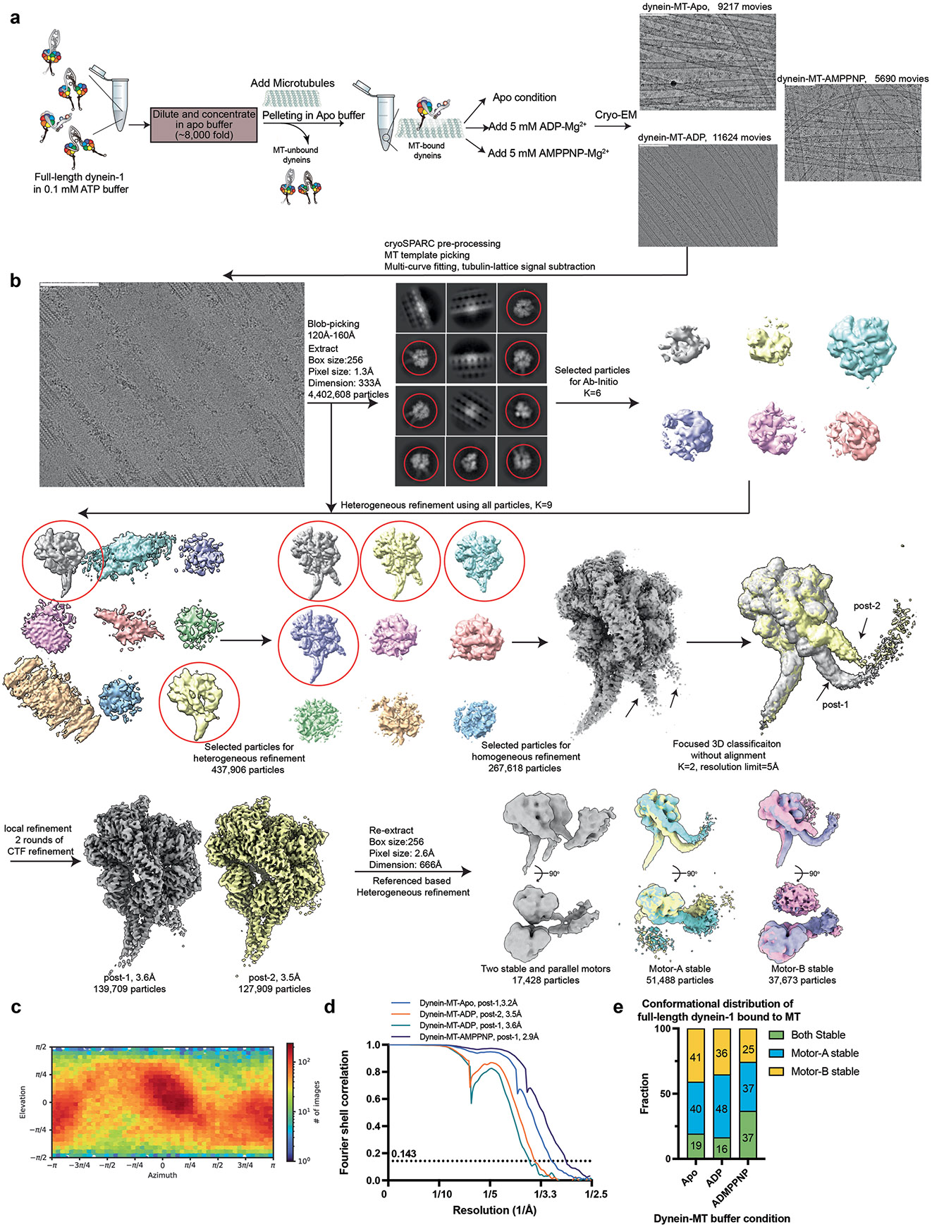
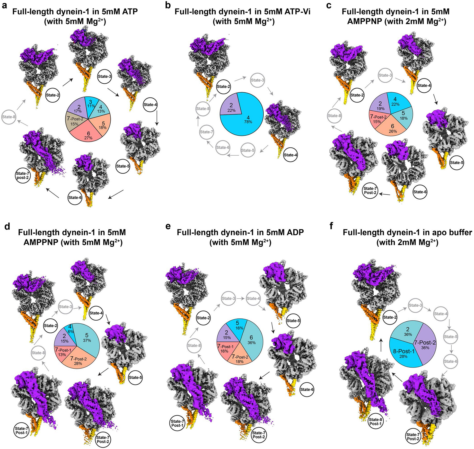
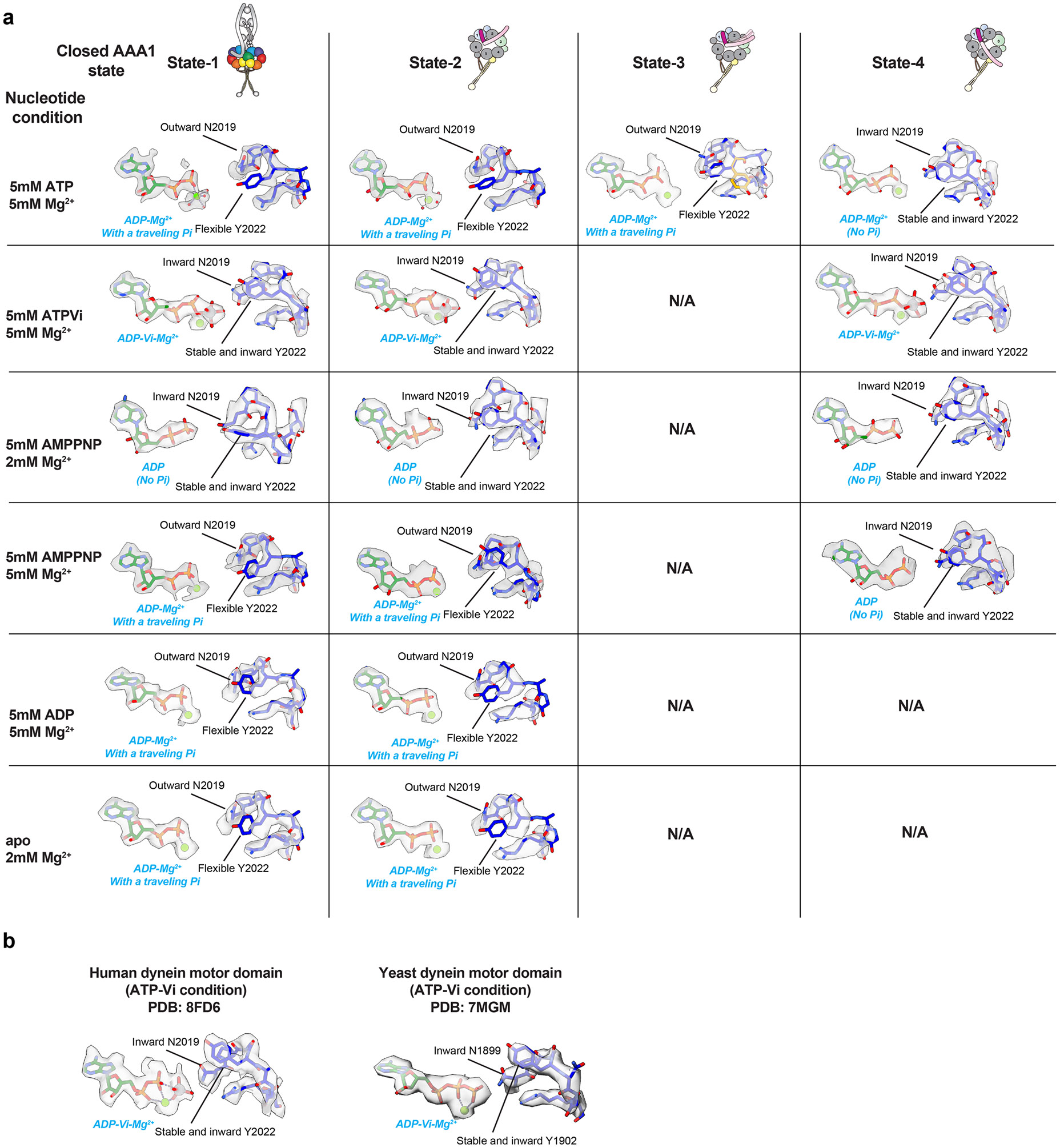
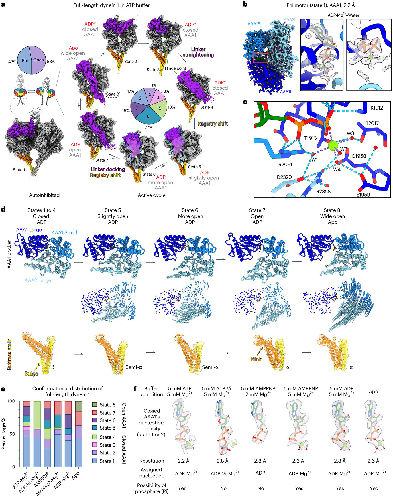
Dynein-driven cargo transport has a pivotal role in diverse cellular activities, central to which is dynein's mechanochemical cycle. Here, we performed a systematic cryo-electron microscopic investigation of the conformational landscape of full-length human dynein 1 in reaction, in various nucleotide conditions, on and off microtubules. Our approach reveals over 40 high-resolution structures, categorized into eight states, providing a dynamic and comprehensive view of dynein throughout its mechanochemical cycle. The described intermediate states reveal mechanistic insights into dynein function, including a 'backdoor' phosphate release model that coordinates linker straightening, how microtubule binding enhances adenosine triphosphatase activity through a two-way communication mechanism and the crosstalk mechanism between AAA1 and the regulatory AAA3 site. Our findings also lead to a revised model for the force-generating powerstroke and reveal means by which dynein exhibits unidirectional stepping. These results improve our understanding of dynein and provide a more complete model of its mechanochemical cycle.


Conflict of interest statement

Competing interests: The authors declare no competing interests.

Figures

Extended Data Fig. 1 ∣
Extended Data Fig. 1 ∣. Cryo-EM data processing of full-length human dynein-1 in the presence of 5 mM ATP.
(a) A representative negative staining micrograph (total 100 micrographs) of purified full-length human dynein-1 and corresponding 2D class averages. (b) A typical cryo-EM micrograph (total 33,302 micrographs) and the flowchart of cryo-EM image processing. (c) Image processing of full-length phi dynein and tail region. (d, e) Orientation distribution of phi dynein and a representative state from open dynein. (f, g) The Fourier shell correlation (FSC) curves of the motor domain in different states and the tail domain. Datasets for dynein in other nucleotide conditions (ATP-Vi, AMPPNP, ADP, apo) were processed similarly.
Extended Data Fig. 2 ∣
Extended Data Fig. 2 ∣. Local resolution analysis and identification of bound nucleotides in AAA1, AAA3, and AAA4 in different states from the dynein-ATP dataset.
The color scheme for the motor domain is consistent with Fig. 4.
Extended Data Fig. 3 ∣
Extended Data Fig. 3 ∣. Cryo-EM data processing of full-length human dynein-1 bound to MTs.
(a) Flowchart of sample preparation and typical cryo-EM micrographs of dynein bound to MTs in apo, ADP, and AMPPNP conditions. The number of micrographs is shown in the panel. (b) Microtubule signal subtraction and image processing flowchart. (c) Orientation distribution of dynein-MT-ADP reconstruction. (d) FSC curves of four MT-bound motors. (e) Plot for the full-length dynein bound to MTs in different conformations (two stable heads, one stable trailing head, and one stable leading head).
Extended Data Fig. 4 ∣
Extended Data Fig. 4 ∣. Cryo-EM analysis of dynein in ATP-Vi and AMPPNP conditions.
(a) Cartoon schematic depicting the experimental method for cryo-EM analysis of full-length dynein in different nucleotide conditions. (b) Typical cryo-EM micrographs of dynein in different nucleotide conditions, with total number of micrographs shown in the panel. (c, d, e) FSC curves and local resolution analysis of dynein in ATP-Vi, AMPPNP-low Mg2+, and AMPPNP-high Mg2+ conditions.
Extended Data Fig. 5 ∣
Extended Data Fig. 5 ∣. Cryo-EM analysis of dynein in ADP and apo conditions and dynein-bound to microtubules.
(a, b) FSC curves and local resolution analysis of dynein in ADP and apo conditions. (c) FSC curves and local resolution analysis of dynein-bound to microtubules.
Extended Data Fig. 6 ∣
Extended Data Fig. 6 ∣. The conformational landscapes of active cycle motor domains in different nucleotide conditions.
(a-f) Conformation landscapes of motor domains in ATP, ATP-Vi, AMPPNP, ADP, and apo conditions. Arrows indicate mechanochemical pathways. Percentages indicate the proportion of particles in each state. The missing states in each condition are shown in grey. Phi dynein is not included in this figure.
Extended Data Fig. 7 ∣
Extended Data Fig. 7 ∣. Cryo-EM densities of the nucleotide and sensor-I loop in various closed AAA1 states.
(a) From this work. (b) From previously published structures (PDB:8FD6 ref. , 7MGM ref. 78). The conformations of N2019 and Y2022 are highlighted.
Extended Data Fig. 8 ∣
Extended Data Fig. 8 ∣. Structural comparisons of MT-bound and -unbound dynein motors.
(a) Comparison between MT-dynein in apo condition with state-8 motor domain. (b) Comparison between MT-dynein in ADP condition with state-7 motor domains. (c) Comparison between MT-dynein in AMPPNP condition with state-7 motor domains. The nucleotide states in AAA1, AAA3, and AAA4 and the linker docking mode are listed. The MT-bound dynein motor domains (from linker to C-terminus) were used as references for structural fitting in ChimeraX.
Extended Data Fig. 9 ∣
Extended Data Fig. 9 ∣. Systematic analysis of linker-ring interactions among four MT-bound motor domains.
(a) Cartoon and molecular models showing the interaction interfaces including linker-AAA2L, linker-AAA5L, and linker-AAA3-4-connector helix. (b-e) Comparison of linker docking modes between dynein-1 and outer-arm dynein.
Extended Data Fig. 10 ∣
Extended Data Fig. 10 ∣. Structural survey of motor domains and statistical analysis of crosstalk between AAA1 and AAA3.
(a) Flowchart of structural survey of 56 motor domain structures. (b) Statistical graphs showing how AAA1 communicates to AAA3, linker and MTBD. (c) Statistical graphs showing how AAA3 communicates to AAA1, linker and MTBD. (d) Cartoon model summarizing communication between AAA1, AAA3, linker and MTBD.
Fig. 1 ∣
Fig. 1 ∣. Cryo-EM analysis of full-length human dynein 1 off and on MTs.
a,b, Schematic of cryo-EM pipeline for determination of dynein structures in reaction with ATP without MTs (a) or with MTs in different nucleotide conditions (b). c, Cryo-EM reconstruction of phi dynein with the 3.8–7.0-Å tail region and 2.2-Å motor domain. d, Representative cryo-EM reconstructions of the motor domain in different states from open dynein. The motor ring, linker, buttress and stalk are colored gray, purple, orange and yellow, respectively. e, Cryo-EM map of full-length two-headed dynein bound to MTs in ADP conditions at 10-Å resolution, with locally refined motor domains at 2.9–3.6-Å resolution. f, High-resolution features are shown by the representative density of amino acid residues and ligands.
Fig. 2 ∣
Fig. 2 ∣. The conformational landscape of MT-unbound dynein motor domains in reaction.
a, Eight major states of the dynein motor domain during the catalytic cycle divided into autoinhibited states (left) and states in the active cycle (right). Arrows indicate the proposed catalytic pathway supported by the literature and apparent AAA+ ring conformations. Small arrows show the possibility of reverse pathways. Percentages indicate the proportion of particles in each state. The apo AAA1 pocket state (state 8) is present at a very low proportion in the presence of ATP but is highly represented in apo conditions (the structure obtained in the apo condition is shown). The linkers from states 4 and 6 are low-pass-filtered to 12 Å and shown at low contours. Only one representative subclass in each state is shown. The color scheme is consistent with Fig. 1d. b, Cryo-EM map of AAA1 pocket from 2.2-Å phi motors. AAA1L, AAA1S and AAA2L are colored blue, light blue and sky blue, respectively. Right, carved transparent gray density is displayed for ADP (green), Mg2+ (light green) and water molecules (violet). c, Residue side chains involved in hydrogen-bonding networks (dashed lines) at the AAA1 active site are displayed and colored by heteroatom. d, Cartoon models showing the AAA1 dynamics and the coupled buttress stalk conformational states. The isolated AAA1 pockets from different states are aligned on AAA1L. Vector maps depicting pairwise Cα interatomic distances between the AAA1 domains of adjacent states depict the 3D trajectories during the pocket-opening process. The buttress stalk regions are highlighted to show how the stalk-CC registries are coupled to motor ring states. e, Plot depicting the proportions of the various motor domain conformations from full-length dynein 1 in different nucleotide conditions. f, EM densities for nucleotides bound to the closed AAA1 pockets in indicated nucleotide conditions.
Fig. 3 ∣
Fig. 3 ∣. The release of Pi from AAA1 through a molecular backdoor mechanism.
a, Comparison of molecular models of closed AAA1 pockets in different states and nucleotide conditions. The sensor I loop exhibits conformational heterogeneity when dynein is in the 5 mM ATP condition. b, Cryo-EM densities of sensor I loop in closed and open AAA1 pockets. c, Structural comparison of the bound nucleotide and the sensor I loop in closed AAA1 states. Transparent cryo-EM densities of the sensor I loop are displayed in isosurface mode. The conformations of two residues, N2019 and Y2022, are highlighted. d, A slice through the structures of the AAA1 pocket near the nucleotide-binding site, shown in surface representation. e, Backdoor model for Pi release from dynein AAA1 controlled by N2019 and Y2022. f, High-resolution 3D classification of the active site in the phi motor, showing the potential Pi group in various states of escape. g, Conservation analysis of the sensor I loop. h, Plot (mean ± s.d., along with individual data points) depicting relative ATPase rates (in the absence or presence of 2 μM MTs) for artificially dimerized yeast dynein motor domain fragments (from left to right, n = 6/5, 6/4 and 6/6 independent replicates from assays minus/plus MTs). Y1902 in yeast dynein corresponds to Y2022 in human dynein. i,j, Representative kymographs (i) and plots (j; mean ± s.d., along with individual data points) depicting indicated motility parameters determined from a single-molecule motility assay (from left to right, n = 508/597, n = 148/880 and n = 679/837 processive/nonprocessive dynein molecules from three independent experiments). MT polarity was determined with a wild-type dynein fragment (Methods). P values in h were calculated using an ordinary one-way analysis of variance (ANOVA) followed by a Tukey’s multiple-comparison test. P values in j were calculated using a Kruskal–Wallis ANOVA followed by a Dunn’s multiple-comparison test (for run length) or an ANOVA followed by a Dunnett’s multiple-comparison test (for velocity). k, Summary of the updated nucleotide state among different closed AAA1 conformations. Note that the presence of a traveling Pi is based on the sensor I loop conformations. N/A, not applicable.
Fig. 4 ∣
Fig. 4 ∣. The mechanism for two-way communication between AAA1 and the MTBD.
a, Cartoon models illustrating the overall motor ring conformations from different states, each with varying degrees of the AAA1 pocket opening. The linker domain is hidden for better visibility of the motor ring states. b, Adjacent models were aligned using the entire motor domain and then vector maps were generated that depict pairwise Cα interatomic distances (for every third residue) between the AAA domains of adjacent states. The length of the lines is proportional to the calculated interatomic distances. The major movements and directions are highlighted by dashed areas and arrows. c, The cartoon model illustrates the signal propagation directions from AAA1 to the stalk-CC in two distinct phases. d, Changes in dynein–MT-binding affinity (y axis), as assessed by buttress stalk conformations, as a consequence of AAA1 pocket conformations in the different states (x axis). e, Cartoon model illustrating the communication from the MTBD to AAA1 through the stalk-CC. f, Cartoon model depicting the design of the α registry-locked dynein motor domain mutant. g, Cryo-EM reconstructions of the α registry mutant in ATP or ATP-Vi conditions. In both conditions, the motor domains exhibit only open AAA1 pockets. As expected, the buttress stalk conformation is consistent with a high MT-binding affinity.
Fig. 5 ∣
Fig. 5 ∣. The mechanism for ATPase stimulation by MTs and dynein’s unidirectional stepping along MTs.
a, High-resolution cryo-EM maps of MT-bound dynein motor domains in different nucleotide conditions. b,c, Cartoon model depicts the mechanism of dynein mechanochemical cycle acceleration by MTs. MT binding rapidly induces the stalk-CC registry from the semi-α to α registry, thereby forcing the AAA1 pocket to adopt an open state (b). MT binding, by stabilizing the stalk-CC registry, allows the AAA1 pocket to adopt a wide open state, which promotes ADP release. Pie graphs show the distribution of dynein in the apo condition with or without MTs (c). d, Dynein adopting the post 2 linker docking mode intrinsically favors a forward step according to previous OAD–MT structures (PDB 7K58 and 7K5B (ref. 54)). Aligning dyneins by their N termini (to the right of the models in the picture) show that the post 2 dynein motor for both OAD and dynein 1 adopts a forward-stepping pattern with respect to the post 1 dyneins. e, Proposed mechanism for dynein unidirectional stepping along MTs. (i) Dynein is bound to MTs in the apo state in post 1 mode (after completing a step). (ii) Upon ATP binding and/or hydrolysis, dynein unbinds MTs with a bent linker. (iii) Following Pi release from AAA1, dynein with a straight but undocked linker (states 4–6) has low to intermediate MT-binding affinity and is searching for a new tubulin-binding site. (iv-b) The preferred post 2 mode in ATP conditions (denoted by a star) creates a net bias toward the minus end of MTs and allows the MTBD to take an 8-nm forward step. Note that adopting a post 1 mode upon MT rebinding (iv-a) leads to a 0-nm step. The cartoon model from state 4 (from (iii)) is shown in gray in (iv-a) and (iv-b) for comparison. (v) Switching from a post 2 to a post 1 mode and ADP release leads to an 8-nm forward movement of the entire dynein complex.
Fig. 6 ∣
Fig. 6 ∣. Model for the mechanochemical cycle of full-length human dynein 1.
The motor domains in phi dynein adopt an autoinhibited state and are capable of ATP hydrolysis and Pi release but not ADP release. (1) The mechanochemical cycle of the active motor states starts with the apo AAA1 state. (2) ATP binding and/or hydrolysis induces pocket closure and triggers MT release. The linker changes from a straight, stably docked configuration to a bent, stably docked conformation. (3) Release of Pi through the backdoor channel triggers the straightening of the linker. (4) A semi-α registry of the stalk-CC helps dynein for MT rebinding. (5) Linker docked at post 2 site provides a net bias toward the minus end of MTs and dynein rebinds to MTs. (6a,6b) The linker docking mode changes from post 2 to post 1 and ADP is released, thus completing the cycle.

References

    1. King SM The dynein microtubule motor. Biochim. Biophys. Acta 1496, 60–75 (2000). - PubMed
    1. Reck-Peterson SL, Redwine WB, Vale RD & Carter AP The cytoplasmic dynein transport machinery and its many cargoes. Nat. Rev. Mol. Cell Biol 19, 382–398 (2018). - PMC - PubMed
    1. Torisawa T. & Kimura A. The generation of dynein networks by multi-layered regulation and their implication in cell division. Front Cell Dev. Biol 8, 22 (2020). - PMC - PubMed
    1. Ricolo D, Castro-Ribera J. & Araujo SJ Cytoskeletal players in single-cell branching morphogenesis. Dev. Biol 477, 22–34 (2021). - PubMed
    1. Klena N. & Pigino G.Structural biology of cilia and intraflagellar transport. Annu. Rev. Cell Dev. Biol 38, 103–123 (2022). - PubMed

LinkOut - more resources