. 2025 Apr 22;15(1):13883.
doi: 10.1038/s41598-025-98974-1.

Artificial intelligence real-time automated recognition of the gastric antrum cross-sectional area and motility rhythm via bedside ultrasound: a pilot study

Affiliations

Artificial intelligence real-time automated recognition of the gastric antrum cross-sectional area and motility rhythm via bedside ultrasound: a pilot study

Tongjuan Zou et al. Sci Rep. .

Abstract

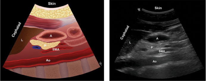
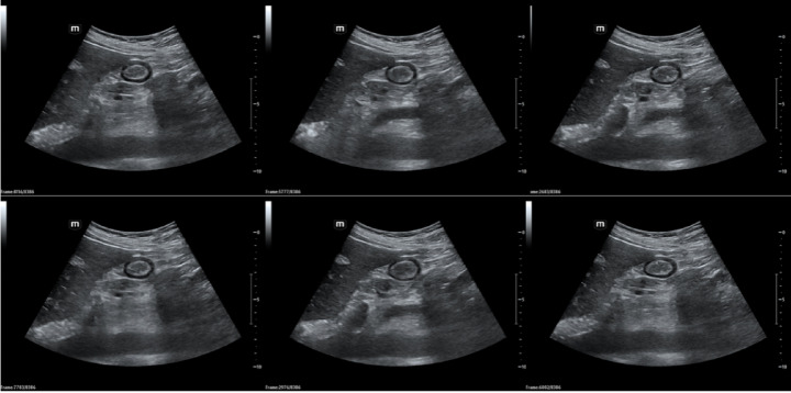
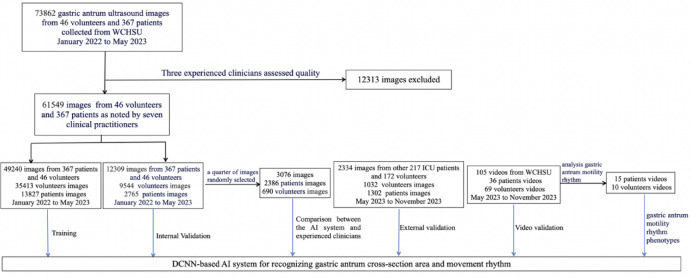
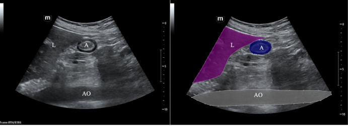
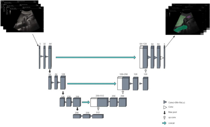
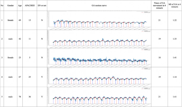
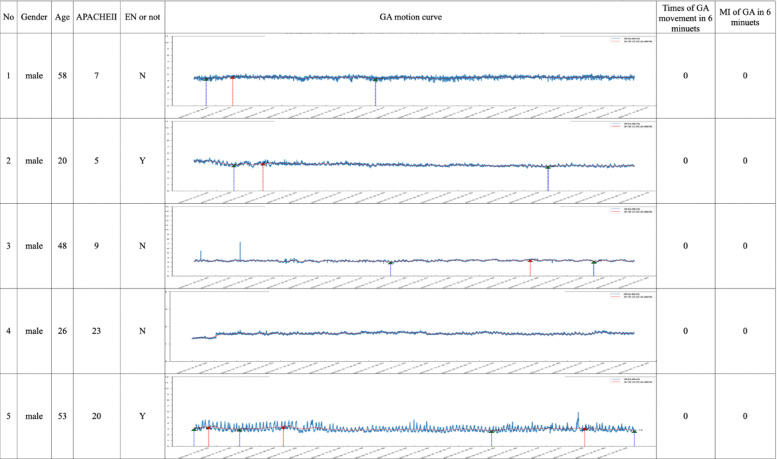
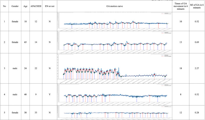
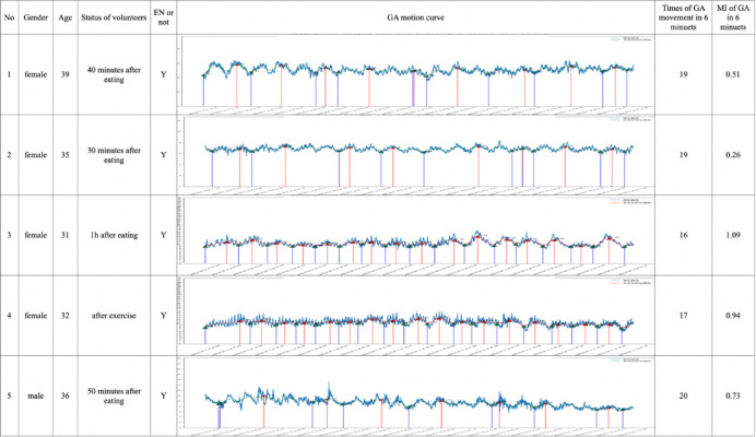
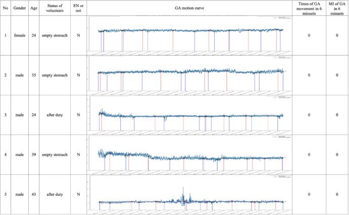
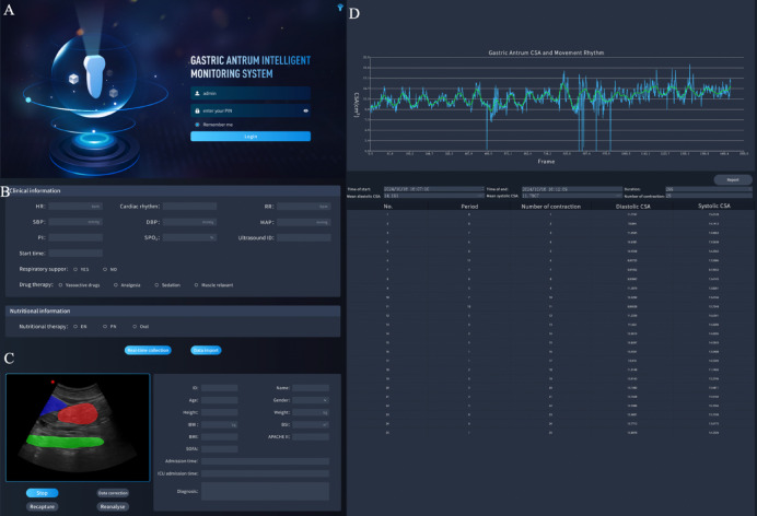
The cross-sectional area (CSA) of the gastric antrum and its motility rhythm reflects the gastrointestinal function of critically ill patients. Monitoring the CSA and motility rhythm is crucial but remains time-consuming and operator dependent. This study aimed to develop an artificial intelligence (AI) system for real-time automated recognition of the gastric antrum CSA and motility rhythm using bedside ultrasound. Gastric antrum ultrasound videos were prospectively collected from West China Hospital to establish training and validation datasets. The AI system's predictions were validated against senior clinicians' annotations to assess accuracy. Additionally, videos were collected to evaluate the performance of the AI system. The antrum motility rhythms of patients and volunteers were preliminarily classified to lay the foundation for the subsequent establishment of gastrointestinal motility rhythm phenotypes in critically ill patients. A total of 907 videos (620 patients and 287 volunteers) were included to develop and validate the AI system from January 2022 to November 2023. 49,240 images were used as training datasets to train the model's ability to locate and segment gastric antrum ultrasound images. The remaining 12,309 images were used as the internal validation dataset, achieving a mean dice coefficient (mDice) of 87.36% and an mean intersection over union (mIOU) of 77.56%. For the external validation dataset, 2334 images were used, resulting in mDice and mIOU values of 86.82% and 76.26%, respectively. Moreover, the AI system demonstrated robust performance in video cut frame analysis, achieving a mDice of 90.23% and a mIOU of 85.16% across 105 videos. The intraclass correlation coefficient (ICC) between human operators and the AI model was good (ICC (2, K): 0.813, 95% CI 0.728-0.871). In terms of antrum motility rhythm phenotypes, we identified several distinct patterns, such as regular movement, minimal movement, and irregular movement, reflecting different statuses, such as fasting, postmeal, postexercise, and postduty. We developed an AI system that is comparable to experienced clinicians in identifying the gastric antrum and measuring its CSA. Furthermore, the system can generate a curve representing the rhythm of antrum movement, reflecting the varying statuses of patients and volunteers. This system may optimize enteral nutrition (EN) protocols by reducing clinicians' workload and minimizing operator dependence.

Keywords: Artificial intelligence; Bedside ultrasound; Gastric antrum; Motility rhythm.


Conflict of interest statement

Declarations. Competing interests: The authors declare no competing interests. Consent for publication: All co-authors have provided consent for publications.

Figures

Fig. 1
Fig. 1
(A) Anatomical map of the gastric antrum; (B) Ultrasound image of the antrum. The antrum is located between the left lobe of the liver (anteriorly) and the pancreas (posteriorly) at the level of the aorta. A gastric antrum, L liver, P pancreas, SMA superior mesenteric artery, Ao abdominal aorta.
Fig. 2
Fig. 2
The gastric antrum video data were collected by professional medical staff and extracted via interval frames. After strict quality control, a total of 63,883 images were obtained.
Fig. 3
Fig. 3
Flowchart of training and validation of the DCNN-based AI system. DCNN deep convolutional neural network, AI artificial intelligence.
Fig. 4
Fig. 4
Example diagram of gastric antrum labeling under the guidance of a professional doctor. Unlabeled ultrasound images are shown on the left and labeled ultrasound images are shown on the right. A gastric antrum, L liver, Ao abdominal aorta.
Fig. 5
Fig. 5
The training process of the gastric antrum segmentation model is as follows: the classical 2D U-Net structure first obtains low-resolution features representing global information through multiple downsampling layers and then generates high-resolution features through upsampling. By using lateral connections, features of different scales, which have been downsampled multiple times, are reintegrated into the input of the upsampling layer in a residual manner to retain more detailed feature information.
Fig. 6
Fig. 6
The gastric antrum motion curve of patients in 6 min (rhythmic movement). Rhythmic movement: the antrum contraction and diastole rhythm were regular, and the antrum movement curve exhibited a sinusoidal waveform. The blue curve is the original curve, and the red curve is the fit curve. EN enteral nutrition, MI motility index, GA gastric antrum.
Fig. 7
Fig. 7
The gastric antrum motion curve of patients in 6 min (Antral hypomotility). Antral hypomotility: there was almost no change in the antrum area, and the antrum movement curve was straight. (Respiratory motion artifacts contributed to minor antrum area fluctuations in the curve). The blue curve is the original curve, and the red curve is the fit curve. EN enteral nutrition, MI motility index.
Fig. 8
Fig. 8
The gastric antrum motion curve of patients (Nonrhythmic movement). Nonrhythmic movement: the area of gastric antrum changed irregularly, and the movement curve of gastric antrum alternated between straight line and sine wave. The blue curve is the original curve, and the red curve is the fit curve. EN enteral nutrition, MI motility index, GA gastric antrum.
Fig. 9
Fig. 9
The gastric antrum motion curve of volunteers in 6 min (Rhythmic movement). Rhythmic movement: the antrum contraction and diastole rhythm were regular, and the antrum movement curve was sine wave. The blue curve is the original curve, and the red curve is the fit curve. EN enteral nutrition, MI motility index, GA gastric antrum.
Fig. 10
Fig. 10
The gastric antrum motion curve of volunteers (Antral hypomotility). Antral hypomotility: there was almost no change in the antrum area, and the antrum movement curve was straight. The curve in the figure was due to the change in the antrum area caused by respiratory movement). The blue curve is the original curve, and the red curve is the fit curve. EN enteral nutrition, GA gastric antrum.
Fig. 11
Fig. 11
Gastric antrum intelligent monitoring system. (A) Login screen; (B) clinical information entry interface, and there are two ways to collect ultrasound images in real time or import previous ultrasound images; (C) identification of the gastric antrum (red), liver (blue) and AO (green); (D) gastric antrum motion curve and the CSA of the gastric antrum. MI motility index, CSA cross-sectional area, AO abdominal aorta.

Similar articles

Cited by

References

    1. Peng, F. et al. Best evidence summary for prevention and management of enteral feeding intolerance in critically ill patients. J. Clin. Nurs.33(3), 781–796 (2024). - PubMed
    1. Blaser, A. R., Starkopf, J., Kirsimägi, Ü. & Deane, A. M. Definition, prevalence, and outcome of feeding intolerance in intensive care: A systematic review and meta-analysis. Acta Anaesthesiol. Scand.58(8), 914–922 (2014). - PubMed
    1. Liu, R. et al. Feeding intolerance in critically ill patients with COVID-19. Clin. Nutr.41(12), 3069–3076 (2022). - PMC - PubMed
    1. Chen, X., Chen, F., Zhao, Q., Zhang, L. & Liu, Z. Ultrasonographic measurement of antral area for estimating gastric fluid volume in pregnant women. J. Clin. Anesth.53, 70–73 (2019). - PubMed
    1. Kaydu, A. & Gokcek, E. Preoperative assessment of ultrasonographic measurement of antral area for gastric content. Med. Sci. Monit.24, 5542–5548 (2018). - PMC - PubMed

LinkOut - more resources