Case Reports
. 2025 Jul;31(7):2174-2178.
doi: 10.1038/s41591-025-03656-0. Epub 2025 Apr 22.

Antisense oligonucleotide treatment in a preterm infant with early-onset SCN2A developmental and epileptic encephalopathy

Affiliations
Case Reports

Antisense oligonucleotide treatment in a preterm infant with early-onset SCN2A developmental and epileptic encephalopathy

Matias Wagner et al. Nat Med. 2025 Jul.

Abstract

Early-onset SCN2A developmental and epileptic encephalopathy is caused by SCN2A gain-of-function variants. Here we describe the clinical experience with intrathecally administered elsunersen, a gapmer antisense oligonucleotide targeting SCN2A, in a female preterm infant with early-onset SCN2A developmental and epileptic encephalopathy, in an expanded access program. Before elsunersen treatement, the patient was in status epilepticus for 7 weeks with a seizure frequency of 20-25 per hour. Voltage-clamp experiments confirmed impaired channel inactivation and increased persistent current consistent with a gain-of-function mechanism. Elsunersen treatment demonstrated a favorable safety profile with no severe or serious adverse events reported after 19 intrathecal administrations over 20 months. After administration in combination with sodium channel blockers, status epilepticus was interrupted intermittently and ultimately ceased after continued dosing. A >60% reduction in seizure frequency corresponding to five to seven seizures per hour was observed, which has been sustained during follow-up until the age of 22 months. These data provide preliminary insights on the safety and efficacy of elsunersen in a preterm infant. Additional investigation on the benefits of elsunersen in clinical trials is warranted.


Conflict of interest statement

Competing interests: Elsunersen was provided by Praxis Precision Medicines within an expanded access program with authors being employees of Praxis Precision Medicines (S.F., R.H., H.J., W.M., B.S., S.P. and M.So.). The other authors declare no competing interests.

Figures

Fig. 1
Fig. 1. Location and functional impact of the A1329D Nav1.2 channel mutation.
a, Side view of the 3D structure of Nav1.2 highlighting the A1329 residue (arrow) residing in the α-helical intracellular linker between transmembrane segments S4 and S5 of domain III (S4–5DIII) (dashed boxed area). b, Zoomed-in views highlighting the amino acid residues before (left) and after (right) in silico mutagenesis. All residues shown in stick representation are within 5 Å of the A1329 or D1329 residues. In the mutant channel, D1329 forms polar interactions with Q1479 and F1489. The D1329–F1489 interaction is likely to affect the binding of the I1488–F1489–M1490 (IFM) inactivation motif to its receptor site, resulting in delayed inactivation and persistent current. c, Representative action potential firing of the axon initial segment hybrid compartment model incorporating WT or A1329D Nav1.2 channel variant in response to stepwise increase in the stimulus current amplitude (6, 8, 10, 14 or 18 pA) in DAPC experiments. The dashed lines indicate the 0 mV level. Action potentials (bottom) on an expanded timescale (upward deflections), corresponding to action potentials enclosed in dashed boxes elicited by 10 pA stimulus current (top), and associated scaled input sodium currents (downward deflections). Note the apparent increase of the action potential width in the presence of A1329D and the associated increase of the inward sodium current component (arrows) compared with WT. The dashed lines indicate the 0 mV and 0 pA levels, respectively. In all experiments, the virtual sodium conductance of the axon initial segment (AIS) model was set to zero (gNav1.6 = 0), whereas the virtual Kv channel conductance was set to 2 (twice the original gKv). In all DAPC experiments, the original (nonscaled) external sodium current INav1.2, the scaled external INav1.2, the membrane potential (Vm), and gKv were simultaneously recorded. d, Input–output relationships for WT and A1329D variants. Data are represented as mean ± s.e.m.; n, the number of experiments between parentheses. The asterisks indicate statistically significant differences in the presence of A1329D relative to WT (*P < 0.05, two-way ANOVA, followed by Dunnett’s post-hoc test; see individual P values in Supplementary Table 3).
Fig. 2
Fig. 2. The patient clinical course after introduction of the elsunersen treatment regimen and effects on seizures.
a, The antiseizure medication (ASM) regimen including high-dose sodium channel blockers and introduction of the elsunersen dosing regimen. b, The associated reduction in seizure frequency. A total of seven elsunersen (intrathecal) doses were administered between 13 March 2023 and 29 September 2023 (30.5 mg total). PB, phenobarbital; PHT, phenytoin; LEV, levetiracetam; LCM, lacosamide; OXC, oxcarbazepine; MDZ, midazolam; CBZ, carbamazepine; LDC, lidocaine; LTG, lamotrigine; ASO, antisense oligonucleotide, here elsunersen; CLB, clobazam; GBP, gabapentin; DZP, diazepam; ESL, eslicarbazepine. Corresponding aEEG traces are displayed in Supplementary Fig. 1a–g.
Extended Data Fig. 1
Extended Data Fig. 1. Prenatal fetal MRI showing arthrogryposis and large basal ganglia.
A and B Prenatal fetal MRI at 29 + 2 weeks of gestational age shows prominent basal ganglia but otherwise normal gyration and myelination. C and D Imaging of the extremities shows hyperextended wrists and fisting with crossing of fingers.
Extended Data Fig. 2
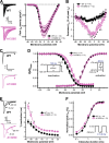
Extended Data Fig. 2. Biophysical properties of the A1329D Nav1.2 channel mutation as determined by voltage clamp experiments.
A Peak sodium current (INa) density-voltage relationships. Currents were elicited from a holding potential of −120 mV by depolarizing voltage steps of 40 ms duration in 5 mV increment in the voltage range between −80 and +70 mV (voltage protocol shown in right inset in D). Left: representative INa traces in the voltage range between −80 and +20 mV; note that only the first 10-ms of the traces are shown. B Persistent inward INa-voltage relationships. Persistent current amplitude was determined as the inward current amplitude 40 ms after depolarization (arrows). Dotted lines indicate 0-pA level. C representative WT and A1329D INa traces elicited by −10 mV depolarizations. D Activation was determined from current-voltage relationships shown in A, whereas inactivation was determined from a holding potential of −120 mV, using 100-ms conditioning steps (between −120 and +10 mV) followed by a 20-ms test pulse to −5 mV to reveal the available current (arrow), at 0.1 Hz (voltage protocol shown in left inset). Voltage dependence of activation and inactivation, respectively, were obtained by measuring macroscopic currents and fitting the observed voltage dependence of the normalised conductance (G/Gmax) to a Boltzmann equation as follows: GGmax=1[1+e(VV0.5)/k], where V is the membrane voltage, V0.5 represents the voltage for half-maximal activation or inactivation (V0.5,act or V0.5,inact, respectively), and k is the slope factor. Conductance (G) was calculated using the equation G = I/(V - Vrev), where Vrev represents the Na⁺ reversal potential. Normalized conductance values (G/Gmax) were plotted against the membrane potential to generate activation curves. E Dependence of the time course of INa inactivation on the membrane potential. Left: Representative WT and A1329D INa traces elicited by −25 and −5 mV voltages. Note the slower time course of A1329D INa relative to WT. The fast time constants (τf) were obtained by fitting a double-exponential equation to the inactivating segment of individual INa traces as follows: IImax=Afet/τf+Aset/τs, where t is time, Af and As are the fractions of the fast and slow inactivation components, and τf and τs are the time constants of the fast and slow inactivating components, respectively. F Recovery from fast inactivation was evaluated using a paired-pulse voltage protocol from a holding potential of −120 mV (inset protocol). The first pulse inactivated the channels, followed by a second pulse to measure the fraction of current that recovered from inactivation after inter-pulse intervals of increasing duration. The time constants of recovery (τ) were determined by fitting the data with a single exponential function, as follows: IImax=1et/τ, where t is the time between the P1 and P2 test pulses. Data are represented as mean ± SEM; n, the number of experiments are shown between parentheses. Asterisks indicate statistically significant differences in the presence of A1329D relative to WT (*P < 0.05). The parameters of the fits and the results of the statistical evaluation are displayed in Supplementary Table 1.
Extended Data Fig. 3
Extended Data Fig. 3. Simulated concentration-time profile of PRAX-222 in lumbar CSF.
Simulated median (90% PI) PRAX-222 concentration-time profile in lumbar CSF following IT-L administration of titrated PRAX-222 dose amounts to the single EAP participant overlaid upon the observed data. Conc.: concentration; CSF: cerebrospinal fluid; EAP: Emergency Access Program; IT-L: intrathecal lumbar; LLOQ: lower limit of quantification; PI: prediction interval.
Extended Data Fig. 4
Extended Data Fig. 4. Simulated concentration-time profile of PRAX-222 in the cerebral cortex.
Simulated median PRAX-222 concentration-time profile in the frontal/motor/temporal cortex following IT-L administration of titrated PRAX-222 dose amounts to the single EAP participant Conc.: concentration; CSF: cerebrospinal fluid; EAP: Emergency Access Program; IT-L: intrathecal lumbar; LLOQ: lower limit of quantification; PI: prediction interval.
Extended Data Fig. 5
Extended Data Fig. 5. Treatment regimen covering the extended observational period.
A total of 94 mg elsunersen (intrathecal) doses were administered between until the age of 21 months. Abbreviations: CBD: cannabidiol, ESL: eslicarbazepine DZP: diazepam, GBP: gabapentin, CLB: clobazam, ASO: antisense oligonucleotide, here elsunersen, LTG: lamotrigine, LDC: lidocaine, CBZ: carbamazepine, MDZ: midazolam, OXC: oxcarbazepine, LCM: lacosamide, LEV: levetiracetam, PHT: phenytoin, PB: phenobarbital.
Extended Data Fig. 6
Extended Data Fig. 6. cMRIs at the age of 5 weeks (a), 17 weeks (b) and 36 weeks (c and d).
cMRI shows progressive symmetrical atrophy of the cerebral cortex sparing the basal ganglia, the thalamus and the cerebellum.
Extended Data Fig. 7
Extended Data Fig. 7. Chemical Structure of PRAX-222 sodium (elsunersen sodium).
PRAX-222 sodium (elsunersen sodium) is a 20-mer single stranded 2’-O-methoxyethyl (MOE) gapmer oligonucleotide with mixed phosphorothioate (n = 13) and phosphodiester (n = 6) backbone linkages. The nucleotide sequence is 5’-CCACGACATATTTTTCTACA-3’.

References

    1. Wolff, M. et al. Genetic and phenotypic heterogeneity suggest therapeutic implications in SCN2A-related disorders. Brain140, 1316–1336 (2017). - PubMed
    1. Hedrich, U. B. S., Lauxmann, S. & Lerche, H. SCN2A channelopathies: mechanisms and models. Epilepsia60, S68–S76 (2019). - PubMed
    1. Berkovic, S. F. et al. Benign familial neonatal–infantile seizures: characterization of a new sodium channelopathy. Ann. Neurol.55, 550–557 (2004). - PubMed
    1. Liao, Y. et al. SCN2A mutation associated with neonatal epilepsy, late-onset episodic ataxia, myoclonus, and pain. Neurology75, 1454–1458 (2010). - PubMed
    1. Howell, K. B. et al. SCN2A encephalopathy: a major cause of epilepsy of infancy with migrating focal seizures. Neurology85, 958–966 (2015). - PMC - PubMed

Publication types

MeSH terms

Substances