. 2025 Apr 22;31(1):145.
doi: 10.1186/s10020-025-01210-9.

Pemafibrate modulates peroxisome proliferator-activated receptor alpha and prevents alcohol-associated liver disease in rats

Affiliations

Pemafibrate modulates peroxisome proliferator-activated receptor alpha and prevents alcohol-associated liver disease in rats

Takashi Saito et al. Mol Med. .

Abstract

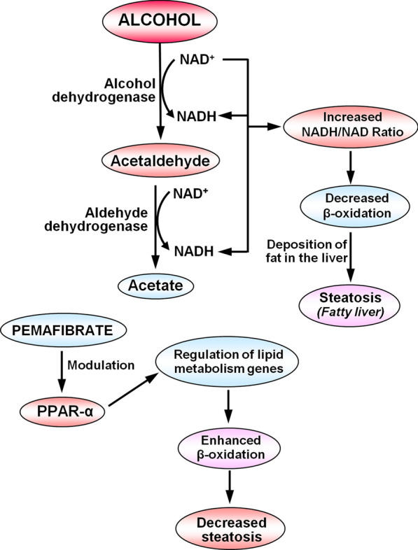
Background and aims: Alcohol-associated liver disease (ALD) with steatosis or steatohepatitis that could progress to liver cirrhosis is a common problem in chronic alcohol consumption. Pemafibrate is a novel, highly specific peroxisome proliferator-activated receptor-α (PPARα) modulator, which regulates the expression of the target genes related to lipid and glucose metabolism. Here, we evaluated the effect of pemafibrate to prevent ALD and steatosis in rats.

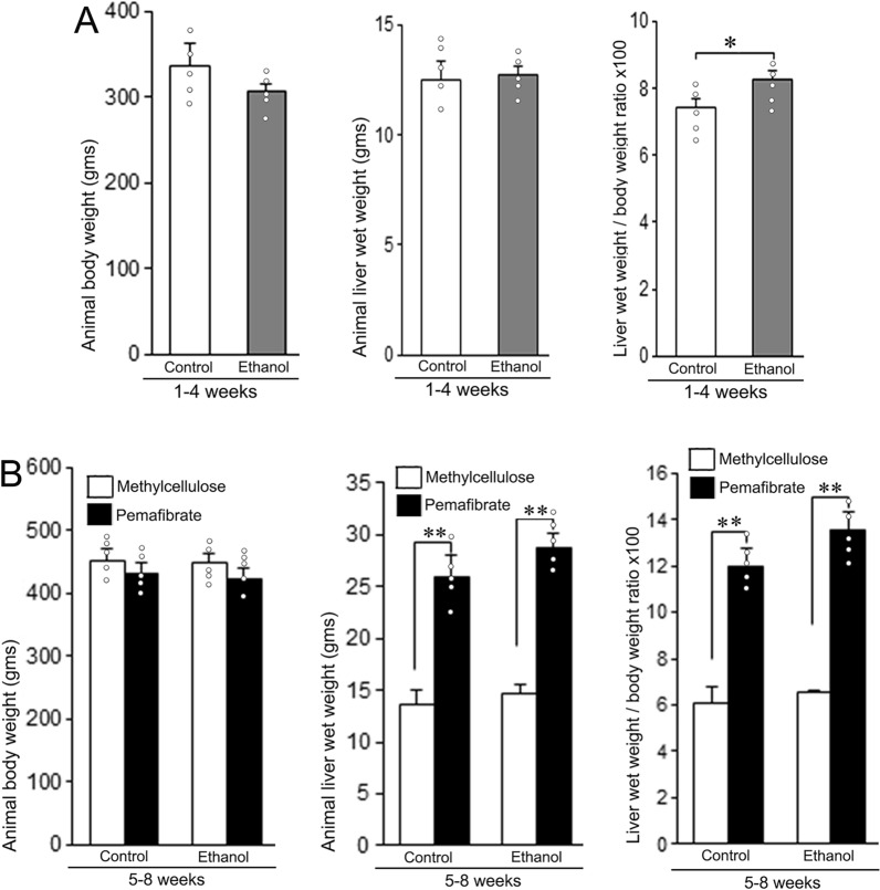
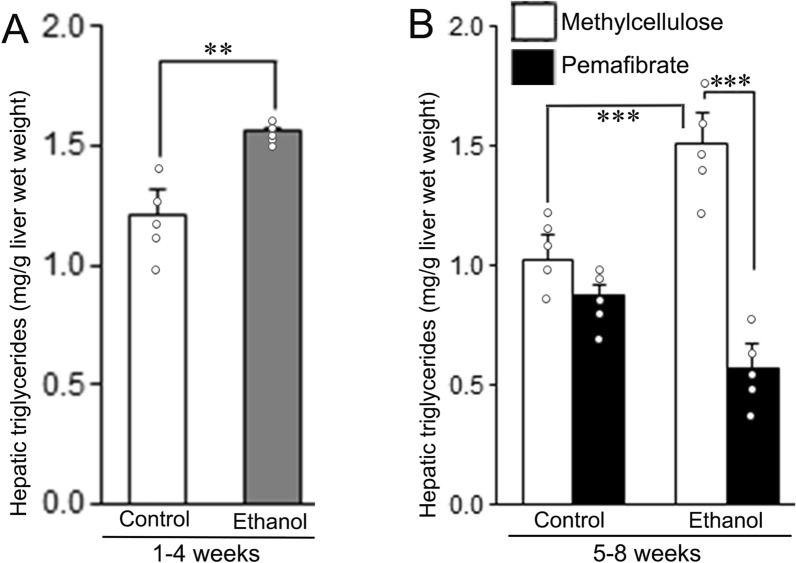
Methods: The animals were treated with liquid diet containing ethanol (36% of total calories) or an isocaloric carbohydrate diet for 4 weeks. Subsequently, both groups were fed with either 0.5% aqueous methylcellulose solution (MC) or MC containing 0.3 mg/kg body weight of pemafibrate orally twice a day along with the liquid diet for another 4 weeks. A set of animals were sacrificed at the 4th week before the start of pemafibrate treatment and the remaining animals at the end of 8 weeks. Blood and liver samples were collected for biochemical and histopathological evaluations.

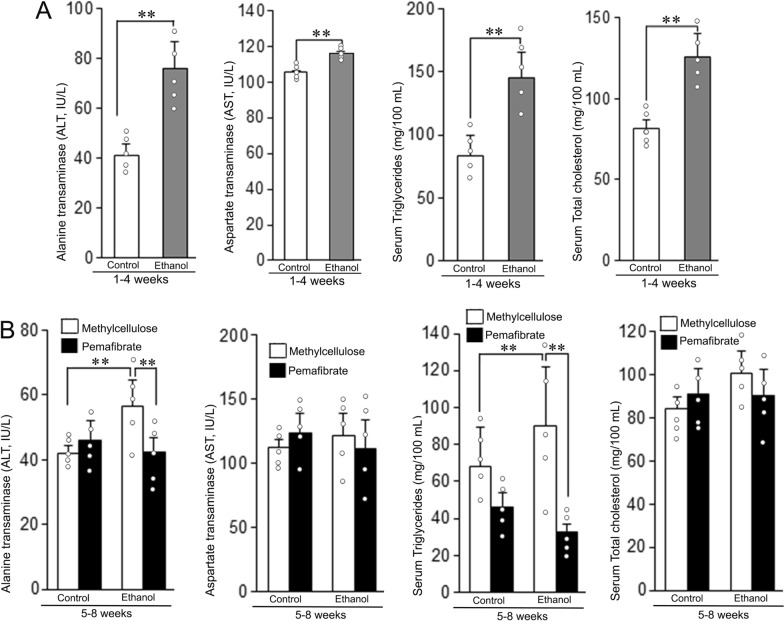
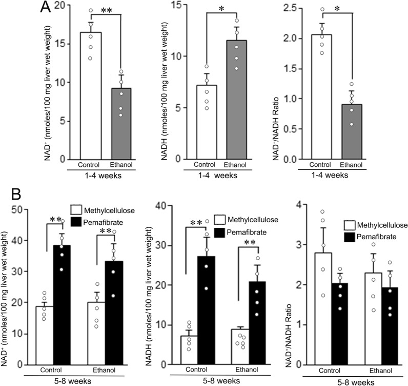
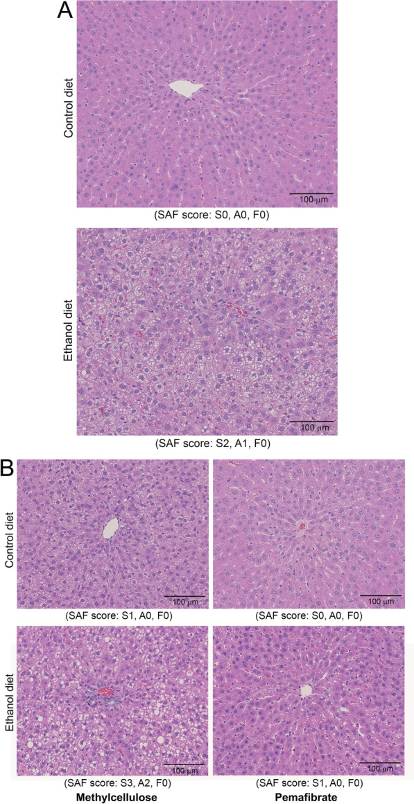
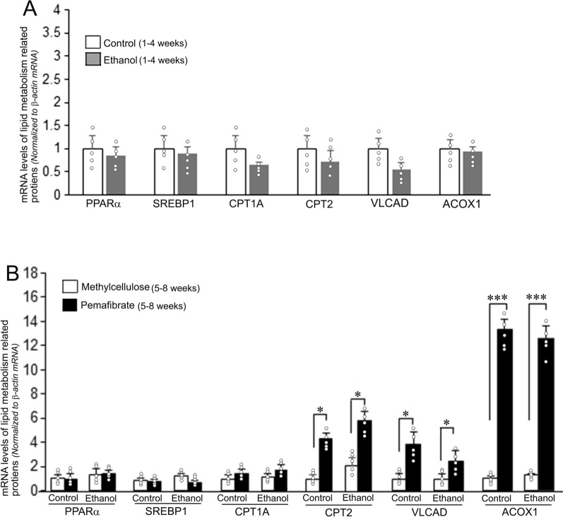
Results: Treatment with pemafibrate prevented inflammation and steatosis in the hepatic tissue. Furthermore, pemafibrate administration markedly increased hepatic NAD and NADH levels, reduced both serum and hepatic triglyceride levels, and upregulated the expression of molecules involved in lipid metabolism.

Conclusions: The results of the present study demonstrated that pemafibrate modulates target genes related to hepatic lipid metabolism and prevents deposition of fat globules in the liver during chronic alcohol feeding in rats. Therefore, pemafibrate could be used as a potent therapeutic agent to prevent steatosis and related adverse events in ALD.

Keywords: Alcohol-associated liver disease; PPARα; Pemafibrate; Steatohepatitis; Steatosis.


Conflict of interest statement

Declarations. Ethics approval and consent to participate: The animal experiments were carried out as per the Guide for the Care and Use of Laboratory Animals published by the US National Institutes of Health (NIH Publication No. 86-23, revised 1996). The animal experimental protocol was approved by the Animal Care and Use Committee of Kanazawa Medical University on the Ethics of use and care of experimental animals (#2022-29). Consent for publication: Not applicable. Competing interests: The authors declare no competing interests.

Figures

Fig. 1
Fig. 1
Body weight, liver wet weight, and liver wet weight to body weight ratio of animals fed with control diet or ethanol diet with methylcellulose or pemafibrate. A Animals fed with control or ethanol diet for 4 weeks. There was no alteration in either body weight or liver weight. The liver weight to body weight ratio was significantly higher compared to the control group. B Animals treated with methylcellulose or pemafibrate for 4 weeks. The liver weight and liver weight to body weight ratio of both control and ethanol groups treated with pemafibrate increased significantly compared to the animals administered with methycellulose. Data are mean ± SD (N = 5). *P < 0.05 and **P < 0.01
Fig. 2
Fig. 2
Serum levels of alanine aminotransferase (ALT), aspartate aminotransferase (AST), triglycerides (TG), and total cholesterol in the animals fed with control diet or ethanol diet with methylcellulose or pemafibrate. A Animals fed with control or ethanol diet for 4 weeks. Serum levels of ALT, AST, TG, and cholesterol were significantly increased in the ethanol group compared to the control group. B Animals treated with methylcellulose or pemafibrate for 4 weeks. Serum levels of ALT and TG were significantly increased in the ethanol group of animals administered with methylcellulose compared to the similarly treated controls, which were markedly reduced after the treatment with pemafibrate. Data are mean ± SD (N = 5). **P < 0.01
Fig. 3
Fig. 3
Levels of NAD, NADH, and NAD/NADH ratio in the livers of rats fed with control diet or ethanol diet with methylcellulose or pemafibrate. A Animals fed with control or ethanol diet for 4 weeks. In the ethanol group, hepatic NAD content decreased and NADH content increased compared with the respective control group, resulting in a significant decrease of NAD+/NADH ratio. B Animals treated with methylcellulose or pemafibrate for 4 weeks. Hepatic NAD+ and NADH content in both the control and ethanol groups treated with pemafibrate increased significantly compared to the respective methylcellulose group. Hepatic NAD+/NADH ratio in both the control and ethanol groups significantly reduced compared to the respective methylcellulose group. Data are mean ± SD (N = 5). *P < 0.05 and **P < 0.01
Fig. 4
Fig. 4
Hematoxylin and Eosin staining of rat livers fed with control diet or ethanol diet and after treatment with methylcellulose or pemafibrate. A Animals fed with control diet or ethanol diet for 4 weeks. The rats fed with the control diet did not show any histopathological alterations. Significant fatty degeneration, mild hepatic inflammation, and ballooning of hepatocytes were present in rat livers fed with ethanol diet. B Animals treated with methylcellulose or pemafibrate for 4 weeks. Mild fatty degeneration was observed in the control group of animals administered with methylcellulose, which were absent in pemafibrate treated group. Extensive fatty degeneration with deposition of large fat globules, moderate hepatic inflammation, and ballooning of hepatocytes were present in the ethanol group. Treatment with pemafibrate prevented significant deposition of fat globules in the hepatic tissue, hepatic inflammation, and ballooning of hepatocytes. However, small and intermittent fat globules were present. All histopathological images are original magnification, ×100
Fig. 5
Fig. 5
Hepatic triglycerides content in the livers of rats fed with control diet or ethanol diet and after treatment with methylcellulose or pemafibrate. A Animals fed with control diet or ethanol diet for 4 weeks. There was an increase of hepatic triglyceride content in the ethanol group. B Animals treated with methylcellulose or pemafibrate for 4 weeks. Hepatic triglyceride levels were significantly decreased in pemafibrate treated control and ethanol groups compared to the respective methylcellulose group. Data are mean ± SD (N = 5). *P < 0.05 and ***P < 0.001
Fig. 6
Fig. 6
mRNA levels of lipid metabolism related molecules in the livers of rats fed with control diet or ethanol diet with methylcellulose or pemafibrate. A Animals fed with control or ethanol diet for 4 weeks. There was no significant difference in the expression rate of hepatic lipid metabolism related genes between the control and ethanol treated groups. B Animals treated with methylcellulose or pemafibrate for 4 weeks. The mRNA levels of CPT2, VLCAD, and ACOX1 were significantly increased in animals treated with pemafibrate in both the control and ethanol groups compared to the rats treated with methylcellulose in the respective groups. Data are mean ± SD (N = 5). *P < 0.05 and ***P < 0.001
Fig. 7
Fig. 7
Levels of lipid metabolism related proteins in the livers of rats fed with control diet or ethanol diet with methylcellulose or pemafibrate. A, B Animals fed with control or ethanol diet for 4 weeks. A Western blots for proteins involved in lipid metabolism. Five samples were run for each molecule. B Quantification of Western blot images. There was no difference in the rate of expression of proteins involved in hepatic lipid metabolism between the control and ethanol groups. Data are mean ± SD of 5 independent samples. C, D) Animals treated with methylcellulose or pemafibrate for 4 weeks. C Western blot images of protein molecules related to lipid metabolism. D Quantification of Western blot images. Protein levels of PPARα, CPT1A, CPT2, VLCAD, and ACOX1 were significantly increased in pemafibrate treated rats of both the control and ethanol groups compared to the animals administered with methylcellulose in the respective groups. The Western blot images were quantified using the Gel-Pro analyzer software. Data are mean ± SD (N = 5). *P < 0.05, **P < 0.01, and ***P < 0.001
Fig. 8
Fig. 8
Schematic presentation of the metabolism of ethanol and the related increase of NADH/NAD ratio leading to decreased β-oxidation of fatty acids and subsequent deposition of fat in the liver (steatosis). Pemafibrate binds to PPAR-α and modulates the expression of genes involved in lipid metabolism that results in enhanced β-oxidation, thus decreasing plasma triglyceride levels and hepatic steatosis

References

    1. Azouaoui D, Choinière MR, Khan M, Sayfi S, Jaffer S, Yousef S, Patten DA, Green AE, Menzies KJ. Meta-analysis of NAD(P)(H) quantification results exhibits variability across mammalian tissues. Sci Rep. 2023;13(1):2464. 10.1038/s41598-023-29607-8. - PMC - PubMed
    1. Bedossa P, Poitou C, Veyrie N, Bouillot JL, Basdevant A, Paradis V, Tordjman J, Clement K. Histopathological algorithm and scoring system for evaluation of liver lesions in morbidly obese patients. Hepatology. 2012;56(5):1751–9. 10.1002/hep.25889. - PubMed
    1. Blair HA. Pemafibrate: first global approval. Drugs. 2017;77(16):1805–10. 10.1007/s40265-017-0818-x. - PubMed
    1. Bougarne N, Weyers B, Desmet SJ, Deckers J, Ray DW, Staels B, De Bosscher K. Molecular actions of PPARα in lipid metabolism and inflammation. Endocr Rev. 2018;39(5):760–802. 10.1210/er.2018-00064. - PubMed
    1. Chen X, Xu Y, Denning KL, Grigore A, Lu Y. PPARα agonist WY-14,643 enhances ethanol metabolism in mice: role of catalase. Free Radic Biol Med. 2021;169:283–93. 10.1016/j.freeradbiomed.2021.04.018. - PMC - PubMed

MeSH terms

Substances

LinkOut - more resources