Review
. 2025 Apr 22;23(1):192.
doi: 10.1186/s12964-025-02042-0.

Mitochondrial DNA signals driving immune responses: Why, How, Where?

Affiliations
Review

Mitochondrial DNA signals driving immune responses: Why, How, Where?

Luca Giordano et al. Cell Commun Signal. .

Abstract

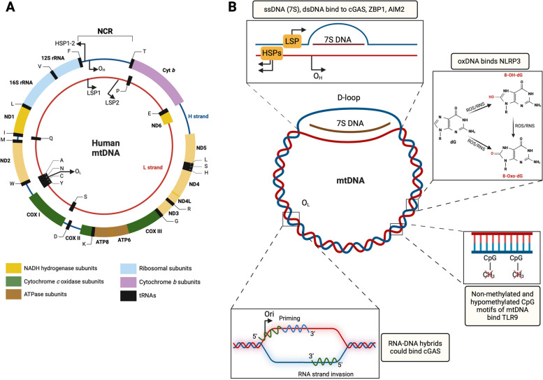
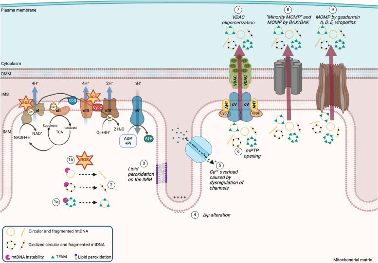
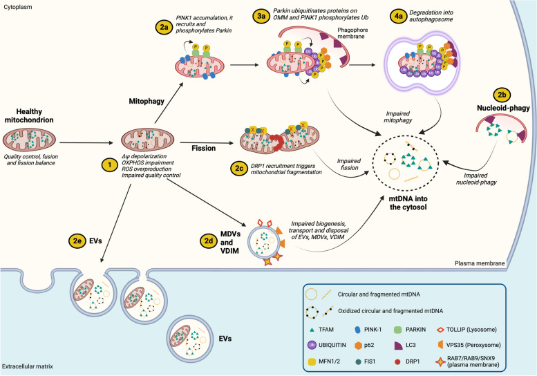
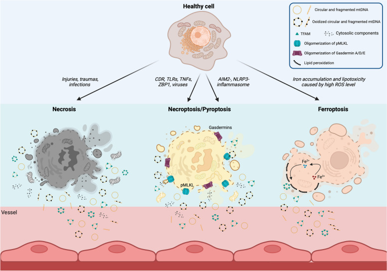
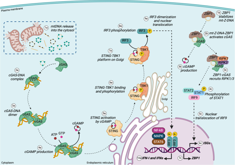
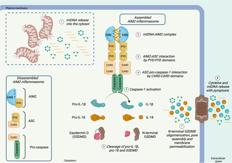
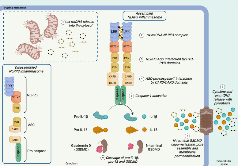
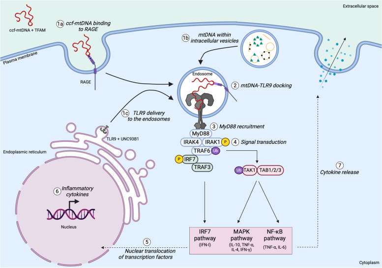
There has been a recent expansion in our understanding of DNA-sensing mechanisms. Mitochondrial dysfunction, oxidative and proteostatic stresses, instability and impaired disposal of nucleoids cause the release of mitochondrial DNA (mtDNA) from the mitochondria in several human diseases, as well as in cell culture and animal models. Mitochondrial DNA mislocalized to the cytosol and/or the extracellular compartments can trigger innate immune and inflammation responses by binding DNA-sensing receptors (DSRs). Here, we define the features that make mtDNA highly immunogenic and the mechanisms of its release from the mitochondria into the cytosol and the extracellular compartments. We describe the major DSRs that bind mtDNA such as cyclic guanosine-monophosphate-adenosine-monophosphate synthase (cGAS), Z-DNA-binding protein 1 (ZBP1), NOD-, LRR-, and PYD- domain-containing protein 3 receptor (NLRP3), absent in melanoma 2 (AIM2) and toll-like receptor 9 (TLR9), and their downstream signaling cascades. We summarize the key findings, novelties, and gaps of mislocalized mtDNA as a driving signal of immune responses in vascular, metabolic, kidney, lung, and neurodegenerative diseases, as well as viral and bacterial infections. Finally, we define common strategies to induce or inhibit mtDNA release and propose challenges to advance the field.

Keywords: Circulating cell-free DNA; DNA-sensing receptors; Inflammation; Innate immunity; Mitochondria; Mitochondrial DNA.


Conflict of interest statement

Declarations. Competing interests: The authors declare no competing interests.

Figures

Fig. 1
Fig. 1
Mitochondrial genome and its immunogenic features. A Mitochondrial genome is a circular DNA of 16.569 base pairs, with outer heavy (H) and inner light (L) strands. It encodes for 13 proteins: NADH-dehydrogenase subunit (ND) 1, ND2, ND3, ND4, ND4L, ND5, ND6, cytochrome b (cyt b), cytochrome c oxidoreductase (COX) I, COXII, COXIII, ATP synthase subunit (ATPase) 6 and ATPase 8; 22 tRNAs (T, L, S, H, R, G, K, D, W, M, I, L, V, F, and P, E, S, A, N, C, Y, Q); 2 rRNA (12S, 16S). The non-coding region (NCR) includes the displacement loop (D-loop). OH and OL are the origins of replication on the H and L strands, respectively, whereas transcription starts from the heavy strand promoters (HSP1 and HSP2) and light strand promoters (LSP1 and LSP2). Directions of replication and transcription are indicated by arrows. B The mitochondrial genome harbors immunogenic features. A single-stranded DNA (7S) is formed during mtDNA replication but is not terminated, forming a three-stranded D-loop structure that could be the main source of cytosolic mtDNA. 7S may bind the cyclic guanosine-monophosphate-adenosine-monophosphate synthase (cGAS), Z-DNA binding protein 1 (ZBP1), and absent in melanoma 2 (AIM2). Deoxyguanosines (dG) of mtDNA are easily oxidized by reactive oxygen and nitrogen species (ROS and RNS) to 8-hydroxy deoxyguanosines (8-OH-dG) and 8-oxo-deoxyguanosines (8-Oxo-dG). Oxidized mtDNA binds NOD-, LRR-, and PYD- domain-containing protein 3 receptor (NLRP3) and cGAS. Mitochondrial DNA, like bacterial DNA, harbors non-methylated/hypomethylated CpG sequences that are docked by the toll-like receptor 9 (TLR9). RNA–DNA hybrids are generated during mtDNA transcription and the initial phase of the replication. Transcription starts near OH (located 100 bp from LSP) by the mitochondrial RNA polymerase, generating an RNA–DNA hybrid that also primes the replication from OH. Similarly, RNA–DNA hybrids are generated at OL. During the strand invasion, RNA–DNA hybrids could potentially activate cGAS
Fig. 2
Fig. 2
Mechanisms of mitochondrial DNA release into the cytosol. (1a) Loss of function of proteins involved in the mtDNA replication and transcription (like TFAM, PolG, TOP3a) causes mtDNA instability. (1b) An impaired electron transport system promotes electron leakage and generates superoxide anion, which is converted into additional reactive oxygen species (ROS). Mitochondrial DNA instability and ROS overproduction induce (2) mtDNA fragmentation, linearization, and oxidation. ROS overproduction indirectly promotes (3) cardiolipin and phosphatidylethanolamine oxidation (major lipid components of the IMM) and causes (4) alteration of the mitochondrial membrane potential (∆Ψmt). Depolarization or hyperpolarization of ∆Ψmt dysregulate (5) mitochondrial carriers, resulting in Ca2+ overload in the mitochondrial matrix and consequent permeabilization of the IMM. The whole mitochondrial nucleoids and/or fragmented mtDNA are released into the cytosol by IMM permeabilization and by pores formed mainly by three different mechanisms: (6) mitochondrial permeability transition pore (mPTP) opening induced by transient short-lived stresses; (7) VDAC oligomerization. Importantly, mPTP and VDAC mainly cooperate in the extrusion of mtDNA into the cytosol. Their persistent activation could also trigger apoptosis; (8) oligomerization of BAX/BAK occurring meanwhile apoptotic caspases are inactive; (9) oligomerization of gasdermin A, or D, or E by inflammasomes. The last (8-9) two mechanisms lead also to the mitochondrial outer membrane permeabilization (i.e., MOMP). A mechanism named (8) “minority MOMP” occurs when only a subgroup of mitochondria releases mtDNA by BAX/BAK oligomerization. (9) Bacteria and viruses also use specific proteins (Ply, viroporins) to create pores on the IMM and OMM, favouring mtDNA release. *Of note, the exact composition of the mPTP is still debated, and how mPTP releases mtDNA is unknown. Here, we have illustrated the mPTP mainly as reported by Bonora and colleagues (59) to give the reader an indication of the complexity of the mPTP structure. Furthermore, VDAC and BAX/BAK may directly interact with mPTP (57), facilitating mtDNA by cooperative mechanisms. IMM and OMM, inner and outer mitochondrial membrane, respectively. IMS, intermembrane space; cI-V, mitochondrial complex I-V; TCA, tricarboxylic acid cycle; ∆Ψmt, mitochondrial membrane potential; voltage-dependent anion channel, VDAC; ANT, adenine nucleotide translocator; CypD, cyclophilin D
Fig. 3
Fig. 3
Cytosolic and extracellular release of mitochondrial DNA is caused by dysfunctional mitophagy, nucleoid-phagy, fission, and remodeling of the mitochondrial membranes. Mitochondria undergo a continuous cycle of fusion, fission, and mitophagy. Dysregulation of these processes causes mtDNA release into the cytosol and extracellularly. Mitophagy is a specific form of autophagy and a common route to remove and recycle damaged mitochondria. Two mechanisms of mitophagy are well known: non-receptor-dependent and receptor-dependent. The release of mtDNA has been described mainly by dysfunctional non-receptor-dependent mitophagy. In basal conditions, PTEN-induced kinase 1 (PINK1) is localized to the outer mitochondrial membrane as a cleaved inactive form. (1) Decreased ∆Ψmt prevents PINK1 cleavage, and (2a) promotes its accumulation, that favours (3a) Parkin recruitment. Parkin is an E3 ubiquitin (Ub) ligase that ubiquitinates several outer membrane proteins that are further phosphorylated by PINK1. The phospho-ubiquitinated chains (p-Ub) serve as an “eat me” signal for the recruitment of the autophagic machinery, including the adaptor protein p62 (optineurin and calcium-binding and coiled-coil domain 2, not shown). (4a) They interact with the microtubule-associated protein light chain (LC3), allowing the formation of a molecular bridge that encapsulates the mitochondrion in a phagophore membrane (autophagosome). Impaired or overload mitophagy causes mtDNA leaking from the autophagosome. (2b) Nucleoid-phagy is a form of autophagy that selectively degrades cytosolic mtDNA bound to the mitochondrial transcription factor A (TFAM). It depends on the LC3-interacting region 2 (LIR2) motif of TFAM that is recognized by LC3B, which, in turn, mediates the encapsulation and further degradation of mtDNA in the autophagosome. Impaired nucleoid-phagy allows the cytosol to retain mtDNA that has already escaped from mitochondria. (2c) Fission occurs on the contact site between the mitochondria and endoplasmic reticulum, and it is mainly regulated by dynamin-related protein (DRP1) and fission protein 1 (FIS1). DRP1 is recruited from the cytosol to the outer mitochondrial membrane, where it oligomerizes, forming a ring that constricts and splits the mitochondrion. FIS1 avoids mitochondrial fusion by blocking the mitochondrial fusion protein 1 and 2 (MFN1, MFN2) and optic-atrophy-1 (OPA1). Impaired fission causes the release of mtDNA into the cytosol. (2d) Mitochondrial-derived vesicles (MDVs, 60-150 nm), vesicles derived from the inner mitochondrial membrane (VDIM) and (2e) extracellular vesicles (EVs, 50-300 nm) are generated during physiological and stress conditions. The budding of the MDVs occurs at both inner and outer mitochondrial membranes and is regulated by several proteins, including the mitochondrial Rho GTPase 1 and DRP1 (not shown). MDVs are involved in intracellular quality control, allowing the degradation of irreparable proteins, lipids, and mtDNA of no yet depolarized mitochondria. The destination of the MDVs depends on the protein decoration of the outer membrane. Toll-interacting protein (TOLLIP), vacuole sorting-associated protein 35 (VSP35), Ras-associated protein 7 and 9 (RAB7/RAB9), and sorting nexin 9 (SNX9) guide the MDV toward endosomes/lysosomes, peroxisomes, and plasma membrane, respectively. VDIM have been recently reported as generated by herniation of the inner mitochondrial membrane through the oligomerization of the voltage-dependent anion channel (VDAC), and are dependent on the ROS-dependent calcium release from lysosomes. EVs are involved in intercellular communication and could transfer whole mitochondria or mitochondrial content, including mtDNA, through the extracellular matrix. The biogenesis and the regulation of the MDVs and EVs are not completely elucidated. However, impaired biogenesis and signaling to govern the destination of MDVs and EVs favours the release of mtDNA also within the cytosol
Fig. 4
Fig. 4
Mechanisms of mitochondrial DNA release in the extracellular environments by cell death. Mitochondrial DNA is released in the extracellular matrix, including serum, mainly by the following cell deaths: necrosis, necroptosis, pyroptosis, and ferroptosis. These mechanisms of cell death have the same outcome: the permeabilization of the plasma membrane (PM) and its rupture by which mtDNA is released. Necrosis is an uncontrolled cell death induced by several injuries, traumas, and infections that suddenly cause loss of membrane integrity, allowing the discharge of intracellular content, including mtDNA. Necroptosis is initiated by ligands activating cell death (CDR), specific toll-like receptors (TLR) like FAS, tumor necrosis factors (TNFs), ligands for Z-DNA binding protein (ZBP1), and viral infections. The necroptotic cascade terminates with the oligomerization of phosphorylated mixed lineage kinase domain-like protein (pMLKL) that forms pores on the PM. Pyroptosis is similar to necroptosis, but its cascade signaling is triggered by the AIM2- or NLRP3- inflammasomes, that are activated by pathogen- and damage-associated molecular patterns (PAMPs and DAMPs). The signaling leads to the primary permeabilization of the PM by oligomerization of A, D or E gasdermins with consequent release of IL-1b and IL-18. The complete permeabilization of the PM also allows the release of mtDNA. Ferroptosis is a regulated cell death caused by iron overload that induces lipid peroxidation by the Fenton reaction (H2O2+Fe2+) and lipoxygenases. It is triggered by reactive oxygen species (ROS) not well neutralized by antioxidant defenses (decreased intracellular glutathione and activity of glutathione peroxidase). Lipid peroxidation permeabilizes the mitochondrial and plasma membranes, allowing the formation of pores and micelles by which cytosolic components and mtDNA leaking from the mitochondria are discharged
Fig. 5
Fig. 5
Mitochondrial DNA activates cGAS-cGAMP-STING and ZBP-cGAS pathways. Mitochondrial DNA (fragments, whole nucleoids, and oxidized mtDNA) released into the (1a) cytosol binds cGAS, forming a (2a) cGAS-DNA complex that (3a) after dimerization converts (4a) ATP and GTP in 2’,3’ cyclic GAMP (cGAMP) dinucleotide. cGAMP is a second messenger that activates (5a) the stimulator of interferon gene (STING), an endoplasmic reticulum (ER)-associated receptor with a binding domain that faces the cytosol. (6a) cGAMP-STING binds to tank-binding kinase 1 (TBK1), and TBK1 phosphorylates STING. (7a) On the STING-TBK1 platform in the Golgi, the C-terminal tail of STING binds the interferon regulatory factor 3 (IRF3) and (8a) phosphorylates it. (9a) Phosphorylated IRF3 dimerizes and translocates to the nucleus, where (10a) it promotes the expression of the type I interferon (IFN-I) and interferon stimulating genes (ISGs). Other transcription factors activated by STING are NF-kB, MAPK, and STAT6. (1b) ZBP1 stabilizes mt-Z-DNA released into the cytosol that facilitates the interaction of ZBP1 with cGAS. (2b) The DNA-protein complex activates cGAS, which catalyzes the (3b) production of cGAMP and also recruits (3c) the receptor-interacting serine/threonine-protein kinase 1 (RIPK1) and RIPK3 forming a multiprotein complex. (4c) These two kinases phosphorylate the signal transducer and activator of transcription 1 (STAT1) that activates (5c) the nuclear translocation of the interferon regulatory factor 9 (IRF9). (6c) IRF9 promotes the expression of the interferon-stimulating genes (ISGs), potentiating the IFN-I response. (7c) IFN-I per se positively regulates the transcription of ZBP1 by the signal transduction through type I interferon receptor
Fig. 6
Fig. 6
Mitochondrial DNA triggers the assembly of the inflammasome by AIM2. The absent in melanoma 2 (AIM2)-inflammasome is disassembled in physiological conditions. AIM2 protein contains two domains: the hematopoietic interferon-inducible nuclear domain (HIN) and the pyrin domain (PYD). (1) Mitochondrial DNA that leaks into the cytosol binds the HIN domain of AIM2, allowing its (2) dimerization. (3) AIM2 dimer interacts with the apoptosis-associated speck-like protein (ASC) by PYD domains. (4) ASC recruits and triggers pro-caspase-1 through the caspase activation and recruitment domains (CARD). (5) Active caspase-1 cleaves (6) pro-IL-1ß and pro-IL-18 in IL-1ß and IL-18, respectively, and generates the N-terminal domain of gasdermin D (GSDMD). (7) The latter oligomerizes and formes pores on the plasma membrane, by which (8) cytokines and other cytoplasmatic proteins are released in the extracellular environments. Protracted activation of AIM2-inflammasome leads to cell lysis by pyroptosis, which favours mtDNA release into the extracellular matrix
Fig. 7
Fig. 7
Mitochondrial DNA promotes the assembly of the inflammasome by NLRP3. The NOD-, LRR-, and PYD- domain-containing protein 3 receptor (NLRP3)-inflammasome is disassembled in physiological conditions. NLRP3 protein presents the nucleotide-binding domain leucine-rich repeat (LRR), the central nucleotide-binding domain (NACHT), and the pyrin domain (PYD). (1) Oxidized mtDNA (ox-mtDNA) leaking into the cytosol binds the LRR domain of NLRP3, allowing the (2) dimerization of NLRP3. (3) NLRP3 dimer interacts with the ASC apoptosis-associated speck-like protein (ASC) through PYD domains. (4) ASC recruits and activates pro-caspase-1 through their caspase activation and recruitment domains (CARD). (5) Active caspase-1 (6) cleaves pro-IL-1ß and pro-IL-18 in IL-1ß and IL-18, respectively, and generates the N-terminal domain of gasdermin D (GSDMD). (7) The N-terminal domains of GSDMD oligomerize on the plasma membrane, forming pores that allow (8) the release of cytokine and ox-mtDNA into the extracellular space. Protracted activation of NLRP3-inflammasome leads to cell lysis by pyroptosis, with consequent release of ox-mtDNA into the extracellular matrix
Fig. 8
Fig. 8
Circulating cell-free DNA and mtDNA within the intracellular vesicles trigger the immune response by TLR9. Circulating cell-free mtDNA (ccf-mtDNA) after binding to (1a) RAGE on the plasma membrane is endocytosed and collected into the endosomes. (1b) Similarly, intracellular membranes (mitochondrial-derived vesicles, autophagosomes, products of mitochondrial dynamics) containing mtDNA that escape canonical routes, fuse with endosomes. (1c) TLR9 from the endoplasmic reticulum is packed into COPII vesicles under the control of UNC93B1 and delivered to the endosomes, where (2) it homodimerizes by binding CpG motifs of mtDNA. (3) The TLR9 homodimer recruits myeloid differentiation primary response 88 (Myd88) that successively interacts with the interleukin-1 receptor-associated kinases 4 (IRAK4), triggering signal transduction. (4) IRAK4 phosphorylates IRAK1, driving a signaling cascade that, through the tumor necrosis factor receptor-associated 6 (TRAF6), activates the interferon regulatory factor 7 (IRF7) and TGF-β-activated kinase-1 (TAK1)-TGF-β-activated kinase 1-binding protein 1/2/3 (TAB1/2/3). The downstream signaling cascades lead to the (5) translocation in the nucleus of the transcription factors IRF7, mitogen-activated protein kinases (MAPK), and nuclear factor kappa-light-chain-enhancer of activated B cells (NF-kB). (6) They promote the transcription of pro-inflammatory cytokines (IL-4, IL-6, IL-10, TNF⍺, and interferons), (7) which are further released

Similar articles

Cited by

References

    1. Roger AJ, Muñoz-Gómez SA, Kamikawa R. The Origin and Diversification of Mitochondria. Curr Biol. 2017;27(21):R1177–92. - PubMed
    1. Lang BF, Burger G, O’Kelly CJ, Cedergren R, Golding GB, Lemieux C, et al. An ancestral mitochondrial DNA resembling a eubacterial genome in miniature. Nature. 1997;387(6632):493–7. - PubMed
    1. Spinelli JB, Haigis MC. The multifaceted contributions of mitochondria to cellular metabolism. Nat Cell Biol. 2018;20(7):745–54. - PMC - PubMed
    1. Pak O, Nolte A, Knoepp F, Giordano L, Pecina P, Hüttemann M, et al. Mitochondrial oxygen sensing of acute hypoxia in specialized cells - Is there a unifying mechanism? Biochim Biophys Acta Bioenerg. 2022;1863(8):148911. - PubMed
    1. Löffler M, Fairbanks LD, Zameitat E, Marinaki AM, Simmonds HA. Pyrimidine pathways in health and disease. Trends Mol Med. 2005;11(9):430–7. - PubMed

Substances

LinkOut - more resources