

Prospective and dichotomous study of biomarkers with swept-source OCT and OCT-angiography in naive patients with diabetic macular edema
- PMID: 40264206
- PMCID: PMC12016437
- DOI: 10.1186/s40942-025-00672-7
Prospective and dichotomous study of biomarkers with swept-source OCT and OCT-angiography in naive patients with diabetic macular edema
Abstract
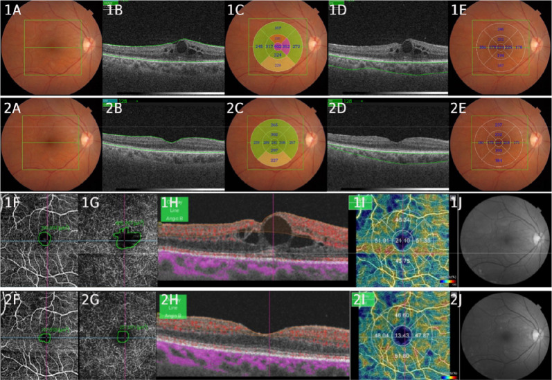
Background: We used state-of-the-art high-resolution retinal imaging to explore the treatment (loading dose of aflibercept) of diabetic macular edema (DME) among treatment-naive patients. Swept-source (SS) OCT and OCT-Angiography (SS-OCTA) were performed, and a dichotomous analysis was conducted to compare responders and treatment-resistant patients (responsive and resistant). Furthermore, treatment responses were evaluated based on the subdivision of choroidal thickness.
Materials and methods: This prospective, noncomparative, interventional case series study examined the following biomarkers: best-corrected visual acuity (BCVA), central macular thickness (CMT), central choroidal thickness (CCT), avascular area of the superficial plexus (AASP), avascular area of the deep plexus (AADP), and vessel density (VD). Data from the baseline and 4-month examinations were compared.
Results: Twenty-eight eyes from 25 patients were included. Significant improvements were observed in BCVA (0.7250 ± 0.23 to 0.3957 ± 0.21; p < 0.000), CMT µm (339.04 ± 66.19 to 265.21 ± 55.75; p < 0.000), CCT µm (221.71 ± 69.69 to 209.07 ± 70.92; p < 0.000), VD (17.90 ± 7.82 to 15.35 ± 5.80; p < 0.038), AASP µm2 (235,374 ± 91,299 to 157,326 ± 77,815; p < 0.000) and AADP µm2 (996,335 ± 1,000,047 to 362,161 ± 277,225; p < 0.000). Dichotomous analysis revealed that 15 patients were responsive (53.57%), and 13 resistant (46.43%). There were no significant differences between any of the pretreatment biomarkers. In the subdivision of choroidal thickness, which ranged from 211 to 270 µm (group 3), we found greater reductions in the CCT, AADP and CD. The choroidal thickness ranged from 181 to 210 µm (group 2): BCVA and AASP exhibited the greatest reductions.
Conclusion: BCVA, CMT, CCT, AASP, AADP and VD were improved after treatment. The pretreatment biomarkers did not predict treatment response between the responsive and resistant. Regarding choroidal stratification, values within the normal range of CCT showed the greatest reductions, indicating that these values may be more responsive to treatment. Notably, this is the first study to analyze biomarkers provided by SS OCT and OCTA, stratify the choroid, and perform a dichotomous analysis.
Keywords: Aflibercept; Biomarkers; Choroidal thickness; Diabetic macular edema; Diabetic retinopathy; OCT angiography; Swept-source OCT.
© 2025. The Author(s).
Conflict of interest statement
Declarations. Ethics approval and consent to participate: This study adhered to the guidelines set forth in Resolution 196/96 by the National Health Council of the Ministry of Health. The research protocol was submitted for evaluation and received formal approval from the Ethics Committee of the Hospital Regional do Câncer da Santa Casa de Misericórdia de Presidente Prudente – SP (CAAE—19386619.1.0000.8247). Competing interests: The authors declare no competing interests.
Figures
Similar articles
-
Vitrectomy in Diabetic Macular Edema:: A Swept-source OCT Angiography Study.Ophthalmol Sci. 2022 Aug 9;2(4):100207. doi: 10.1016/j.xops.2022.100207. eCollection 2022 Dec. Ophthalmol Sci. 2022. PMID: 36385773 Free PMC article.
-
OCT Angiography Biomarkers for Predicting Visual Outcomes after Ranibizumab Treatment for Diabetic Macular Edema.Ophthalmol Retina. 2019 Oct;3(10):826-834. doi: 10.1016/j.oret.2019.04.027. Epub 2019 May 7. Ophthalmol Retina. 2019. PMID: 31227330 Free PMC article.
-
Application of optical coherence tomography angiography in the assessment of diabetic macular edema staging and laser photocoagulation efficacy.Photodiagnosis Photodyn Ther. 2024 Apr;46:104055. doi: 10.1016/j.pdpdt.2024.104055. Epub 2024 Mar 18. Photodiagnosis Photodyn Ther. 2024. PMID: 38508440
-
Optical Coherence Tomography (Angiography) Biomarkers in the Assessment and Monitoring of Diabetic Macular Edema.J Diabetes Res. 2020 Dec 31;2020:6655021. doi: 10.1155/2020/6655021. eCollection 2020. J Diabetes Res. 2020. PMID: 33490283 Free PMC article. Review.
-
Profile of non-responder and late responder patients treated for diabetic macular edema: systemic and ocular factors.Acta Diabetol. 2020 Aug;57(8):911-921. doi: 10.1007/s00592-020-01496-7. Epub 2020 Feb 29. Acta Diabetol. 2020. PMID: 32114642 Review.
References
-
- Glassman AR, Wells JA 3rd, Josic K, Maguire MG, Antoszyk AN, Baker C, Beaulieu WT, Elman MJ, Jampol LM, Sun JK. Five-year outcomes after initial aflibercept, bevacizumab, or ranibizumab treatment for diabetic macular edema (protocol T extension study). Ophthalmology. 2020;127(9):1201–10. 10.1016/j.ophtha.2020.03.021. - PMC - PubMed
-
- Laíns I, Wang JC, Cui Y, Katz R, Vingopoulos F, Staurenghi G, Vavvas DG, Miller JW, Miller JB. Retinal applications of swept source optical coherence tomography (OCT) and optical coherence tomography angiography (OCTA). Prog Retin Eye Res. 2021;84: 100951. 10.1016/j.preteyeres.2021.100951. - PubMed
-
- Podkowinski D, Beka S, Mursch-Edlmayr AS, Strauss RW, Fischer L, Bolz M. A Swept source optical coherence tomography angiography study: imaging artifacts and comparison of non-perfusion areas with fluorescein angiography in diabetic macular edema. PLoS ONE. 2021;16(4): e0249918. 10.1371/journal.pone.0249918. - PMC - PubMed
-
- Rigatto C, Barrett BJ. Randomized controlled trials 5: biomarkers and surrogates/outcomes. Methods Mol Biol. 2021;2249:261–80. 10.1007/978-1-0716-1138-8_15. - PubMed
LinkOut - more resources
Full Text Sources
