. 2025 Apr 23;16(1):201.
doi: 10.1186/s13287-025-04320-7.

Sildenafil promotes osteogenic differentiation of human mesenchymal stem cells and inhibits bone loss by affecting the TGF-β signaling pathway

Affiliations

Sildenafil promotes osteogenic differentiation of human mesenchymal stem cells and inhibits bone loss by affecting the TGF-β signaling pathway

Menglong Hu et al. Stem Cell Res Ther. .

Abstract

Background: Osteoporosis, a common bone disorder, is primarily managed pharmacologically. However, existing medications are associated with non-trivial side-effects. Sildenafil, which already finds many clinical applications, promotes angiogenesis and cellular differentiation. Osteoporotic patients often exhibit a reduced intraosseous vasculature and impaired cellular differentiation; sildenafil may thus usefully treat osteoporosis.

Methods: Here, the effects of sildenafil on the osteogenic differentiation of human mesenchymal stem cells (hMSCs) were explored, as were the molecular mechanisms in play. We treated hMSCs with varying concentrations of sildenafil and measured cell proliferation and osteogenic differentiation in vitro. We used a mouse model of subcutaneous ectopic osteogenesis to assess sildenafil's effect on hMSC osteogenic differentiation in vivo. We also explored the effects of sildenafil on bone loss in tail-suspended (TS) and ovariectomized (OVX) mice. Mechanistically, we employed RNA-sequencing to define potentially relevant molecular pathways.

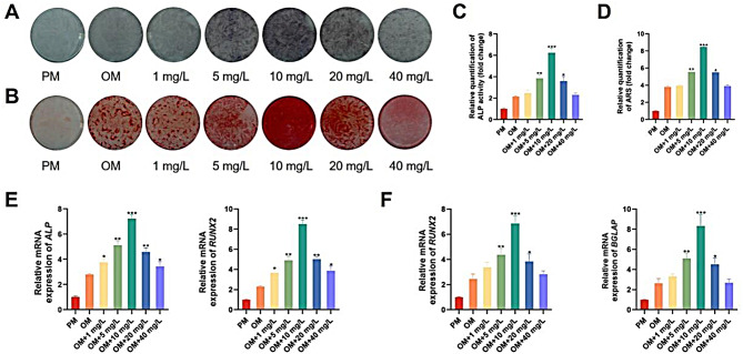
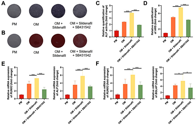
Results: The appropriate concentrations of sildenafil significantly enhanced osteogenic hMSC differentiation; the optimal sildenafil concentration may be 10 mg/L. Sildenafil mitigated osteoporosis in OVX and TS mice. The appropriate concentrations of sildenafil probably promoted hMSC osteogenic differentiation by acting on the transforming growth factor-β (TGF-β) signaling pathway.

Conclusions: In conclusion, sildenafil enhanced hMSC osteogenic differentiation and inhibited bone loss. Sildenafil may usefully treat osteoporosis. Our findings offer new insights into the physiological effects of the medicine.

Keywords: Mesenchymal stem cells; Osteogenesis; Osteoporosis; Sildenafil; TGF-β signaling pathway.


Conflict of interest statement

Declarations. Ethics approval and consent to participate: The study titled “The Effects and Mechanisms of Sildenafil in the Treatment of Osteoporosis” was approved by the Laboratory Animal Welfare and Ethics Committee of the Biomedical Ethics Committee at Peking University (Date: 16. 02. 2023, No. LA2023198). The specific animal experimental protocol for this study was pre-designed prior to the research. All surgeries were performed under anesthesia, and all efforts were made to minimize animal suffering. hBMSCs and hASCs were obtained from ScienCell Company (USA). ScienCell Company has confirmed that there was initial ethical approval for collection of human cells, and that the donors had signed informed consent. Consent for publication: Not applicable. Competing interests: The authors declare no conflicts of interest.

Figures

Fig. 1
Fig. 1
The appropriate concentrations of sildenafil enhanced the proliferation and migration of hBMSCs in vitro. A. Chemical structure of sildenafil; B. hBMSC growth curve derived using the CCK-8 assay; C, D. The transwell assay was employed to investigate how sildenafil at different concentrations affected hBMSC migration; E, F. The morphologies and quantitative analyses of the scratch assay migration areas used to explore the effects of sildenafil at different concentrations on hBMSC migration. The data are means ± standard deviations. *p < 0.05, **p < 0.01, and ***p < 0.001 compared to the PM group. N ≥ 3. Transwell assay image [scale bar]: 100 μm. Scratch assay image [scale bar]: 500 μm. hBMSCs, human bone marrow-derived mesenchymal stem cells
Fig. 2
Fig. 2
The appropriate concentrations of sildenafil enhanced osteogenic differentiation of hBMSCs in vitro. A. ALP staining revealing the effects of sildenafil at different concentrations on the osteogenic differentiation of hBMSCs; B. ARS staining revealing the effects of sildenafil at different concentrations on the osteogenic differentiation of hBMSCs; C. Quantification of ALP staining; D. Quantification of ARS staining; E. The relative levels of mRNAs encoding RUNX2 and ALP in hBMSCs as revealed by qRT-PCR after 7 days of osteogenic induction; F. The relative levels of mRNAs encoding RUNX2 and BGLAP in hBMSCs after 14 days of osteogenic induction. The data are means ± standard deviations. *p < 0.05, **p < 0.01, and ***p < 0.001 compared to the OM group. hBMSCs, human bone marrow-derived mesenchymal stem cells; ALP, alkaline phosphatase; ARS, alizarin red S; RUNX2, runt-related transcription factor 2; BGLAP, bone gamma-carboxyglutamate protein; qRT-PCR, quantitative real-time reverse transcription polymerase chain reaction; OM, osteogenic medium
Fig. 3
Fig. 3
Sildenafil at 10 mg/L promoted osteogenic differentiation of hBMSCs in the subcutaneous ectopic osteogenesis model. A. H&E staining of the PM and the PM + sildenafil groups; B. Masson trichrome staining of the PM and the PM + sildenafil groups; C. Immunohistochemical staining for OCN of the PM and the PM + sildenafil groups; D. Statistical analysis of the heterotopic bone area/tissue area (%) in the H&E staining of the PM and PM + sildenafil groups; E. Statistical analysis of the collagen area/tissue area (%) in the Masson staining of the PM and PM + sildenafil groups; F. Statistical analysis of the relative IOD in the immunohistochemical staining for OCN of the PM and PM + sildenafil groups. [scale bars]: 500 μm (10x) and 100 μm (40x). hBMSCs, human bone marrow-derived mesenchymal stem cells; H&E, hematoxylin and eosin; PM, proliferation medium; OCN, osteocalcin
Fig. 4
Fig. 4
Sildenafil at 10 mg/L alleviated osteoporosis in OVX mice. A. Micro-CT images, H&E staining and Masson staining of the sham + PBS, sham + sildenafil, OVX + PBS, and OVX + sildenafil groups; B-G. The BMD, BV/TV, Tb.N, Tb.Th, BS/BV, and Tb.Sp values of the of sham + PBS, sham + sildenafil, OVX + PBS, and OVX + sildenafil groups; H, I. The expression levels of the bone formation-related serum markers BALP and P1NP in the sham + PBS, sham + sildenafil, OVX + PBS, and OVX + sildenafil groups. The data are means ± standard deviations. *p < 0.05, **p < 0.01, and ***p < 0.001. n ≥ 3. H&E staining image [scale bar]: 500 μm. OVX, ovariectomized; micro-CT, micro-computed tomography; PBS, phosphate balanced solution; H&E, hematoxylin and eosin; BMD, bone mineral density; BV/TV, bone volume fraction; Tb.N, trabecular number; Tb.Th, trabecular thickness; BS/BV, bone surface area/bone volume; Tb.Sp, trabecular separation; BALP, bone alkaline phosphatase; P1NP, procollagen type 1 N-terminal propeptide
Fig. 5
Fig. 5
Sildenafil at 10 mg/L alleviated osteoporosis in TS mice. A. Micro-CT images, H&E staining and Masson staining of the sham + PBS, sham + sildenafil, suspension + PBS, and suspension + sildenafil groups; B-G. The BMD, BV/TV, Tb.N, Tb.Th, BS/BV, and Tb.Sp values of the sham + PBS, sham + sildenafil, suspension + PBS, and suspension + sildenafil groups; H, I. The expression levels of the bone formation-related serum markers BALP and P1NP in the sham + PBS, sham + sildenafil, suspension + PBS, and suspension + sildenafil groups. The data are means ± standard deviations. *p < 0.05, **p < 0.01, and ***p < 0.001. n ≥ 3. H&E staining image [scale bar]: 500 μm. TS, tail-suspended; micro-CT, micro-computed tomography; PBS, phosphate balanced solution; H&E, hematoxylin and eosin; BMD, bone mineral density; BV/TV, bone volume fraction; Tb.N, trabecular number; Tb.Th, trabecular thickness; BS/BV, bone surface area/bone volume; Tb.Sp, trabecular separation; BALP, anti-bone alkaline phosphatase; P1NP, type I N terminal propeptide
Fig. 6
Fig. 6
Sildenafil may promote osteogenic differentiation of hBMSCs by modulating the TGF-β signaling pathway. A. The Venn diagram of co-expressed genes in the OM and OM + sildenafil groups; B. The volcano plot of differentially expressed genes in the OM and OM + sildenafil groups; C. GO gene enrichment maps; D. The KEGG enrichment scatter plot; E-H. qRT-PCR analysis of the relative levels of mRNAs encoding TGF-β1, TGF-β2, TGF-βR1, and TGF-βR2 in hBMSCs; I. Western blots revealing the expression levels of TGF-β1, TGF-βR2, p-TGF-βR2, Smad2/3, p-Smad2/3, and GAPDH. Full-length blots are presented in Supplementary Figures (Fig. S8-12), the samples derived from the same experiment and that blots were processed in parallel; J-L. Quantification of TGF-β1, p-TGF-βR2/ TGF-βR2, and p-Smad2/3/ Smad2/3 expression. The data are means ± standard deviations. *p < 0.05, **p < 0.01, and ***p < 0.001 compared to OM. N ≥ 3. hBMSCs, human bone marrow-derived mesenchymal stem cells; OM, osteogenic medium; GO, Gene Ontology; KEGG, Kyoto Encyclopedia of Genes and Genomes; qRT-PCR, quantitative real-time reverse transcription polymerase chain reaction; TGF-β1, transforming growth factor-β1; TGF-β2, transforming growth factor-β2; TGF-βR1, transforming growth factor-β type I receptor; TGF-βR2, transforming growth factor-β type II receptor; p-TGF-βR2, phospho- transforming growth factor-β type II receptor; p-Smad2/3, phospho-Smad2/3; GAPDH, glyceraldehyde-3-phosphate dehydrogenase; OM, osteogenic media
Fig. 7
Fig. 7
Sildenafil promoted osteogenic differentiation of hBMSCs by activating the TGF-β signaling pathway. A. ALP staining revealing the effects of sildenafil and the TGF-β pathway inhibitor SB431542 on the osteogenic differentiation of hBMSCs; B. ARS staining revealing the effects of sildenafil and the TGF-β pathway inhibitor SB431542 on the osteogenic differentiation of hBMSCs; C. Quantification of ALP staining; D. Quantification of ARS staining; E. The relative levels of mRNAs encoding RUNX2 and ALP in hBMSCs as revealed by qRT-PCR after 7 days of osteogenic induction; F. The relative levels of mRNAs encoding RUNX2 and BGLAP in hBMSCs after 14 days of osteogenic induction. The data are means ± standard deviations. *p < 0.05, **p < 0.01, and ***p < 0.001. hBMSCs, human bone marrow-derived mesenchymal stem cells; ALP, alkaline phosphatase; ARS, alizarin red S; RUNX2, runt-related transcription factor 2; BGLAP, bone gamma-carboxyglutamate protein; qRT-PCR, quantitative real-time reverse transcription polymerase chain reaction; OM, osteogenic medium; TGF-β, transforming growth factor-β
Fig. 8
Fig. 8
Sildenafil promotes osteogenic differentiation of hMSCs and suppresses bone loss by affecting TGF-β signaling. The appropriate concentrations of sildenafil enhanced the proliferation and migration of hMSCs in vitro, and promoted osteogenic differentiation of hMSCs both in vitro and in vivo. In vivo, 10 mg/L sildenafil reduced bone loss in both OVX and TS mice. Sildenafil may promote osteogenic differentiation of hBMSCs by influencing the TGF-β signaling pathway. hMSCs, human mesenchymal stem cells; TGF-β, transforming growth factor-β; OVX, ovariectomized; TS, tail-suspended; hBMSCs, human bone marrow-derived mesenchymal stem cells

Similar articles

References

    1. Li Z, Li D, Chen R, Gao S, Xu Z, Li N. Cell death regulation: A new way for natural products to treat osteoporosis. Pharmacol Res. 2023;187:106635. - PubMed
    1. Salari N, Ghasemi H, Mohammadi L, Behzadi MH, Rabieenia E, Shohaimi S, et al. The global prevalence of osteoporosis in the world: a comprehensive systematic review and meta-analysis. J Orthop Surg Res. 2021;16:609. - PMC - PubMed
    1. Foessl I, Dimai HP, Obermayer-Pietsch B. Long-term and sequential treatment for osteoporosis. Nat Reviews Endocrinol. 2023;19:520–33. - PubMed
    1. Peng J, Lai ZG, Fang ZL, Xing S, Hui K, Hao C, et al. Dimethyloxalylglycine prevents bone loss in ovariectomized C57BL/6J mice through enhanced angiogenesis and osteogenesis. PLoS ONE. 2014;9:e112744. - PMC - PubMed
    1. Chen Y, Huang Y, Li J, Jiao T, Yang L. Enhancing osteoporosis treatment with engineered mesenchymal stem cell-derived extracellular vesicles: mechanisms and advances. Cell Death Dis. 2024;15:119. - PMC - PubMed

MeSH terms

Substances

LinkOut - more resources