Meta-Analysis
. 2025 Apr 22;21(1):12.
doi: 10.1186/s12993-025-00274-1.

Imaging-validated correlates and implications of the pathophysiologic mechanisms of ageing-related cerebral large artery and small vessel diseases: a systematic review and meta-analysis

Affiliations
Meta-Analysis

Imaging-validated correlates and implications of the pathophysiologic mechanisms of ageing-related cerebral large artery and small vessel diseases: a systematic review and meta-analysis

Joseph A Ackah et al. Behav Brain Funct. .

Abstract

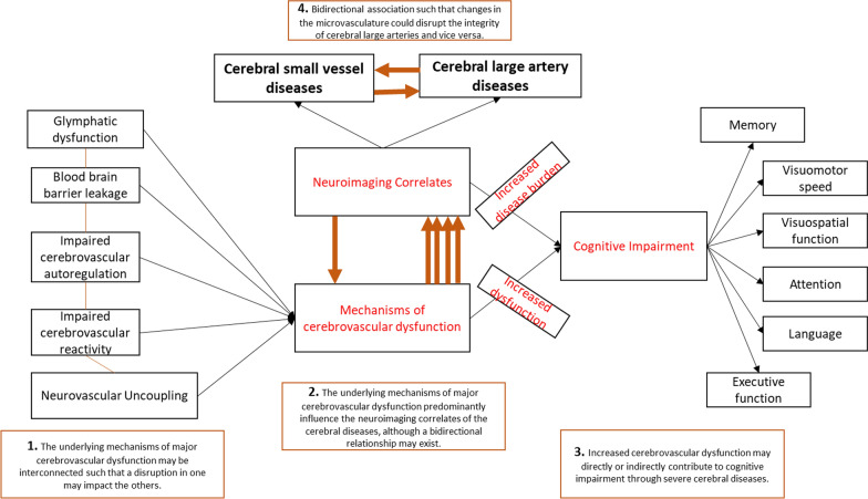
Background: Cerebral large artery and small vessel diseases are considered substrates of neurological disorders. We explored how the mechanisms of neurovascular uncoupling, dysfunctional blood-brain-barrier (BBB), compromised glymphatic pathway, and impaired cerebrovascular reactivity (CVR) and autoregulation, identified through diverse neuroimaging techniques, impact cerebral large artery and small vessel diseases.

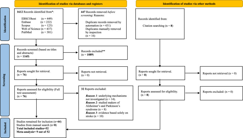
Methods: Studies (1990-2024) that reported on neuroradiological findings on ageing-related cerebral large artery and small vessel diseases were reviewed. Fifty-two studies involving 23,693 participants explored the disease mechanisms, 9 studies (sample size = 3,729) of which compared metrics of cerebrovascular functions (CF) between participants with cerebral large artery and small vessel diseases (target group) and controls with no vascular disease. Measures of CF included CVR, cerebral blood flow (CBF), blood pressure and arterial stiffness.

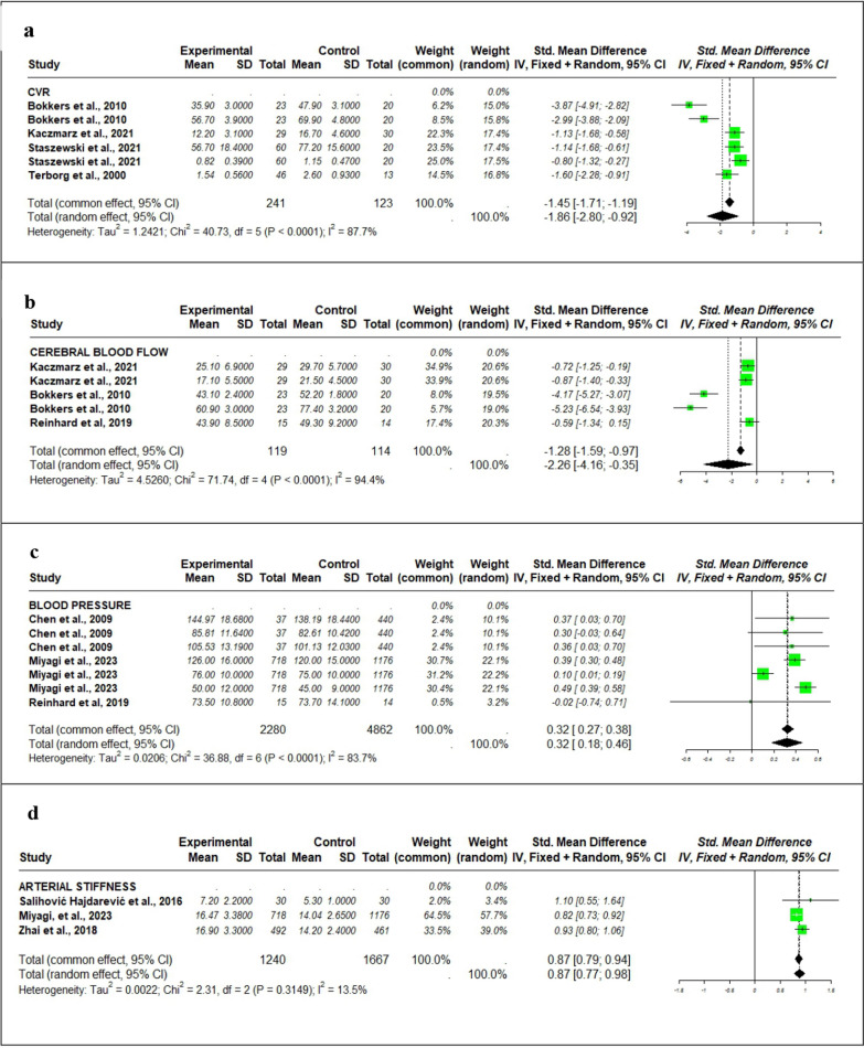
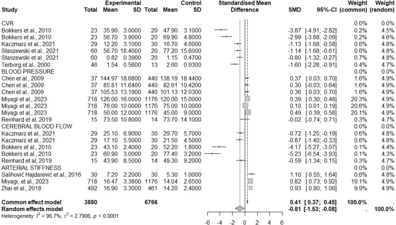
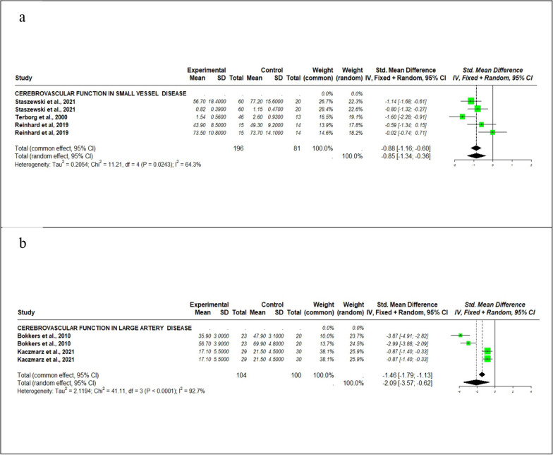
Results: The findings from 9 studies (sample size = 3,729, mean age = 60.2 ± 11.5 years), revealed negative effect sizes of CVR [SMD = - 1.86 (95% CI - 2.80, - 0.92)] and CBF [SMD = - 2.26 (95% CI - 4.16, - 0.35)], respectively indicating a reduction in cerebrovascular functions in the target group compared to their controls. Conversely, there were significant increases in the measures of blood pressure [SMD = 0.32 (95% CI 0.18, 0.46)] and arterial stiffness [SMD = 0.87 (95% CI 0.77, 0.98)], which signified poor cerebrovascular functions in the target group. In the combined model the overall average effect size was negative [SMD = - 0.81 (95% CI - 1.53 to - 0.08), p < 0.001]. Comparatively, this suggests that the negative impacts of CVR and CBF reductions significantly outweighed the effects of blood pressure and arterial stiffness, thereby predominantly shaping the overall model. Against their controls, trends of reduction in CF were observed exclusively among participants with cerebral large artery disease (SMD = - 2.09 [95% CI: - 3.57, - 0.62]), as well as those with small vessel diseases (SMD = - 0.85 [95% CI - 1.34, - 0.36]). We further delineated the underlying mechanisms and discussed their interconnectedness with cognitive impairments.

Conclusion: In a vicious cycle, dysfunctional mechanisms in the glymphatic system, neurovascular unit, BBB, autoregulation, and reactivity play distinct roles that contribute to reduced CF and cognitive risk among individuals with cerebral large artery and/or small vessel diseases. Reduction in CVR and CBF points to reductions in CF, which is associated with increased risk of cognitive impairment among ageing populations ≥ 60 years.

Keywords: Ageing; Cerebral small vessel disease; Cognitive; Large artery disease; Neuroimaging; Pathophysiology.


Conflict of interest statement

Declarations. Competing interests: The authors declare no competing interests.

Figures

Fig. 1
Fig. 1
PRISMA flow diagram for search and study selection
Fig. 2
Fig. 2
Distribution of studies by countries. Overall, forest plot. A negative overall SMD indicates a reduction in effect size, while a positive SMD indicates an increase. This figure demonstrates an overall reduction in cerebrovascular function in participants with cerebral large artery and small vessel diseases compared to the control group without vascular disease
Fig. 3
Fig. 3
Forest plot assessing each metric of cerebrovascular function. From (a) and (b), the negative effect sizes of CVR [MSD = − 1.86 (95% CI − 2.80, − 0.92)] and cerebral blood flow [SMD = − 2.26 (95% CI − 4.16, − 0.35)], respectively, indicate a reduction in cerebrovascular functions in individuals with cerebral large artery and small vessel diseases compared to their control group with no vascular disease. Conversely, (c) and (d) respectively show positive effect sizes of blood pressure [MSD = 0.32 (95% CI 0.18, 0.46)] and arterial stiffness [MSD = 0.87 (95% CI 0.77, 0.98)] indicating an increase in these metrics, which signify poor cerebrovascular functions. Forest plot for subgroup analysis. CVR metrics significantly reduced in individuals with with cerebral large artery and small vessel diseases compared to the control group without vascular disease. Whiles, blood pressure and flow were elevated but this increase was insignificantly observed between the two groups. There were significant declines in cerebrovascular functions specifically noted in individuals with cerebral large artery disease (SMD= − 2.09 [95% CI − 3.57, − 0.62]
Fig. 4
Fig. 4
Forest plot for combined metrics of cerebrovascular functions. The forest plot shows measures of CVR and cerebral blood flow were reduced in the target group compared to their control. Conversely, blood pressure and arterial stiffness were increased in the target group compared to their control group. There is indication that the negative effects of CVR and cerebral blood flow predominantly influenced the overall negative effect size of the random effects model in the combined measures of CVR, blood pressure, cerebral blood flow, and arterial stiffness
Fig. 5
Fig. 5
Subgroup analysis according disease subtype. Using the measures of cerebral blood flow and CVR as the measures of cerebrovascular function in subgroup analyses: (a) Shows an overall negative effect size [SMD = − 0.85 (95% CI − 1.34, − 0.36)], which signifies a reduction in cerebrovascular functions in participants presenting exclusively with small vessel diseases. (b) Shows an overall negative effect size [SMD = − 2.09 (95% CI − 3.57, − 0.62)], which signifies a reduction in cerebrovascular functions in participants presenting exclusively with large arterial diseases. Schematic model for cerebral large artery and small vessel diseases. A simple diagram that could serve as a guide to explore the interconnectedness of underlying mechanisms implicated in cerebral large artery and small vessel diseases
Fig. 6
Fig. 6
Schematic diagram of study results

Similar articles

References

    1. Li H, Cao W, Zhang X, et al. BOLD-fMRI reveals the association between renal oxygenation and functional connectivity in the aging brain. Neuroimage. 2018;2019(186):510–7. 10.1016/j.neuroimage.2018.11.030. - PubMed
    1. Iadecola C. The neurovascular unit coming of age: a journey through neurovascular coupling in health and disease. Neuron. 2017;96(1):17–42. 10.1016/j.neuron.2017.07.030. - PMC - PubMed
    1. Rodriguez Lara F, Toro AR, Pinheiro A, et al. Relation of MRI-visible perivascular spaces and other MRI markers of cerebral small vessel disease. Brain Sci. 2023;13(9):1–22. 10.3390/brainsci13091323. - PMC - PubMed
    1. Tian Y, Zhao M, Chen Y, Yang M, Wang Y. The underlying role of the glymphatic system and meningeal lymphatic vessels in cerebral small vessel disease. Biomol. 2022;12(6):748. 10.3390/BIOM12060748. - PMC - PubMed
    1. Xu X, Wang B, Ren C, et al. Age-related impairment of vascular structure and functions. Aging Dis. 2017;8(5):590–610. 10.14336/AD.2017.0430. - PMC - PubMed

MeSH terms

LinkOut - more resources