. 2025 Apr 21;13(4):e70469.
doi: 10.1002/ccr3.70469. eCollection 2025 Apr.

Vagus Nerve Schwannoma: A Case Report and Literature Review

Affiliations

Vagus Nerve Schwannoma: A Case Report and Literature Review

Kato Ronald et al. Clin Case Rep. .

Abstract

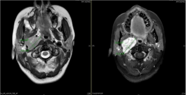
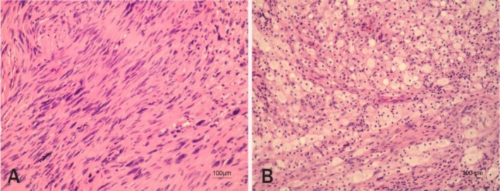
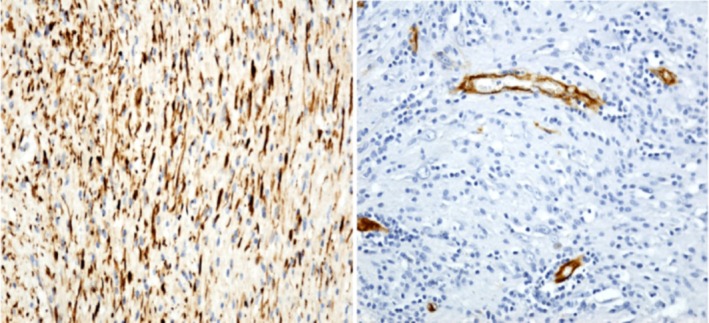
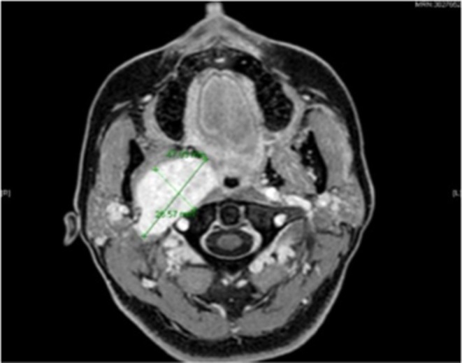
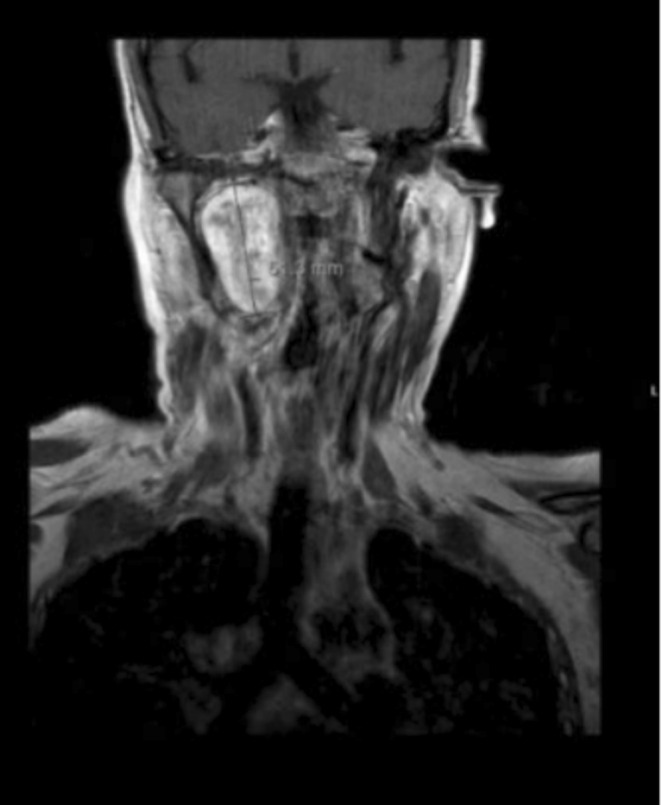
Vagus schwannomas are rare, benign masses that arise in the cervical region but can develop anywhere along the Vagus nerve. In most cases, patients present in their third to sixth decades, with neck swelling and hoarseness; herein, we discuss a case report of a 41-year-old female who presented with slurred speech and locking of her tongue and was diagnosed with Vagus schwannoma. The patient had an inability to speak properly and locking of her tongue during speech, with pain to the right side of the neck as Intermittent, prompted by turning her head to the right and shooting pain to her shoulder, which is short-lived, with a sensation of difficulty swallowing and talking that is alleviated by moving her head to a neutral position. On examination, there was mild neck stiffness, fasciculations of the tongue, and left deviation of the tongue with inability to swallow. Blood pressure was 128/70 mmHg with 42 bpm bradycardia, which was persistent on several readings, with a small right neck mass measuring 1 cm × 2 cm, non-tender on palpation. Imaging studies showed a well-defined ovoid lesion centered in the right retrostyloid parapharyngeal space measuring 3.3 × 2.1 × 3.8 cm with central T2 hyperintensities representing a nerve sheath tumor that appears stable in size and morphology. The histology report typically displayed a characteristic pattern with two distinct tissue types, Antoni A and Antoni B, with tightly packed spindle cells arranged in palisades around a central verocay body and a looser arrangement of myxoid matrix. Immunohistochemistry showed lesional cells had strong immunopositivity for S100 and were negative for CD34. Hence, the conclusion of a Vagus nerve schwannoma. Patient was sent to the radiation oncology team and was started on Cyberknife 25 Gy treatment in five fractions, which were completed and sent for re-evaluation and follow-up. There was no surgical intervention in this case due to the delicate anatomical location of the mass; hence, Cyberknife radiation was the best option for treatment. Vagus schwannomas are rare, benign masses that usually develop in the cervical region but can arise anywhere along the Vagus nerve. Patients may be asymptomatic, but in most cases, they present in their third to sixth decades; hence, they should be considered in patients with otherwise unexplained bradycardia, with a history of dysphagia, slurred speech, and cervical masses.

Keywords: Vargus nerve; benign; bradycardia; schwannoma; tumor.


Conflict of interest statement

The authors declare no conflicts of interest.

Figures

FIGURE 1
FIGURE 1
(A) Right without contrast and (B) left with contrast. Avidly enhancing well‐defined ovoid lesion centered in the right retrostyloid parapharyngeal space measuring 3.3 × 2.1 × 3.8 cm (AP × TV × CC) with central T2 hyperintensities representing a nerve sheath tumor that appears stable in size and morphology. This lesion results in splaying of the right ICA/ECA. Posterior displacement and flattening of the right internal jugular vein are noted. Mild mass effect on the right aspect of the oropharynx results in mild narrowing of the airway. Mass effect on the masticator space and parapharyngeal space occurs without evidence of invasion.
FIGURE 2
FIGURE 2
(A) Right and (B) left: Biopsy was performed and showed typically a characteristic pattern with two distinct tissue types, Antoni A and Antoni B, respectively. Antoni A areas showed tightly packed spindle cells arranged in palisades around a central verocay body with elongated nuclei displaced in concentric rows around eosinophilic material, while Antoni B areas exhibited a looser arrangement of cells within a myxoid matrix with detached fragments of skeletal muscle and fragments of fibroadipose and vascular tissue, with a conclusion of the impression of a nerve schwannoma.
FIGURE 3
FIGURE 3
(A) (Right) showing positive for S100 and (B) (left) showing negative for CD34.
FIGURE 4
FIGURE 4
Showed a redemonstration of a T2 hyperintense, lobulated, well‐defined lesion within the right retro‐styloid parapharyngeal space measuring 4.2 × 2.5 × 4.5 cm but that has increased in comparison to the prior MRI. When it measured approximately 3.4 × 1.9 × 3.7 cm and measured similarly.
FIGURE 5
FIGURE 5
Showing a similar mass effect on internal and external carotid arteries, right‐sided mass effect on the oropharynx, and narrowing of the right internal jugular vein that is posterolaterally displaced by the mass and avidly enhancing well‐defined lobulated T2 hyperintensity in the right aspect of the lateral tongue representing a schwannoma with mass effect on the regional vessels without evidence of thrombus.

Similar articles

References

    1. Lin H. L., Yu S. Y., and Lig K., “Extracranial Head and Neck Schwannomas: A Study of the Nerve Origin,” European Archives of Oto‐Rhino‐Laryngology 268 (2011): 1343–1347, 10.1007/s00405-011-1491-4. - DOI - PMC - PubMed
    1. Biswas D., Marnane C. N., Mal R., and Baldwin D., “Extracranial Head and Neck Schwannomas—A 10‐Year Review,” Auris Nasus Larynx 34, no. 3 (2007): 353–359, 10.1016/j.anl.2007.01.006. - DOI - PubMed
    1. Langer E. and Del Negro A., “Schwannomas in the Head and Neck; Retrospective Analysis of 21 Patients and Review of Literature,” São Paulo Medical Journal 125 (2007): 220–222, 10.1590/S1516-31802007000400005. - DOI - PMC - PubMed
    1. Lahoti B. K., Kaushal M., Garge S., and Aggarwal G., “Extra Vestibular Schwannomas; A Two‐Year Experience,” Indian Journal of Otolaryngology and Head & Neck Surgery 63 (2011): 305–309, 10.1007/s12070-011-0154-5. - DOI - PMC - PubMed
    1. Ferner R. E. and O'Doherty M. J., “Neurofibroma and Schwannoma,” Current Opinion in Neurology 15, no. 6 (2002): 679–684, 10.1097/01.wco.0000044763.39452.aa. - DOI - PubMed

LinkOut - more resources