. 2025 Apr 18:20:5039-5057.
doi: 10.2147/IJN.S506830. eCollection 2025.

Heme Oxygenase-1 Modulates Macrophage Polarization Through Endothelial Exosomal miR-184-3p and Reduces Sepsis-Induce Lung Injury

Affiliations

Heme Oxygenase-1 Modulates Macrophage Polarization Through Endothelial Exosomal miR-184-3p and Reduces Sepsis-Induce Lung Injury

Wei Chen et al. Int J Nanomedicine. .

Abstract

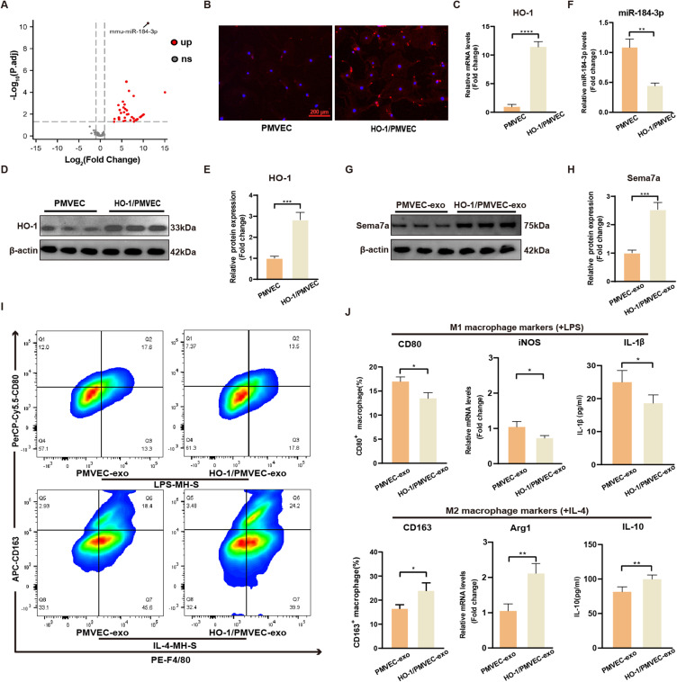
Introduction: Pulmonary microvascular endothelial cells (PMVECs) are notably implicated in the pathogenesis of sepsis-induced lung injury. Exosomes derived from PMVECs facilitate intercellular communication among various cell types, especially crosstalk with macrophages. Heme oxygenase-1 (HO-1), an early stress-responsive enzyme with inherent protective functions, has been implicated in acute lung injury (ALI) mitigation. But research on the mechanism of HO-1 in macrophage polarization via PMVEC exosomes in sepsis-induced lung injury is lacking.

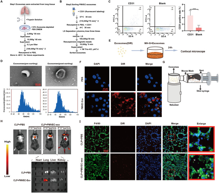
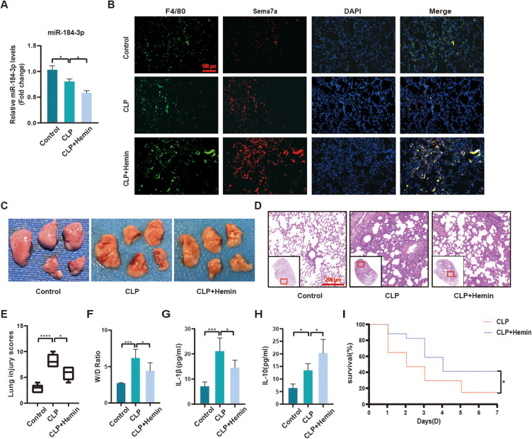
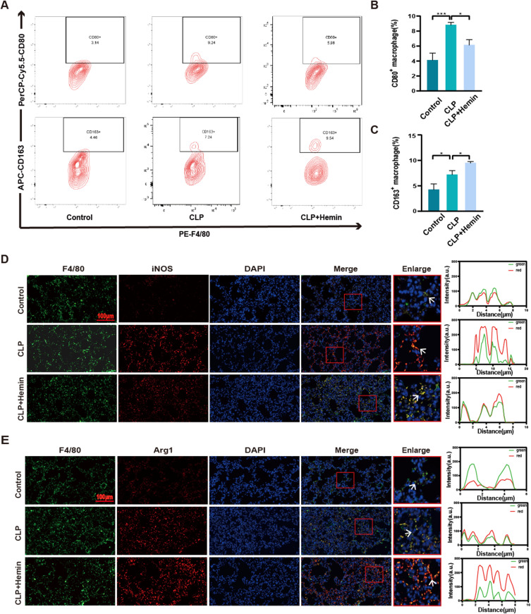
Methods: To investigate the role of HO-1 in the interaction between endothelial cells and macrophages, HO-1 knockout mouse model were established. Exosomes from PMVECs were isolated, and differential expression of microRNA (miRNA) was determined by sequencing. An in vitro co-culture system involving Murine Alveolar Macrophage Cell Line (MH-S cells) and HO-1/ PMVECs-derived exosomes (HP-exos) was used to investigate the underlying mechanisms. To further verify the involvement of HO-1 in intercellular communication through exosomal miRNA in vivo, the level of pulmonary inflammation was evaluated, and the polarization of pulmonary macrophages was analyzed.

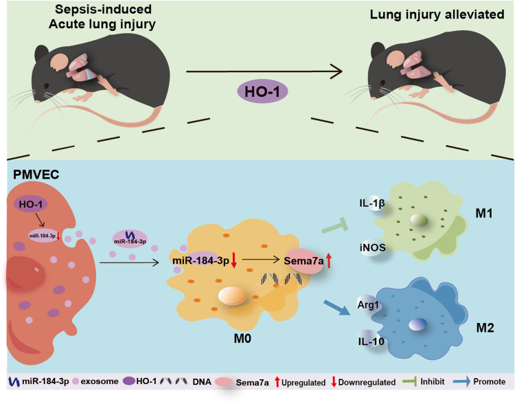
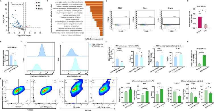
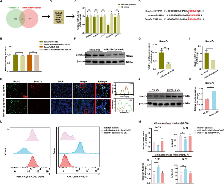
Results: The results showed that miR-184-3p was significantly downregulated in HP-exos, and supplementation of miR-184-3p enhanced the polarization of M1 macrophages, thus intensifying lung inflammation. HO-1 regulates the polarization of macrophages by regulating endothelial exosomes. Overexpression of HO-1 downregulates miR-184-3p, which negatively regulates Semaphorin 7A (Sema7a), which attenuated M1 type macrophages (M1) polarization and augmented M2 type macrophages (M2) polarization, thereby partially mitigating lung injury and inflammation.

Conclusion: Collectively, we elucidated a novel potential therapeutic mechanism that HO-1 alleviate inflammation by modulating the M1/M2 ratio in sepsis-induced ALI by regulating miR-184-3p/Sema7a expression.

Keywords: MiRNA; acute lung injury; exosome; macrophage polarization; sepsis.


Conflict of interest statement

The author(s) report no conflicts of interest in this work.

Figures

None
Graphical abstract
Figure 1
Figure 1
Exosome extraction, identification and phagocytosis by macrophages (A) Extraction of exosomes from lung tissue. (B) Schematic of the sorting process for PMVEC-derived exosomes. (C) Flow cytometric analysis for the isolation of CD31+ exosomes (n=6). (D) Characterization of exosomes isolated from lung tissue/PMVEC via TEM (scale bar=100 nm) and NTA. (E) Experimental schematic for macrophage capture of PMVEC-derived exosomes. (F) Observation of exosome phagocytosis by macrophages using confocal microscopy (scale bar=30 μm, n=3). (G) Schematic of the custom-built mouse nebulizer. (H) Utilization of small animal imaging technology demonstrated increased fluorescent intensity. (I) Immunofluorescence staining displaying F4/80 (green), DIR (red), and DAPI (blue) with a scale bar of 100 μm. Data are presented as mean±SD and statistically analyzed using Student’s t-test. ***P<0.001.
Figure 2
Figure 2
Effects of miR-184-3p overexpression on macrophage M1/M2 polarization (A) Volcano plot illustrating differentially expressed genes (n=3). (B) GO analysis of pathways enriched by miR-184-3p target genes. (C) Nano-flow cytometric detection of exosomal markers CD63 and CD81. (D) Quantitative analysis of miR-184-3p expression levels (n=6). (E) Macrophages were co-cultured with PMVEC-derived exosomes treated with LPS or PBS for 24 h to assess changes in miR-184-3p expression levels (n=6). (F) Co-culture with PMVEC-derived exosomes treated with LPS or PBS, the counts of M1 (CD80+) and M2 (CD163+) macrophages was quantified using flow cytometry. (G) RT-qPCR was utilized to measure the expression of iNOS and Arg1 (n=6). ELISA assays quantified the levels of IL-1β and IL-10 in the culture supernatant (n=5). (H) Comparative expression levels of miR-184-3p in NC mimic and miR-184-3p mimic groups (n=6). (I-J) Proportions of F4/80+ CD80+ and F4/80+ CD163+ macrophages assessed via flow cytometry (n=3), RT-qPCR was employed to evaluate the expression of iNOS and Arg-1 (n=6), ELISA quantification of IL-1β and IL-10 levels in the supernatant (n=5). Data are presented as mean ± SD and analyzed using Student’s t-test. *P<0.05, **P<0.01, ***P<0.001.
Figure 3
Figure 3
MiR-184-3p negatively regulates Sema7a to promote M1 and inhibit M2 polarization of macrophages. (A-B) Predictive analysis using starBase 3.0 identified 261 candidate genes potentially targeted by miR-184-3p. Intersection with 198 inflammation-related from DisGeNET yielded five candidate genes. (C) RT-qPCR analysis of these five candidate genes was performed in MH-S cells transfected with either miR-184-3p mimic or NC mimic (n=6). (D-E) Co-transfection of miR-184-3p and PmirGLO-sema7a-WT/MUT reporter plasmids into 293T cells revealed significant differences in the expression levels of the sema7a-WT wild-type luciferase reporter gene compared to the NC group (n=3). (F-G) Western blot analysis and protein quantification were employed to assess Sema7a levels. (H) Immunofluorescence staining was used to visualize F4/80 (green), Sema7a (red), and DAPI (blue) across various groups (scale bar=100 μm, n=3). (I) RT-qPCR was conducted to measure Sema7a expression (n=6). (J-K) Western blot analysis were conducted to assess Sema7a levels. (L) Counts of CD80+ and CD163+ macrophages were determined via flow cytometry. (M) mRNA levels of iNOS and Arg-1 (n=6), ELISA quantified the secretion levels of IL-1β and IL-10 in the supernatants (n=6). Data are presented as mean±SD and analyzed using Student’s t-test and one-way ANOVA. *P<0.05, **P<0.01.
Figure 4
Figure 4
Regulation of Macrophage Polarization by HO-1-Modified PMVECs via Downregulation of miR-184-3p. (A) Volcano plot illustrating differentially expressed genes (n=3). (B) Immunofluorescence labeling for HO-1. (C) mRNA expression levels of HO-1 (n=6). (D-E) Western blot analysis and protein quantification for HO-1. (F) Expression levels of miR-184-3p (n=6). (G-H) Western blot analysis and protein quantification for Sema7a. (I-J) Proportions of F4/80+ CD80+ and F4/80+ CD163+ macrophages assessed via flow cytometry (n=3). Expression levels of iNOS and Arg-1 (n=6), ELISA quantification of IL-1β and IL-10 secretion levels in supernatants (n=5). Data are presented as mean±SD and analyzed using Student’s t-test. *P<0.05, **P<0.01, ***P<0.001, ****P<0.0001.
Figure 5
Figure 5
HO-1 agonist Hemin reduces lung tissue inflammation by regulating miR-184-3p. (A) Expression levels of miR-184-3p (n=6). (B) Immunofluorescence staining showing F4/80 (green), Sema7a (red), and DAPI (blue) across various groups, (scale bar=100 μm, n=3). (C) Representative photograph of mouse lung tissues illustrating injury severity (n=3). (D) Histopathological alterations in lung sections, highlighted by H&E staining (scale bar=200 μm, n=3). (E) Semi-quantitative assessment of lung tissue damage using lung injury scores via the Mann–Whitney U-test. (F) Measurement of lung wet/dry weight ratio (W/D) in rats. (G-H) ELISA quantification of IL-1β and IL-10 levels in BALF supernatant (n=5). (I) Survival enhancement in CLP mice treated with Hemin (n=15). Data expressed as mean±SD, analyzed using Student’s t-test and one-way ANOVA. *P<0.05, ***P<0.001, ****P<0.0001.
Figure 6
Figure 6
Regulatory effects of HO-1 agonist Hemin on macrophage polarization in lung tissues. (A-C) Flow cytometric analysis to determine the ratio of F4/80+CD80+ to F4/80+CD163+ macrophages (n=3). (D-E) Immunofluorescence staining for F4/80 (green), iNOS (red), Arg1 (red), and DAPI (blue) in different groups (scale bar=100 μm, n=3). Data expressed as mean±SD, analyzed using Student’s t-test and one-way ANOVA. *P<0.05, ***P<0.001.

Similar articles

References

    1. Singer M, Deutschman CS, Seymour CW. et al. The third international consensus definitions for sepsis and septic shock (Sepsis-3). JAMA. 2016;315(8):801–810. doi:10.1001/jama.2016.0287 - DOI - PMC - PubMed
    1. Zhang J, Muri J, Fitzgerald G, et al. Endothelial lactate controls muscle regeneration from ischemia by inducing M2-like macrophage polarization. Cell Metab. 2020;31(6):1136–53.E7. doi:10.1016/j.cmet.2020.05.004 - DOI - PMC - PubMed
    1. Wang Z, Wang Z. Park7 interacts with p47phox to direct NADPH oxidase-dependent ROS production and protect against sepsis. Front Immunol. 2023;14:1209438. doi:10.3389/fimmu.2023.1209438 - DOI - PMC - PubMed
    1. Lin F-Y, Song C, Zeng Y-J, et al. Canagliflozin alleviates LPS-induced acute lung injury by modulating alveolar macrophage polarization. Int Immunopharmacol. 2020;88:106969. doi:10.1016/j.intimp.2020.106969 - DOI - PubMed
    1. Vassiliou AG, Kotanidou A, Dimopoulou I, Orfanos SE. Endothelial damage in acute respiratory distress syndrome. Int J mol Sci. 2020;21(22):8793. doi:10.3390/ijms21228793 - DOI - PMC - PubMed

MeSH terms