

How Does the Mandible Age? Comprehensive Artificial Intelligence-Assisted Shape Analysis in the White Population
- PMID: 40264908
- PMCID: PMC12011572
- DOI: 10.1097/GOX.0000000000006650
How Does the Mandible Age? Comprehensive Artificial Intelligence-Assisted Shape Analysis in the White Population
Abstract
Background: Mandible contour significantly influences facial appearance, framing the lower facial silhouette. Redefining mandibular contour is key for facial and neck rejuvenation. Yet, there is limited facial aging research across different lifespans and sexes. Here, we utilize artificial intelligence and advanced 3-dimensional (3D) analysis to elucidate mandibular aging patterns in male and female subjects.
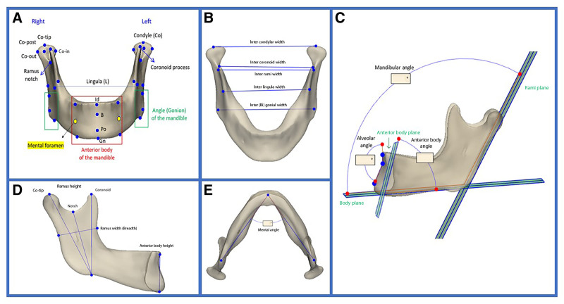
Methods: A retrospective analysis of facial computed tomography scans in White patients was conducted, categorizing subjects into 3 age groups (20-79 y) and stratifying them by sex. Artificial intelligence-assisted segmentation into 3D mandibles was done in Mimics v.25, and statistical shape modeling was used to create an average mandible for each group. Volume and linear measurements were assessed via 3D overlays.
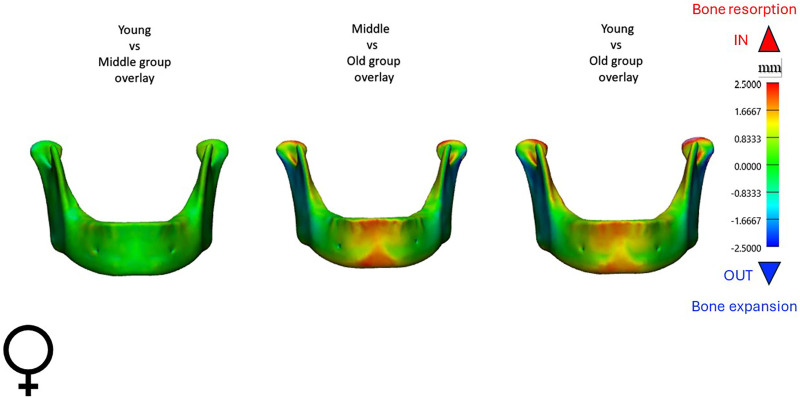
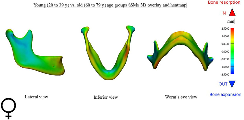
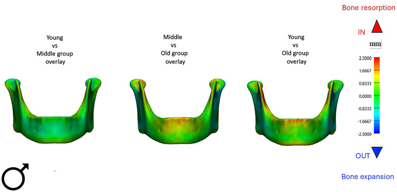
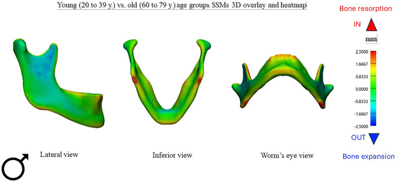
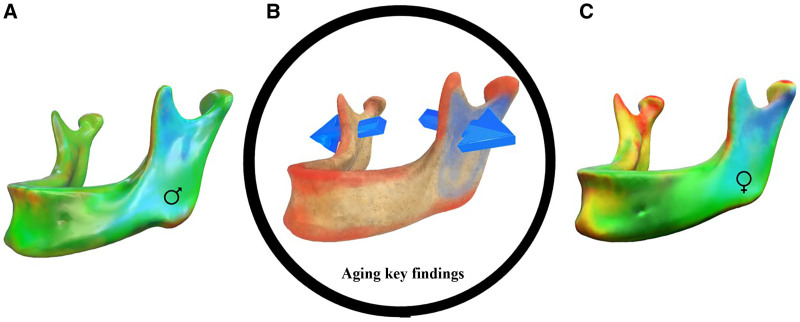
Results: Analysis of 280 mandibles demonstrated statistically significant aging changes in both sexes. Ramus height showed a marked decrease with age, by approximately 5.3 mm in women and 4.2 mm in men (P < 0.001). Interrami and intercondylar widths increased by a mean of 4-5 mm (P < 0.01). Women exhibited an increase in mandibular angle (P < 0.01), and bony resorption over the chin compared to men, who exhibited concentrated bone resorption at the gonion projection.
Conclusions: Mandibular aging, independent of tooth loss, exhibits specific bone remodeling patterns by sex. Posteriorly, mandibular widths increase in both sexes, whereas ramus height decreases. Women experience more resorption at the anterior alveolar surface and chin than men. Statistical shape modeling effectively visualizes these patterns on a population level, bridging the gap between traditional aging research and current understanding.
Copyright © 2025 The Authors. Published by Wolters Kluwer Health, Inc. on behalf of The American Society of Plastic Surgeons.
Conflict of interest statement
The authors have no financial interest to declare in relation to the content of this article.
Figures
Similar articles
-
Mandibular Gender Dimorphism: The Utility of Artificial Intelligence and Statistical Shape Modeling in Skeletal Facial Analysis.Aesthetic Plast Surg. 2024 Nov;48(21):4272-4279. doi: 10.1007/s00266-024-04300-x. Epub 2024 Aug 26. Aesthetic Plast Surg. 2024. PMID: 39187587
-
Aging of the mandible and its aesthetic implications.Plast Reconstr Surg. 2010 Jan;125(1):332-342. doi: 10.1097/PRS.0b013e3181c2a685. Plast Reconstr Surg. 2010. PMID: 20048624
-
A novel measurement method for the morphology of the mandibular ramus using homologous modelling.Dentomaxillofac Radiol. 2015;44(8):20150062. doi: 10.1259/dmfr.20150062. Dentomaxillofac Radiol. 2015. PMID: 26143939 Free PMC article.
-
Aging of the facial skeleton: aesthetic implications and rejuvenation strategies.Plast Reconstr Surg. 2011 Jan;127(1):374-383. doi: 10.1097/PRS.0b013e3181f95b2d. Plast Reconstr Surg. 2011. PMID: 20871486
-
Facial bone density: effects of aging and impact on facial rejuvenation.Aesthet Surg J. 2012 Nov;32(8):937-42. doi: 10.1177/1090820X12462865. Epub 2012 Sep 24. Aesthet Surg J. 2012. PMID: 23012659
References
-
- Mendelson B, Wong CH. Facial anatomy and aging. In: Neligan PC, Rubin JP, Matarasso A, eds. Plastic Surgery, Vol 2: Aesthetic Surgery, 5th ed. Elsevier; 2024:131–148.
-
- Beer K, Beer J. Overview of facial aging. Facial Plast Surg. 2009;25:281–284. - PubMed
-
- Hirai T, Ishijima T, Hashikawa Y, et al. . Osteoporosis and reduction of residual ridge in edentulous patients. J Prosthet Dent. 1993;69:49–56. - PubMed
-
- Boyde A, Kingsmill VJ. Age changes in bone. Gerodontology. 1998;15:25–34. - PubMed
LinkOut - more resources
Full Text Sources
Miscellaneous