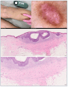
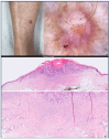
Uncommon Collision Tumors: Dermoscopic and Histopathological Features of Basal Cell Carcinoma Overlying Dermatofibroma
- PMID: 40265341
- PMCID: PMC12015826
- DOI: 10.3390/dermatopathology12020010
Uncommon Collision Tumors: Dermoscopic and Histopathological Features of Basal Cell Carcinoma Overlying Dermatofibroma
Abstract
Dermatofibromas (DFs) represent prevalent benign fibrohistiocytic tumors, typically manifesting as solitary lesions. In the majority of cases, the clinical presentation and dermoscopic and histopathological features of DFs adhere to a characteristic profile. However, DFs may exhibit atypical clinical presentations and, more commonly, histologic attributes, posing challenges in differential diagnosis. Both DFs and basal cell carcinomas (BCCs) are frequently encountered cutaneous lesions, each characterized by distinct clinical and dermoscopic features and microscopic morphology. The simultaneous occurrence of these two entities within the same lesion is rare. DFs have been documented to form collision tumors in conjunction with a spectrum of benign and malignant lesions, encompassing not only BCC but also balloon cell nevus, squamous cell carcinoma (SCC), and melanoma. Alterations in the epidermis overlaying a DF range from simple hyperplasia to the proliferation of basaloid cells. Accurate diagnosis, leading to the complete excision of the lesion, is contingent upon the recognition of dermoscopic criteria, precluding misinterpretation as a benign lesion. We present two cases of collision tumors comprising DF and BCC. This case report underscores the paramount importance of dermoscopy and adherence to dermoscopic criteria in the assessment of collision lesions and the diagnostic process related to cutaneous malignancies.
Keywords: basal cell carcinoma; collision tumors; dermatofibroma; dermoscopy.
Conflict of interest statement
The authors declare no conflict of interest.
Figures


Similar articles
-
Dermoscopic Features of Basal Cell Carcinoma on the Lower Limbs: A Chameleon!Dermatology. 2017;233(6):482-488. doi: 10.1159/000487300. Epub 2018 Mar 22. Dermatology. 2017. PMID: 29566370
-
Typical and atypical dermoscopic presentations of dermatofibroma.J Eur Acad Dermatol Venereol. 2013 Nov;27(11):1375-80. doi: 10.1111/jdv.12019. Epub 2012 Nov 24. J Eur Acad Dermatol Venereol. 2013. PMID: 23176079
-
Desmoplastic Trichoepithelioma: An Uncommon but Diagnostically Problematic Benign Adnexal Tumor.Acta Dermatovenerol Croat. 2019 Dec;27(4):282-284. Acta Dermatovenerol Croat. 2019. PMID: 31969246
-
Clinical and Dermoscopic Patterns of Basal Cell Carcinoma and Its Mimickers in Skin of Color: A Practical Summary.Medicina (Kaunas). 2024 Aug 24;60(9):1386. doi: 10.3390/medicina60091386. Medicina (Kaunas). 2024. PMID: 39336428 Free PMC article. Review.
-
Clinical and Dermoscopic Features Associated With Difficult-to-Recognize Variants of Cutaneous Melanoma: A Systematic Review.JAMA Dermatol. 2020 Apr 1;156(4):430-439. doi: 10.1001/jamadermatol.2019.4912. JAMA Dermatol. 2020. PMID: 32101255
References
-
- Mortimore A., Muir J., Muir J. Basal cell carcinoma in dermatofibroma: Dual diagnosis. Aust. J. Gen. Pract. 2020;49:842–844. - PubMed
Publication types
LinkOut - more resources
Full Text Sources
Research Materials
Miscellaneous

