

Super-Enhancers in Placental Development and Diseases
- PMID: 40265369
- PMCID: PMC12015882
- DOI: 10.3390/jdb13020011
Super-Enhancers in Placental Development and Diseases
Abstract
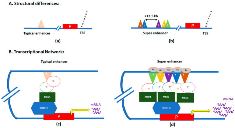
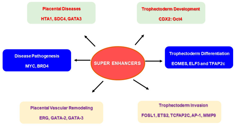
The proliferation of trophoblast stem (TS) cells and their differentiation into multiple lineages are pivotal for placental development and functions. Various transcription factors (TFs), such as CDX2, EOMES, GATA3, TFAP2C, and TEAD4, along with their binding sites and cis-regulatory elements, have been studied for their roles in trophoblast cells. While previous studies have primarily focused on individual enhancer regions in trophoblast development and differentiation, recent attention has shifted towards investigating the role of super-enhancers (SEs) in different trophoblast cell lineages. SEs are clusters of regulatory elements enriched with transcriptional regulators, forming complex gene regulatory networks via differential binding patterns and the synchronized stimulation of multiple target genes. Although the exact role of SEs remains unclear, they are commonly found near master regulator genes for specific cell types and are implicated in the transcriptional regulation of tissue-specific stem cells and lineage determination. Additionally, super-enhancers play a crucial role in regulating cellular growth and differentiation in both normal development and disease pathologies. This review summarizes recent advances on SEs' role in placental development and the pathophysiology of placental diseases, emphasizing the potential for identifying SE-driven networks in the placenta to provide valuable insights for developing therapeutic strategies to address placental dysfunctions.
Keywords: placental development; placental diseases; super-enhancers; transcriptional regulators; trophoblast stem cells.
Conflict of interest statement
The authors declare no conflicts of interest.
Figures
Similar articles
-
Super-enhancer-associated transcription factors collaboratively regulate trophoblast-active gene expression programs in human trophoblast stem cells.Nucleic Acids Res. 2023 May 8;51(8):3806-3819. doi: 10.1093/nar/gkad215. Nucleic Acids Res. 2023. PMID: 36951126 Free PMC article.
-
Super-enhancer-guided mapping of regulatory networks controlling mouse trophoblast stem cells.Nat Commun. 2019 Oct 18;10(1):4749. doi: 10.1038/s41467-019-12720-6. Nat Commun. 2019. PMID: 31628347 Free PMC article.
-
The super-enhancer repertoire in porcine liver.J Anim Sci. 2023 Jan 3;101:skad056. doi: 10.1093/jas/skad056. J Anim Sci. 2023. PMID: 36800318 Free PMC article.
-
[ Super-enhancers. Are they regulators of regulatory genes of development and cancer?].Mol Biol (Mosk). 2015 Nov-Dec;49(6):915-22. doi: 10.7868/S0026898415060051. Mol Biol (Mosk). 2015. PMID: 26710770 Review. Russian.
-
Super enhancers as master gene regulators in the pathogenesis of hematologic malignancies.Biochim Biophys Acta Rev Cancer. 2022 Mar;1877(2):188697. doi: 10.1016/j.bbcan.2022.188697. Epub 2022 Feb 9. Biochim Biophys Acta Rev Cancer. 2022. PMID: 35150791 Review.
References
Publication types
Grants and funding
LinkOut - more resources
Full Text Sources
