

A Case of Canine Hepatitis with Hepatocellular Attack by Non-Neoplastic Perforin-Laden Lymphocytes
- PMID: 40266952
- PMCID: PMC11945539
- DOI: 10.3390/vetsci12030211
A Case of Canine Hepatitis with Hepatocellular Attack by Non-Neoplastic Perforin-Laden Lymphocytes
Abstract
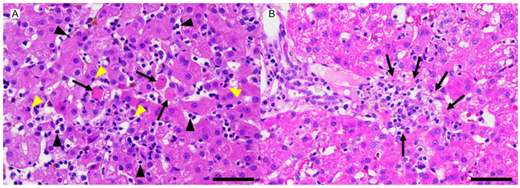
The etiology of canine chronic hepatitis (CH) is unknown, although an autoimmune background has been suggested in some cases of canine CH. An 11 y old spayed female Norwich Terrier showed a marked elevation of liver enzymes with hyperbilirubinemia, regenerative anemia, and thrombocytopenia. A bacterial culture of the surgically excised liver tissue and bile was negative. The histological features of the liver biopsy resembled those of human autoimmune hepatitis except for a paucity of intralesional plasma cells. It was established through immunohistochemistry that CD3-positive perforin-containing T lymphocytes had actively infiltrated the patient's liver causing hepatocellular apoptosis, implying an autoimmune attack on hepatocytes. The patient's general condition improved, with normalization of platelet and serum total bilirubin levels, after immunosuppressive therapy with prednisolone and cyclosporine, whereas liver enzymes did not reach the reference interval. The dog died 11 months after the initiation of immunosuppressive therapy. These pathological findings may be one aspect of autoimmune mediation in canine CH.
Keywords: autoimmune hepatitis; canine chronic hepatitis; hepatocellular apoptosis; immunohistochemistry; immunosuppressive therapy; perforin-containing T lymphocytes.
Conflict of interest statement
The authors declared no potential conflicts of interest with respect to the research, authorship, and/or publication of this article.
Figures


Similar articles
-
Folliculotropic Mycosis Fungoides Associated with Autoimmune Hepatitis.Acta Dermatovenerol Croat. 2017 Oct;25(3):248-250. Acta Dermatovenerol Croat. 2017. PMID: 29252180
-
[Autoimmune hepatitis: two case reports with different clinical courses - case 3/2011].Dtsch Med Wochenschr. 2011 Mar;136(9):436. doi: 10.1055/s-0030-1247622. Epub 2011 Mar 3. Dtsch Med Wochenschr. 2011. PMID: 21374554 German.
-
Perforin and granzyme B lytic protein expression during chronic viral and autoimmune hepatitis.Liver. 1998 Dec;18(6):391-7. doi: 10.1111/j.1600-0676.1998.tb00823.x. Liver. 1998. PMID: 9869393
-
[Autoimmune liver diseases and their overlap syndromes].Praxis (Bern 1994). 2006 Sep 6;95(36):1363-81. doi: 10.1024/1661-8157.95.36.1363. Praxis (Bern 1994). 2006. PMID: 16989180 Review. German.
-
Review article: autoimmune hepatitis -- current management and challenges.Aliment Pharmacol Ther. 2013 Oct;38(8):887-913. doi: 10.1111/apt.12470. Epub 2013 Sep 8. Aliment Pharmacol Ther. 2013. PMID: 24010812 Review.
References
-
- WSAVA Liver Standardization Group . WSAVA Standards for Clinical and Histological Diagnosis of Canine and Feline Liver Disease. Elsevier; Amsterdam, The Netherlands: 2006.
Publication types
LinkOut - more resources
Full Text Sources
