

Novel Soluble apxIVA-Truncated Protein and Its Application to Rapid Detection and Distinction of Actinobacillus pleuropneumoniae Wild-Strain-Infected Samples from Those Vaccinated with apxIV-Partially Deleted Vaccine
- PMID: 40266988
- PMCID: PMC11946594
- DOI: 10.3390/vetsci12030278
Novel Soluble apxIVA-Truncated Protein and Its Application to Rapid Detection and Distinction of Actinobacillus pleuropneumoniae Wild-Strain-Infected Samples from Those Vaccinated with apxIV-Partially Deleted Vaccine
Abstract
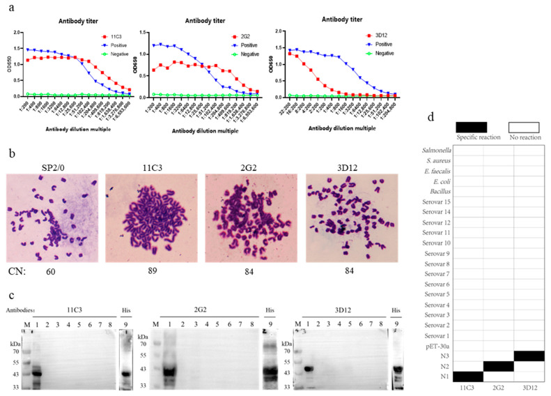
Actinobacillus pleuropneumoniae (APP) is a bacterial pathogen causing porcine pleuropneumonia, causing great economic loss to the global pig industry. Although natural apxIV contributes to the prevention and control of porcine pleuropneumonia, its isolation poses a great challenge, and recombinant soluble apxIV proteins tend to carry large molecular weight tags. The traditional serologic methods tend not to accurately detect the apxIV-partially deleted vaccine (GDV). In this study, we screened the soluble protein apxIVA N2 (756 bp) from six apxIV-truncated proteins and applied it to the enzyme-linked immunosorbent assay (ELISA) and colloidal gold immunochromatographic strip for detecting the samples vaccinated with APP GDV. The results indicate that N2 was close to the natural apxIV protein in terms of structure and function as it only contained a single His (0.86 kDa) tag and a single S (2 kDa) tag. Among the six candidate proteins, N2 exhibited the best performance in distinguishing APP-infected samples from those vaccinated with the APP GDV. Both ELISA and colloidal gold immunochromatographic strips based on this protein exhibited an excellent performance in detecting and distinguishing wild-strain-infected samples from those vaccinated with the subunit vaccine or the GDV. In addition, three monoclonal antibodies against different antigenic epitopes were identified using these truncated proteins. Our studies are of great significance for further research on APP, the differential diagnosis of wild strains and vaccine strains, and pig control breeding, exhibiting a broad application prospect in the on-site diagnosis of APP, particularly in remote areas lacking detection instruments and professionals.
Keywords: Actinobacillus pleuropneumoniae; ELISA; apxIV; colloidal gold immunochromatographic strip; gene-deleted vaccine; rapid detection; wild strain.
Conflict of interest statement
The authors declare no conflicts of interest.
Figures


Similar articles
-
Use of recombinant ApxIV in serodiagnosis of Actinobacillus pleuropneumoniae infections, development and prevalidation of the ApxIV ELISA.Vet Microbiol. 2004 Apr 19;99(3-4):227-38. doi: 10.1016/j.vetmic.2004.01.004. Vet Microbiol. 2004. PMID: 15066725
-
Detection of Actinobacillus Pleuropneumoniae ApxIV Toxin Antibody in Serum and Oral Fluid Specimens from Pigs Inoculated Under Experimental Conditions.J Vet Res. 2017 Dec 6;61(2):163-171. doi: 10.1515/jvetres-2017-0021. eCollection 2017 Jun. J Vet Res. 2017. PMID: 29978069 Free PMC article.
-
[The seroprevalence of Actinobacillus pleuropneumoniae in Swiss pig breeding herds--a study with the ApxIV ELISA].Schweiz Arch Tierheilkd. 2008 Mar;150(3):103-9. doi: 10.1024/0036-7281.150.3.103. Schweiz Arch Tierheilkd. 2008. PMID: 18429500 German.
-
New trends in innovative vaccine development against Actinobacillus pleuropneumoniae.Vet Microbiol. 2018 Apr;217:66-75. doi: 10.1016/j.vetmic.2018.02.028. Epub 2018 Mar 6. Vet Microbiol. 2018. PMID: 29615259 Review.
-
Actinobacillus pleuropneumoniae: The molecular determinants of virulence and pathogenesis.Adv Microb Physiol. 2021;78:179-216. doi: 10.1016/bs.ampbs.2020.12.001. Epub 2021 Jan 25. Adv Microb Physiol. 2021. PMID: 34147185 Review.
Cited by
-
Preparation and Evaluation of Novel Epitope-Based ETEC K88-K99 Bivalent Vaccine.Vet Sci. 2025 Apr 18;12(4):381. doi: 10.3390/vetsci12040381. Vet Sci. 2025. PMID: 40284883 Free PMC article.
References
-
- Stancheva S.G., Frömbling J., Sassu E.L., Hennig-Pauka I., Ladinig A., Gerner W., Grunert T., Ehling-Schulz M. Proteomic and immunoproteomic insights into the exoproteome of Actinobacillus pleuropneumoniae, the causative agent of porcine pleuropneumonia. Microb. Pathog. 2022;172:105759. doi: 10.1016/j.micpath.2022.105759. - DOI - PubMed
-
- Auger E., Deslandes V., Ramjeet M., Contreras I., Nash J.H., Harel J., Gottschalk M., Olivier M., Jacques M. Host-pathogen interactions of Actinobacillus pleuropneumoniae with porcine lung and tracheal epithelial cells. Infect. Immun. 2009;77:1426–1441. doi: 10.1128/IAI.00297-08. - DOI - PMC - PubMed
-
- Rao J., Wei X., Li H., Zhang Z., Liu J., Lian M., Cao W., Yuan L., Dou B., Tian Y., et al. Novel Multiplex PCR Assay and Its Application in Detecting Prevalence and Antibiotic Susceptibility of Porcine Respiratory Bacterial Pathogens in Guangxi, China. Microbiol. Spectr. 2023;11:e0397122. doi: 10.1128/spectrum.03971-22. - DOI - PMC - PubMed
Grants and funding
- 2022YFD1800905/the National Key Research and Development Program of China
- 32273039/the National Natural Science Foundation of China
- (2023) General 076/the achievements Transformation Project of Guizhou Provincial Science and Technology Department
- CARS-35/the China Agriculture Research System of MOF and MARA
LinkOut - more resources
Full Text Sources
