. 2025 Apr 25;11(17):eads4661.
doi: 10.1126/sciadv.ads4661. Epub 2025 Apr 23.

Synaptophysin accelerates synaptic vesicle fusion by expanding the membrane upon neurotransmitter loading

Affiliations

Synaptophysin accelerates synaptic vesicle fusion by expanding the membrane upon neurotransmitter loading

Julia Preobraschenski et al. Sci Adv. .

Abstract

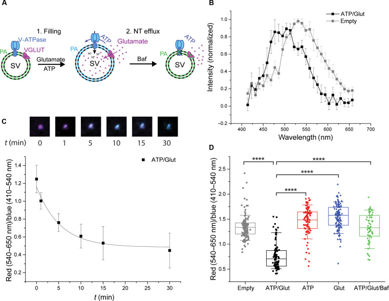
Synaptic transmission is mediated by the exocytotic release of neurotransmitters stored in synaptic vesicles (SVs). SVs filled with neurotransmitters preferentially undergo exocytosis, but it is unclear how this is achieved. Here, we show that during transmitter loading, SVs substantially increase in size, which is reversible and requires synaptophysin, an abundant membrane protein with an unclear function. SVs are larger when synaptophysin is knocked out, and conversely, liposomes are smaller when reconstituted with synaptophysin. Moreover, transmitter loading of SVs accelerates fusion in vitro, which is abolished when synaptophysin is lacking despite near normal transmitter uptake. We conclude that synaptophysin functions as a curvature-promoting entity in the SV membrane, allowing for major lateral expansion of the SV membrane during neurotransmitter filling, thus increasing their propensity for exocytosis.


Figures

Fig. 1.
Fig. 1.. SVs reversibly expand upon Glut loading.
(A) Cartoon illustrating the SV size changes associated with Glut influx and efflux. (B) Time-dependent size changes, measured by DLS, of SVs (upper panel) and proteoliposomes reconstituted with VGLUT1 and TFoF1 (lower panel) upon incubation in 10 mM K-glutamate, 4 mM Mg-ATP, and 4 mM KCl (Glut uptake conditions: ATP/Glut; black line), 4 mM ATP (red line), and 10 mM K-glutamate (Glut; blue line) in 300 mM glycine and 5 mM Hepes (pH 7.4; uptake buffer). The reaction was started by the addition of ATP and Glut. In addition, SV size changes under Glut uptake conditions after addition of Baf were monitored. The time point of addition (10 min) is marked with an arrow. (C) Increase in SV radii, measured by DLS, in the presence of various substrates after an incubation of 10 min at 37°C. (D and E) Representative micrographs of SVs acquired by cryo-EM (scale bars correspond to 50 nm) (D) and histogram and box plot showing the SV diameter determined by cryo-EM (E) under Glut uptake conditions (ATP/Glut; black) and in the presence of Glut ATP and Baf (ATP/Glut/Baf; green). The box in the box plot represents data between 25 and 75%, and the whiskers show data between 10 and 90%. The dot within the box indicates the mean value, and the line indicates the median. The data in the box plot were analyzed using a two-tailed paired t test, ****P < 0.0001. [(B) and (C)] Measured by DLS. (B) n = 3 to 6; (C) n = 2 to 6; (D) n = 2; number of SVs, 602 (ATP/Glut/Baf) and 948 (ATP/Glut).
Fig. 2.
Fig. 2.. Size changes associated with Glut loading affect the lipid order of the SV membrane.
(A) Illustration of changes in lipid order in empty and Glut-filled SVs. (B) Emission spectra of PA dye–labeled, Glut-filled (black squares) and empty (gray squares) SVs. (C) Time course of lipid order changes represented as red (540 to 650 nm)/blue (410 to 540 nm) ratios of PA dye–labeled SVs during a time course of Glut uptake (exposure to ATP/Glut at time 0) with representative corresponding images of a single SV. (D) Box plot representing red (540 to 650 nm)/blue (410 to 540 nm) ratios of PA dye–labeled SVs in the absence of substrates (empty; gray), under Glut-loaded conditions (ATP/Glut; black), and in the presence of ATP (red), Glut (blue), and ATP, Glut, and Baf (ATP/Glut/Baf; green). The box represents data between 25 and 75%, and the line in the box indicates the median. The whiskers show data between 10 and 90%. Individual values are displayed as gray (empty) and black rectangles (ATP/Glut), red circles (ATP), and blue upward-facing (Glut) and green downward-facing (ATP/Glut/Baf) triangles. The data in the box plot were analyzed using a two-tailed paired t test. ****P < 0.0001. (B) n = 7; (C) n = 7; (D) n = 55 to 128.
Fig. 3.
Fig. 3.. SYP KO SVs expand less and are larger than WT SVs.
(A) Schematic depicting Glut filling–associated size changes in WT and SYP KO SVs. (B) Time-dependent expansion of WT (black line) and SYP KO (dark red line) SVs under Glut uptake conditions. Inset: radii of WT and SYP KO SVs after 1-min (black bars) and 10-min (white bars) incubation under Glut uptake conditions. (C) Representative micrographs of empty and filled WT SVs acquired by cryo-EM. Scale bars correspond to 20 nm. (D) Bilayer thickness of empty and Glut-filled WT and SYP KO SVs. (E) Size distribution of isolated WT (black line) and SYP KO (dark red line) mouse SVs analyzed by DLS. (F) Size distribution of synaptophysin-reconstituted (SYP LUVs), VGLUT1-reconstituted (VGLUT1 LUVs), and protein-free (LUVs) liposomes demonstrating that synaptophysin supports the formation of liposomes with a high curvature. (G) Representative micrographs of SVs in mouse hippocampal WT (left panel) and SYP KO (right panel) neurons acquired by electron microscopy. (H) Histogram and box plot (inset) showing the size distribution of WT (black bars and box) and SYP KO (dark red bars and box) SVs in mouse hippocampal neurons imaged and analyzed using electron microscopy. The box in the box plots in (D) and (H) represents data between 25 and 75%, and the whiskers show data between 10 and 90%. The dot within the box indicates the mean value, and the line indicates the median. The data in the box plots in (D) and (H) were analyzed using a two-tailed paired t test. ****P < 0.0001. [(A) to (D)] Analyzed by DLS. (B) n = 2 to 3; (D) 15,829 to 38,238 sites in 15 to 29 SVs per condition. (E) n = 3; (F) n = 3; (H) n = 7 to 9; number of SVs, 1383 (WT) and 1011 (SYP KO).
Fig. 4.
Fig. 4.. NT-filled SVs fuse faster than empty SVs.
(A) Cartoon of the fusion assay of SVs [see (35) for details]. Planar supported bilayers containing the SNARE proteins syntaxin-1A and SNAP-25 were incubated with SVs in the presence of complexin-1, Munc18, and Munc13. Subsequently, ATP and NTs were added as indicated to allow for SV filling for 20 min. Ca2+ (100 μM) was then added to trigger rapid fusion of SVs, while images of single vesicles were acquired on a total internal reflection fluorescence microscope. (B) Cumulative distribution of the fusion delay times of predocked SVs from the time of Ca2+ addition (shown as the percent of docked vesicles at the onset of the reaction). Note that the data for ATP/Glut are shown in both panels for comparison. (C) Rate constants of whole-brain SV fusion calculated by fitting using either one-component (k1; left panel) or two-component first-order kinetic models (k1 and k2) (see Materials and Methods for details). (D) Immunoblot showing the enrichment of VGLUT1 and VGAT, respectively, in fractions obtained by immunoisolation using beads coupled with VGLUT1- and VGAT-specific antibodies. Compared to the starting vesicle fraction (SV), the supernatants from the immunoisolation experiments (SN) reveal selective depletion of the respective vesicle populations. For reference, a blot for the V0a1 subunit of the V-ATPase is shown, which is present on all SVs. (E) Comparison between the fusion kinetics of immunoisolated glutamatergic and GABAergic SVs with those of nondepleted SVs after preloading with the respective NT (all SVs ATP/Glut and all SVs ATP/GABA versus VGLUT1 SVs ATP/Glut and VGAT SVs ATP/GABA). (C) n = 4 to 7. Note that both glutamatergic and GABAergic vesicles displayed similar fast fusion kinetics when incubated with ATP and the respective NT. Open symbols: experiments with immunoisolated SVs; closed symbols: experiments with conventionally isolated SVs.
Fig. 5.
Fig. 5.. In contrast to SVs from WT mice, fusion of SVs from SYP KO mice is not enhanced upon loading with Glut.
Fusion of WT (A) and SYP KO (B) SVs after Glut preloading (ATP/Glut; black squares) in comparison to control conditions [ATP only (ATP; red circles) and Glut only (Glut; blue triangles)]. (C) Rate constants of WT and SYP KO SVs under uptake and control conditions. See Fig. 4 for details of the assay. (C) n = 4 to 9. Note that in these experiments, the two kinetic components could not be clearly resolved anymore, possibly due to differences in the vesicle preparation methods (mouse versus rat).

References

    1. Herman M. A., Ackermann F., Trimbuch T., Rosenmund C., Vesicular glutamate transporter expression level affects synaptic vesicle release probability at hippocampal synapses in culture. J. Neurosci. 34, 11781–11791 (2014). - PMC - PubMed
    1. Rost B. R., Schneider F., Grauel M. K., Wozny C., Bentz C., Blessing A., Rosenmund T., Jentsch T. J., Schmitz D., Hegemann P., Rosenmund C., Optogenetic acidification of synaptic vesicles and lysosomes. Nat. Neurosci. 18, 1845–1852 (2015). - PMC - PubMed
    1. Wojcik S. M., Rhee J. S., Herzog E., Sigler A., Jahn R., Takamori S., Brose N., Rosenmund C., An essential role for vesicular glutamate transporter 1 (VGLUT1) in postnatal development and control of quantal size. Proc. Natl. Acad. Sci. U.S.A. 101, 7158–7163 (2004). - PMC - PubMed
    1. Gracz L. M., Wang W. C., Parsons S. M., Cholinergic synaptic vesicle heterogeneity: Evidence for regulation of acetylcholine transport. Biochemistry 27, 5268–5274 (1988). - PubMed
    1. Bruns D., Riedel D., Klingauf J., Jahn R., Quantal release of serotonin. Neuron 28, 205–220 (2000). - PubMed

LinkOut - more resources