. 2025 Apr 23;139(11):507-526.
doi: 10.1042/CS20241204. Online ahead of print.

Angiotensin II-induced cardiac fibrosis and dysfunction are exacerbated by deletion of cGKI in periostin+ myofibroblasts

Affiliations

Angiotensin II-induced cardiac fibrosis and dysfunction are exacerbated by deletion of cGKI in periostin+ myofibroblasts

Melanie Cruz Santos et al. Clin Sci (Lond). .

Abstract

Differentiation of cardiac fibroblasts (CF) into myofibroblasts (CMFs) is considered a critical event in response to the maladaptive cardiac remodeling triggered by angiotensin II (Ang II). Active CMFs are proliferative and contribute to the production of extracellular matrix and matricellular proteins such as periostin, to myocardial fibrosis and thus muscle stiffness. Although previous studies provided substantial evidence for the antifibrotic signaling elicited by NO/NP-cGMP-cGKI, the role of this axis in modulating CMF function(s) in vivo remains unclear.To address this, Ang II was delivered through osmotic minipumps into tamoxifen-induced CMF-specific cGKI knockout (cmfKO) and littermate control (CTR) male mice. CMF-restricted Cre activity in periostin+ cells resulted in an effective depletion of the cGKI protein observed in myocardial sections and in primary CF/CMF protein lysates obtained from Ang II-and tamoxifen-treated cmfKO. Although both genotypes responded identically to Ang II in terms of blood pressure and cardiac enlargement, cmfKO hearts showed significantly increased cardiomyocyte cross-sectional areas and developed a marked increase in myocardial fibrosis. Moreover, non-invasive echocardiography revealed a structure-related distortion of global systolic function and longitudinal deformation capacity in cmfKO versus CTR. Consistent with the results obtained in vivo, we observed a higher proliferation rate of CF/CMF derived from Ang II-treated cmfKO hearts compared to respective CTR cells as well as an increase in cardiomyocyte apoptosis in the absence of cGKI in periostin+ CMF. Our data confirm that endogenous cGKI function in periostin+ CMFs counteracts the Ang II-induced morphologic and structural changes that impair cardiomyocyte survival ultimately causing loss of heart function in male mice.

Keywords: angiotensin II; cGKI; cGMP; cardiac myofibroblasts; cardiac remodeling; fibrosis; periostin.


Conflict of interest statement

All other authors have explicitly declared that there are no conflicts of interest related to this article.

Figures

Figure 1:
Figure 1:. CMF-specific cGKI-KO mice exhibit a high vulnerability to chronic Ang II exposure.
(A) Genomic PCR analysis demonstrates TAM-mediated cell-specific PostniCre-recombination. DNA isolated from indicated tissues as well as primary CF/CMF of Ang II-treated cmfKO mice was amplified by allele-specific primer sets for Prkg1, encoding murine cGKI. As expected, conversion of the floxed Prkg1 allele ([fl]; 338 bp) to the KO allele ([-]; 250 bp) was almost exclusively detectable in samples obtained from primary CF/CMF cultures, while recombination in the lungs did occur, but at a much lower level. Prior (data not shown) or 28 days upon TAM-induced PostniCre recombination all other tissues tested remained negative for the KO-specific PCR product. (B, C) Representative immunoblot images of primary CF/CMF protein lysates obtained from +TAM and +Ang II-treated CTR mice exhibited a single band, corresponding to the expected molecular weight of cGKI, whereas cGKI immunoreactivity was largely attenuated in CF/CMFs derived from primary cmfKO cultures. GAPDH was utilized as loading control. CM protein lysates obtained from CM-specific cGKI KO (αMHC-Cretg/+ x cGKIfl/fl; CM-cKO) hearts served as negative controls. (C) Quantification of the immunoblot shown in (C) with N = 6 protein samples per genotype. (D) Representative histochemical staining’s of consecutive heart cryosections derived from +TAM-treated CTR and cmfKO mice following 28 days of Ang II treatment. Fibrotic areas were visualized by PSR staining (left panels). Validated antibodies specifically targeting periostin (middle panels) [66] or cGKI (right panels) [67] were used to detect the respective proteins in serial sections adjacent to histologically detectable myocardial fibrosis. While periostin was restricted to the fibrotic cardiac areas in both genotypes, cGKI expression in cmfKO versus CTR hearts was largely depleted within the PSR- and periostin-positive heart area. In total, n = 3 cryosections were evaluated per heart deriving from N = 3 mice per genotype. Scale bar = 100 µm. (E) Kaplan–Meier analysis of CTR and cmfKO mice following either +TAM (CTR, N = 13; cmfKO, N = 12) or +TAM and +Ang II (CTR, N = 39; cmfKO, N = 42) treatment. Overall survival within +TAM-treated groups was not affected by genotype (n.s.), and all mice survived the respective treatment. With chronic Ang II exposure, 85% of CTR mice survived the indicated treatment, whereas the survival of cmfKO mice was 71% and therefore significantly lower compared with the corresponding TAM-treated cmfKO group. (F) Time course of MAP changes in response to Ang II during the first seven days (day = 1–7) after osmotic pump implantation (day = 1) yielded no genotype-related differences although, as expected, the Ang II treatment by itself significantly increased BP compared with the average MAP values obtained during the last 24 h of the basal measurements, i.e., immediately prior to minipump implantation (day = 0). N = 4 unrestrained and awake mice per genotype carrying telemetry implants were monitored prior and during the first seven days of the Ang II infusion. (G) Quantification of the relative increase (compared with basal values) in MAP, systolic and diastolic BP, and pulse pressure (PP) expressed as △mmHg exhibited no genotype-related differences. Statistics: (C, F, and G) Data are represented as mean + or ± SEM with *P<0.05 (C) using an unpaired t-test and (E) the log-rank test (Mantel-Cox test) to assess the survival rate with †P<0.05 revealing differences between the distinct treatments (+TAM versus +TAM+Ang II) within the cmfKO group. (F) Two-way ANOVA followed by Šídák’s multiple comparisons test showed a significant increase (#P<0.001) in BP over time due to the +TAM+Ang II treatment within both the CTR and cmfKO group, but no differences between genotypes (n.s.). (G) Multiple unpaired t-test corrected for multiple comparison using the Holm-Šídák method revealed again no differences (n.s.) on all parameters plotted between the genotypes due to the +TAM+Ang II treatment. Further details concerning statistics are listed together with raw data in Supplementary Table S1. Ang II, angiotensin II; CF, cardiac fibroblast; CMF, cardiac myofibroblast; CTR, control; MAP, mean arterial pressure; PP, pulse pressure; TAM, tamoxifen.
Figure 2:
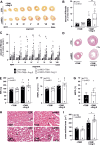
Figure 2:. Ang II provokes greater histopathological changes in cmfKO hearts.
(A) Hearts derived from +TAM and +TAM plus Ang II-treated CTR and cmfKO animals were divided into eight equidistant cardiac segments, ranging from the apex (segment I) to the base of the hearts (segment VIII) and were subsequently stained with PSR to (B) quantify the amount of fibrosis as percentage of the whole myocardial area within each segment. These analyses revealed an overall significantly higher percentage of fibrosis in cmfKO hearts (N = 9) compared with CTR hearts (N = 8), which was due to an elevated (C) amount of collagen depositions in each (I–VIII) of the cmfKO heart segments. Compared with the +TAM-treated groups the +TAM+Ang II treatment exaggerated fibrosis in segments II–VI of the cmfKO compared with CTR. In (C) n = 2–4 consecutive heart slices per segment (I–VIII) per animal (N = 9 cmfKO and N = 8 CTR) were included in the analysis. (D) Representative H&E staining of whole heart cross-sections obtained from +TAM or +TAM+Ang II-challenged CTR and cmfKO mice. Both genotypes showed pronounced hypertrophy development to a comparable extent upon the chronic Ang II exposure (CTR, N = 8; cmfKO, N = 9), as indicated by significant increases in (E) total HW, (F) HW/TL ratio as well as (G) in the percentage increase in HW/TL compared with the corresponding +TAM control groups (CTR, N = 10; cmfKO, N = 9). (H) Representative H&E-staining’s of cardiac cryosections from CTR and cmfKO after either +TAM or +TAM+Ang II treatment. (I) CM cross-sectional areas significantly increased in both genotypes after Ang II infusion (CTR, N = 7; cmfKO, N = 9) compared with their respective control (+TAM) group (CTR, N = 8; cmfKO, N = 6). Ang II-induced hypertrophic CM growth, however, resulted in a greater enlargement in cmfKO compared with CTR hearts. Cross-sectional areas of n = 150 CMs were evaluated per individual heart. Representative cells that were included in the statistics are outlined in black in all panels. Statistics: (B) Two-way ANOVA followed by the two-stage linear step-up procedure of Benjamini, Krieger, and Yekutieli, with statistical significance determined by a false discovery rate (FDR) threshold of q<0.05. Significant differences were found at **P<0.01 between genotypes and at †P<0.05 or #P<0.001 between distinct treatments (+TAM versus +TAM+Ang II) within the same genotype as indicated. (C) Two-way ANOVA followed by the two-stage linear step-up procedure of Benjamini, Krieger, and Yekutieli, with statistical significance determined by a FDR threshold of q<0.05. Significant differences were found at *P<0.05 and **P<0.01 between genotypes in the respective segments and at †P<0.05, §P<0.01, and #P<0.001 for distinct treatments (+TAM versus +TAM+Ang II) within the same segment/genotype. (E, F, and I). Two-way ANOVA followed by Tukey’s multiple comparisons test revealing significant differences at *P<0.05 between genotypes and at †P<0.05, §P<0.01, or #P<0.001 between the distinct treatments (+TAM versus +TAM+Ang II) within the same genotype. (G) Unpaired t-test did not show a difference between the [%] increase in HW/TL between genotypes. Details concerning statistics are listed together with raw data in Supplementary Table S2. Ang II, angiotensin II; CF, cardiac fibroblast; CMF, cardiac myofibroblast; CTR, control; HW, heart weight; TAM, tamoxifen; TL, tibia length.
Figure 3:
Figure 3:. CMF-specific ablation of cGKI resulted in higher CMs cell death after Ang II infusion.
Assessment of CM cell death in heart slices obtained from +TAM- and Ang II-treated CTR and cmfKO animals. (A) Representative images of the TUNEL assay co-stained with troponin I for identification of CMs in both fibrotic and non-fibrotic cardiac regions. Quantification of TUNEL-positive CMs, expressed as a percentage of the total number of CMs, revealed a significantly increased cell death rate in (B) non-fibrotic cardiac regions of the cmfKO hearts (N = 6) in response to prolonged Ang II stimulation compared with the corresponding CTR hearts (N = 5). (C) In contrast, no genotype-related difference could be detected within fibrotic cardiac regions (N = 6 for each genotype) containing proliferating non-CM. (D) If the two previously separately analyzed heart areas are considered together, higher CM cell death was confirmed in cmfKO (N = 12) compared with CTR hearts (N = 11). For each animal, n = 3 non-fibrotic/fibrotic areas, originating from two different heart segments (IV, VI), within n = 6 heart slices comprising an area of 0.15 mm2 were analyzed. Statistics: Data in (B) and (D) are represented as mean  +  SEM with *P<0.05 indicating significant difference between genotypes (unpaired student t-test). Additional information on statistics are presented together with raw data in Supplementary Table S3. Ang II, angiotensin II; CF, cardiac fibroblast; CMF, cardiac myofibroblast; CTR, control; TAM, tamoxifen.
Figure 4:
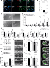
Figure 4:. Loss of CMF-specific cGKI causes CF/CMF hyperproliferation, relieves anti-fibrotic effects on gene transcription by 8-Br-cGMP in vitro, and results in a worsening of LV function in vivo.
(A) Immunofluorescence staining and (B) quantification of the proliferation marker Ki-67. Collagen I antibodies were used in order to localize the fibrotic area containing CMFs. To calculate the proliferative index, the number of Ki-67+ cells was related to defined areas of fibrosis. Analysis of n = 3 areas comprising 0.15 mm2 in distinct heart segments (segment IV–VI) exhibited a significantly increased accumulation of Ki-67+ nuclei in cmfKO (N = 9) versus CTR (N = 7). (C) Representative images acquired from the grid-based proliferation assay. CF/CMF isolated from either CTR or cmfKO mice receiving +TAM+Ang II in vivo for 28 days are shown after t = 120 h in primary culture. (D) During the five-day monitoring period, this assay revealed a significantly enhanced proliferation rate of primary cmfKO (N = 6) versus CTR (N = 8) CF/CMF cells. n = 3 replicates were assessed per genotype per experiment. (E) qRT-PCR analysis of common fibroblast marker transcripts from +TAM and +Ang II-treated CTR and cmfKO hearts. Stimulation of CF/CMF with 8-Br cGMP (8-Br; 1 mM) for 24 h revealed that levels of Acta2, Col1a1, and Fn1, but not of TGFβ1 and Il6, were sensitive to the 8-Br cGMP treatment in CTR cells. In contrast, transcript levels of all pro-fibrotic markers examined did not differ for cmfKO both under basal and stimulated conditions. (F) CTR and cmfKO CF/CMF obtained from +TAM+Ang II in vivo treated mice (28 d) were stimulated in culture using 8-Br for 30 min to induce VASP phosphorylation at Ser239 (pVASPS239). (F) Representative immunoblots and (G) relative densitometric bar graphs with individual data points plotted revealed a significant increase in the pVASPS239 to VASP ratio only in the presence of cGKI, i.e., in CTR samples. (H) Representative M-mode images in the PSLAX view of CTR and cmfKO mice either after TAM injection alone or following additional 28 days of Ang II treatment. While CTR animals (N = 8) displayed no deterioration of global cardiac function after chronic Ang II exposure versus unchallenged CTR animals (N = 8), non-invasive analysis of the global heart function yielded a significant decline in (I) EF and (J) FS in cmfKO mice (N = 9) as compared with Ang II-treated CTR and corresponding TAM-treated cmfKO (N = 7) groups. (K) Regional analysis of wall motion tracking in PSLAX B-mode during systole and diastole is expressed by velocity vectors in representative images obtained from CTR and cmfKO mice subjected to 28 days of Ang II treatment. Both the systolic and the diastolic velocity vectors were shorter in cmfKO heart compared with CTR hearts, indicating an impairment of the ventricular wall motion. Statistics: Data in (B) are represented as mean  +  SEM with **P<0.01 using an unpaired student t-test. (D) Two-way ANOVA with ***P<0.001 (F1,71 = 16.47) showing significant difference for the overall proliferation rate between genotypes followed by Šídák’s multiple comparisons test revealing significant difference at *P<0.05 for 96 h and 120 h between genotypes. (E) Two-way ANOVA followed by Tukey’s multiple comparisons test with †P<0.05 indicating significant difference for the Acta2/Hprt (F1.20 = 7.3) Col1a1/Hprt (F1.18 = 7.540) and the Fn1/Hprt (F1.15 = 5.836) ratios between the distinct treatments (basal versus 8-Br) for the CTR+TAM+Ang II group. (G) Two-way ANOVA followed by Tukey’s multiple comparisons test with *P<0.05 (F1.16 = 7.63) showing significant difference for the pVASP/VASP ratio between genotypes (considering all basal and 8-Br values) and for the 8-Br condition (*P<0.05). Only the values in the CTR group reached the significance level at #P<0.001 for the comparison of the basal versus 8-Br condition. (I, J) Two-way ANOVA followed by Tukey’s multiple comparison test with significant difference *P<0.05 between genotypes and †P<0.05 revealing differences between the distinct treatments (+TAM versus +TAM+Ang II) within the cmfKO group. Further details are listed together with raw data in Supplementary Table S4. 8-Br, 8-Br cGMP; Ang II, angiotensin II; CF, cardiac fibroblast; CMF, cardiac myofibroblast; CTR, control; TAM, tamoxifen; VASP, vasodilator-stimulated phospho-protein.
Figure 5:
Figure 5:. Summarized scheme of the results.
As indicated, the loss of endogenous cGKI function in the cardiac myofibroblasts of male mice with angiotensin II exposure resulted in numerous deleterious effects and dysfunctions at the cellular and organ level (Created with BioRender.com).

References

    1. Jellis, C., Martin, J., Narula, J. and Marwick, T.H. (2010) Assessment of nonischemic myocardial fibrosis. J. Am. Coll. Cardiol. 56, 89–97 10.1016/j.jacc.2010.02.047 - DOI - PubMed
    1. Hinderer, S. and Schenke-Layland, K. (2019) Cardiac fibrosis - a short review of causes and therapeutic strategies. Adv. Drug Deliv. Rev. 146, 77–82 10.1016/j.addr.2019.05.011 - DOI - PubMed
    1. Jiang, W., Xiong, Y., Li, X. and Yang, Y. (2021) Cardiac fibrosis: cellular effectors, molecular pathways, and exosomal roles. Front. Cardiovasc. Med. 8, 715258 10.3389/fcvm.2021.715258 - DOI - PMC - PubMed
    1. Maruyama, K. and Imanaka-Yoshida, K. (2022) The pathogenesis of cardiac fibrosis: a review of recent progress. Int. J. Mol. Sci. 23, 2617 10.3390/ijms23052617 - DOI - PMC - PubMed
    1. Mc Namara, K., Alzubaidi, H. and Jackson, J.K. (2019) Cardiovascular disease as a leading cause of death: how are pharmacists getting involved? Integr. Pharm. Res. Pract. 8, 1–11 10.2147/IPRP.S133088 - DOI - PMC - PubMed

LinkOut - more resources