. 2025 Apr;15(4):e70305.
doi: 10.1002/ctm2.70305.

Ad-E6/7-HR vaccine improves the prophylactic and therapeutic efficacy in HPV-associated cancers

Affiliations

Ad-E6/7-HR vaccine improves the prophylactic and therapeutic efficacy in HPV-associated cancers

Yu Zhang et al. Clin Transl Med. 2025 Apr.

Abstract

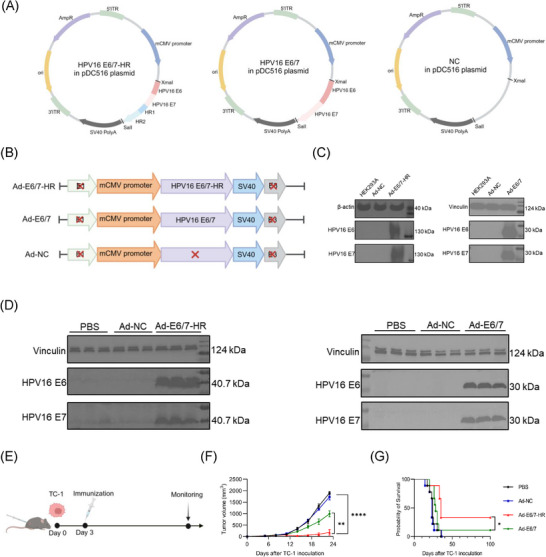
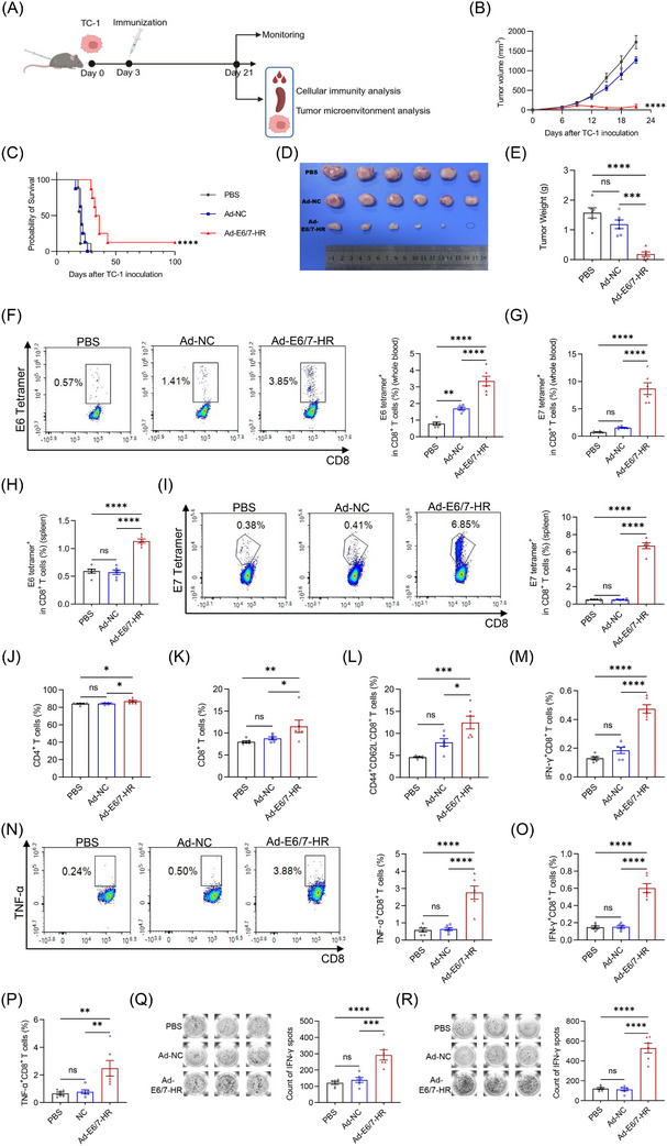
Background: High-risk human papillomavirus (HPV), especially HPV16, is closely correlated with certain cancers. E6 and E7 proteins of HPV16 play critical roles in oncogenesis, making them optimal targets for treating HPV-associated cancers. Here, we engineered an innovative vaccine, Ad-E6/7-HR, designed to evoke immune responses through the incorporation of self-assembling heptad-repeat 1 (HR1) and HR2 originated from Severe acute respiratory syndrome coronavirus 2.

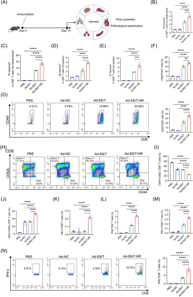
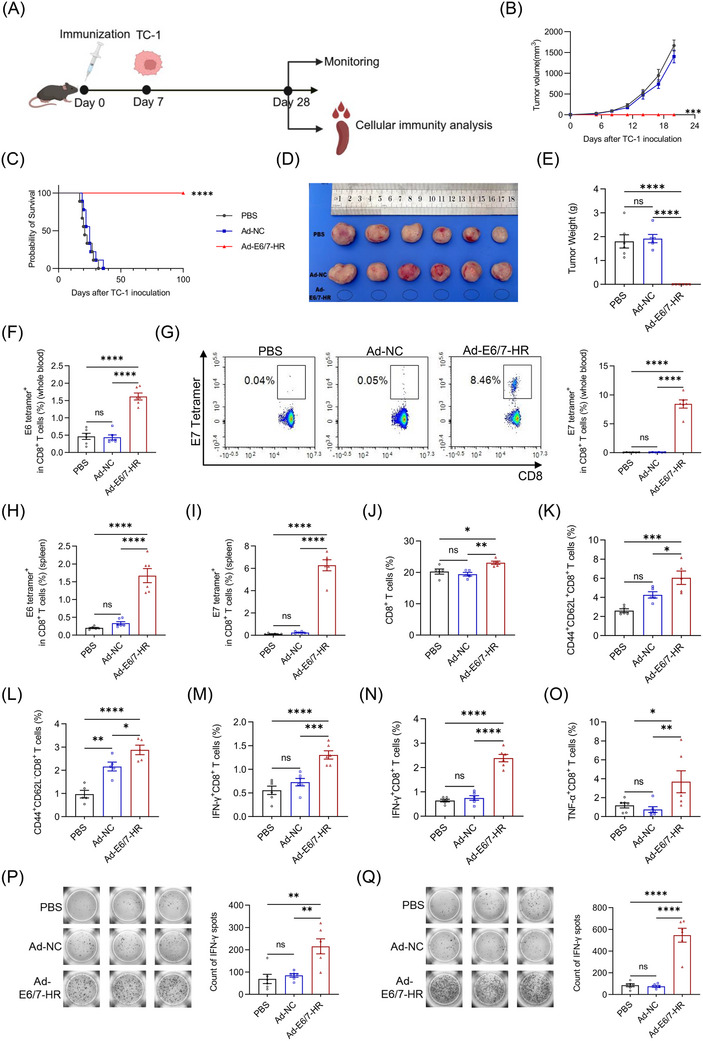
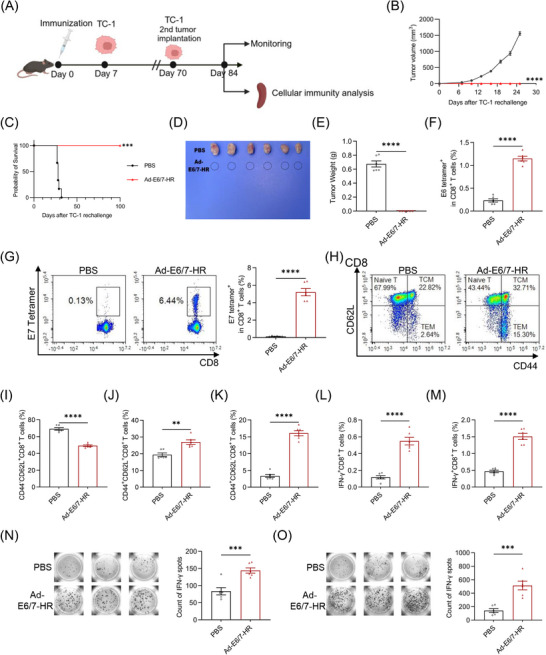
Methods: Ad-E6/7-HR was constructed utilising a replication-defective human adenovirus serotype 5 vector and evaluated its immunogenicity and therapeutic efficacy in murine models. We verified the antitumour efficacy of the vaccine in TC-1 subcutaneous and pulmonary models. Flow cytometry, enzyme-linked immunospot assay, and immunofluorescence staining were used to assess the cellular immunogenicity of Ad-E6/7-HR.

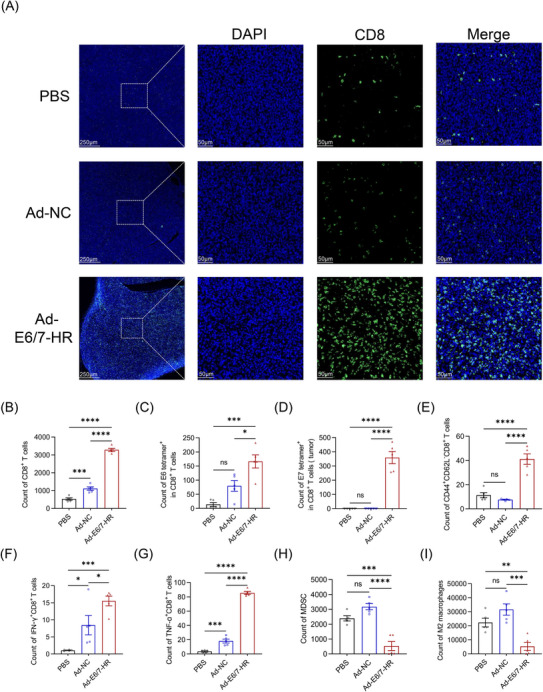
Results: Ad-E6/7-HR induced robust immune responses, significantly increasing antigen-specific CD8+ T cells. The vaccine also enhanced memory T-cell generation and induced potent cytokine secretion, as exemplified by interferon-γ and tumour necrosis factor-α. Ad-E6/7-HR conferred complete protection against tumour growth in the prophylactic model. In therapeutic settings, Ad-E6/7-HR significantly reduced tumour size and improved survival. Furthermore, Ad-E6/7-HR reshaped the tumour microenvironment by increased CD8+ T-cell recruitment and reduced immunosuppressive cells, like myeloid-derived suppressor cells and M2 macrophages, thereby enhancing antitumour immunity.

Conclusions: By targeting HPV16 E6 and E7 proteins and leveraging the self-assembling HR1 and HR2 sequences to enhance immune responses, Ad-E6/7-HR represented a promising candidate for preventing and treating HPV-associated cancers. Further clinical investigation is warranted to evaluate its potential in human trials.

Keywords: adenovirus vaccine; cellular immunity; human‐papillomavirus‐associated cancers; oncology.


Conflict of interest statement

The authors declare no conflict of interest.

Figures

FIGURE 1
FIGURE 1
Design of Ad‐E6/7‐HR vaccine. (A, B) The schematic design of the vaccine. (C) Western blot analysis of expression of HPV16 E6 and E7 in HEK293A cells after transduced with Ad‐E6/7‐HR, Ad‐E6/7, or Ad‐NC. (D) Western blot analysis of expression of HPV16 E6 and E7 in vivo after mice were immunised with PBS, Ad‐NC, Ad‐E6/7‐HR, or Ad‐E6/7. (E) The timeline of the animal experiment. (F, G) Kinetics of tumour growth (F) and overall survival (G) were shown in a therapeutic model. (F), n = 6 per group. (G), n = 9 per group. Data are shown as mean ± SEM and analysed by two‐way ANOVA (F) and Log‐rank test (G). * p < .05, ** p < .01, **** p < .0001.
FIGURE 2
FIGURE 2
Ad‐E6/7‐HR vaccine can induce robust immune responses. (A) Strategy of the experiment. Mice were immunised intramuscularly with 5×10⁹ VP of Ad‐E6/7‐HR, Ad‐E6/7, Ad‐NC, or PBS respectively. (B, C) Percentages of E6 tetramer+ (B) and E7 tetramer+ (C) CD8+ T cells from blood. (D, E) Percentages of E6 tetramer+ (D) and E7 tetramer+ (E) CD8+ T cells in splenocytes. (F) Proportions of activated CD4+ T cells in splenocytes. (G) Representative plot and proportion of activated CD8+ T cells from spleen. (H) Representative plots of CD8+ naïve T cells (CD44 CD62L+) and CD8+ effector memory T cells (TEM, CD44+ CD62L). (I, J) Proportions of CD8+ naïve T cells (I) and CD8+ TEM (J) from spleen. (K, L) Percentages of TNF‐α+ CD8+ (K) and IFN‐γ+ CD8+ (L) T cells stimulated by E6 peptide pool. (M) Percentages of TNF‐α+ CD8+ T cells after stimulated by E7 peptide. (N) Representative plot and percentage of IFN‐γ+ CD8+ T cells after stimulated by E7 peptide. n = 5 per group. Data are shown as mean ± SEM and analysed by one‐way ANOVA with Tukey's multiple comparisons. * p < .05, ** p < .01, *** p < .001, **** p < .0001.
FIGURE 3
FIGURE 3
A single immunisation with Ad‐E6/7‐HR vaccine provides protection in a prophylactic TC‐1 model. (A) The timeline of experiments in the prophylactic model. (B–E) Kinetics of tumour growth (B) (n = 6 per group), overall survival (C) (n = 9 per group), representative images (D), and tumour weight of each group (E) were shown. (F) Percentages of E6 tetramer+ CD8+ T cells from blood. (G) Typical plot and proportion of E7 tetramer+ CD8+ T cells in blood. (H, I) Proportions of E6 tetramer+ (H) and E7 tetramer+ (I) CD8+ T cells in splenocytes. (J) Ratio of CD8+ T cells in the spleen. (K, L) Percentages of CD8+ central memory T cells (TCM, CD44+ CD62L+) (K) and CD8+ TEM (L) in the spleen. (M–O) Percentages of IFN‐γ‐producing and TNF‐α‐producing CD8+ T cells after stimulated by E6 peptide pool (M) or E7 peptide (N, O) separately. (P, Q) Images and quantitative plot of IFN‐γ analysed by ELISPOT stimulated by E6 peptide pool (P) or E7 peptide (Q). (E–Q) n = 6 per group. Data are shown as mean ± SEM and analysed by two‐way ANOVA (B), Log‐rank test (C), and one‐way ANOVA with Tukey's multiple comparisons (E–Q). * p < .05, ** p < .01, *** p < .001, **** p < .0001, ns: not significant.
FIGURE 4
FIGURE 4
Ad‐E6/7‐HR vaccine provides long‐lasting protection from TC‐1 tumours. (A) Mice were inoculated with 2× 10⁵ TC‐1 cells 7 days after immunisation. Mice that survived were injected with 2× 10⁵ TC‐1 cells again on Day 70. Cellular immunity assays were conducted 14 days after rechallenge. (B‐E) Kinetics of tumour growth (B), overall survival (C), representative images (D), and tumour weight of each group (E) were shown. (F) Percentages of E6 tetramer+ CD8+ T cells in splenocytes. (G) Typical plot and proportion of E7 tetramer+ CD8+ T cells in splenocytes. (H) Representative plots of CD8+ naïve T, CD8+ TCM, and CD8+ TEM. (I–K) Proportions of CD8+ naïve T (I), CD8+ TCM (J), and CD8+ TEM (K) from spleen. (L, M) Percentages of IFN‐γ+ CD8+ T cells after stimulated by E6 peptide pool (L) or E7 peptide (M). (N, O) Images and quantitative plot of IFN‐γ analysed by ELISPOT stimulated by E6 peptide pool (N) or E7 peptide (O). n = 6 per group. Data are shown as mean ± SEM and analysed by two‐way ANOVA (B), Log‐rank test (C), and unpaired t‐test (E–G, I–O). ** p < .01, *** p < .001, **** p < .0001.
FIGURE 5
FIGURE 5
A single immunisation with Ad‐E6/7‐HR vaccine provides protection in a therapeutic TC‐1 model. (A) The timeline of experiments in the therapeutic model. (B–E) Kinetics of tumour growth (B) (n = 6 per group), overall survival (C) (n = 9 per group), representative images (D), and tumour weight of each group (E) were shown. (F) Typical plot and ratio of E6 tetramer+ CD8+ T cells from blood. (G) Percentages of E7 tetramer+ CD8+ T cells from blood. (H) Percentages of E6 tetramer+ CD8+ T cells from spleen. (I) Representative plot and ratio of E7 tetramer+ CD8+ T cells from spleen. (J, K) Frequencies of CD4+ (J) and CD8+ T cells (K). (L) Percentages of CD8+ TEM in the spleen. (M) Proportions of IFN‐γ+ CD8+ T cells stimulated by E6 peptide pool. (N) Symbolic plot and ratio of TNF‐α+ CD8+ T cells stimulated by E6 peptide pool. (O, P) Percentages of IFN‐γ+ CD8+ (O) and TNF‐α+ CD8+ (P) T cells stimulated by E7 peptide. (Q, R) Images and quantitative plot of IFN‐γ analysed by ELISPOT stimulated by E6 peptide pool (Q) or E7 peptide (R). (E–R) n = 6 per group. Data are shown as mean ± SEM and analysed by two‐way ANOVA (B), Log‐rank test (C), and one‐way ANOVA with Tukey's multiple comparisons (E–R). * p < .05, ** p < .01, *** p < .001, **** p < .0001, ns: not significant.
FIGURE 6
FIGURE 6
The alterations in the tumour microenvironment. (A) Images of immunofluorescence stained with anti‐CD8 antibodies in the tumour. Scale bar: 250 and 50 µm. (B) Absolute count of CD8+ T cells in the tumour. (C, D) Count of E6 tetramer+ (C) and E7 tetramer+ (D) CD8+ T cells in the tumour. (E) Count of CD8+ TEM in the tumour. (F, G) Count of IFN‐γ+ CD8+ (F) and TNF‐α+ CD8+ (G) T cells. (H) Count of MDSC in the tumour. (I) Count of M2 macrophages in the tumour. (B–I) n = 5 per group. Data are shown as mean ± SEM and analysed by one‐way ANOVA with Tukey's multiple comparisons (B–I). * p < .05, ** p < .01, *** p < .001, **** p < .0001, ns: not significant.

References

    1. Jensen JE, Becker GL, Jackson JB, Rysavy MB. Human papillomavirus and associated cancers: a review. Viruses. 2024;16(5):680. doi:10.3390/v16050680 - DOI - PMC - PubMed
    1. Tian R, Liu J, Fan W, et al. Gene knock‐out chain reaction enables high disruption efficiency of HPV18 E6/E7 genes in cervical cancer cells. Mol Ther Oncolytics. 2022;24:171‐179. doi:10.1016/j.omto.2021.12.011 - DOI - PMC - PubMed
    1. Bhattacharjee R, Das SS, Biswal SS, et al. Mechanistic role of HPV‐associated early proteins in cervical cancer: molecular pathways and targeted therapeutic strategies. Crit Rev Oncol Hematol. 2022;174:103675. doi:10.1016/j.critrevonc.2022.103675 - DOI - PubMed
    1. Williamson AL. recent developments in human papillomavirus (HPV) vaccinology. Viruses. 2023;15(7):1440. doi:10.3390/v15071440 - DOI - PMC - PubMed
    1. Martinez‐Zapien D, Ruiz FX, Poirson J, et al. Structure of the E6/E6AP/p53 complex required for HPV‐mediated degradation of p53. Nature. 2016;529(7587):541‐545. doi:10.1038/nature16481 - DOI - PMC - PubMed

LinkOut - more resources