. 2025 Apr 23;11(1):84.
doi: 10.1038/s41531-025-00916-1.

Midbrain and pallidal iron changes identify patients with REM sleep behaviour disorder and Parkinson's disease

Affiliations

Midbrain and pallidal iron changes identify patients with REM sleep behaviour disorder and Parkinson's disease

Erind Alushaj et al. NPJ Parkinsons Dis. .

Abstract

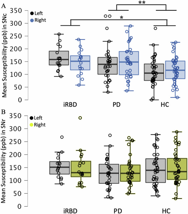
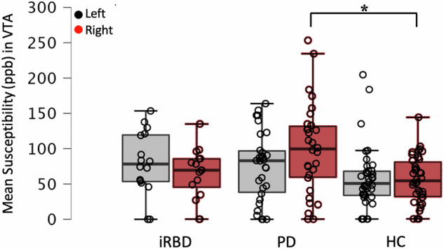
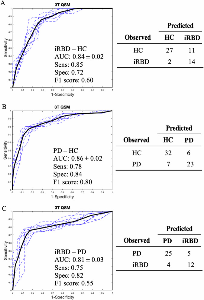
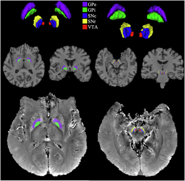
Idiopathic REM sleep behaviour disorder (iRBD) is considered a prodromal form of Parkinson's Disease (PD), potentially exhibiting similar patterns of neurodegeneration, such as brain iron changes. We investigated midbrain and pallidal iron using quantitative susceptibility mapping (QSM) in 16 iRBD patients, 30 PD patients, and 38 age-matched healthy controls (HCs) with 3T MRI. QSM revealed elevated substantia nigra pars compacta (SNc) mean susceptibility in both iRBD and PD patient groups compared to HCs, though iRBD and PD QSM measures did not differ. There were no SN pars reticulata group differences. Mean susceptibility was reduced for PD relative to iRBD and HCs in the globus pallidus externa (GPe). Furthermore, mean susceptibility was reduced for PD relative to iRBD in the GP interna (GPi). GPe/GPi mean susceptibility decreased with PD subgroup motor severity. Consistent with this, QSM in left GPi and MDS-UPDRS-III scores correlated negatively in PD patients, as well as in iRBD and PD patients combined. PD patients also evidenced higher mean susceptibility in the right ventral tegmental area (VTA) compared to iRBD and HCs, consistent with later VTA degeneration. RBD symptomatology did not correlate with QSM values. Combining SNc, GPe, GPi, and VTA QSM values, we distinguished iRBD-HCs, PD-HCs, and iRBD-PD patients at single-subject levels (0.84, 0.86, and 0.81 accuracies), using ROC curve analyses with repeated k-folds cross-validation. Using 3T MRI, QSM values in SNc, GPe, GPi, and VTA demonstrate promise as investigational measures and diagnostic/progression biomarkers of prodromal and early PD.


Conflict of interest statement

Competing interests: The authors declare no competing interests.

Figures

Fig. 1
Fig. 1. Mean susceptibility of SNc and SNr.
Mean susceptibility of the A SNc and B SNr for iRBDs, PDs, and HCs. Boxplots show left and right hemisphere mean susceptibility in ppb from QSM at 3T. A Significant differences were found between groups in SNc with both iRBD and PD patients showing increased mean susceptibility relative to HCs, reflecting higher iron deposition. B No significant group differences were found in the SNr. niRBD = 16, nPD = 30, nHC = 38. *p < 0.05, **p < 0.01.
Fig. 2
Fig. 2. Mean susceptibility of GPe and GPi.
Mean susceptibility of the A GPe and B GPi for iRBDs, PDs, and HCs. Boxplots show left and right hemisphere mean susceptibility in ppb from QSM at 3T. A Significant differences were found between groups in the GPe with HC and iRBD patients both showing higher mean susceptibility relative to PDs, reflecting higher iron deposition. B Significant group differences were found in the GPi with iRBD patients showing higher susceptibility than PDs. A: niRBD = 15, nPD = 27, nHC = 38. B: niRBD = 16, nPD = 29, nHC = 38. *p < 0.05.
Fig. 3
Fig. 3. Mean susceptibility of the VTA for iRBDs, PDs, and HCs.
Boxplots show left and right hemisphere mean susceptibility in ppb from QSM at 3T. Significant differences were found between groups in the right VTA only with PD patients showing higher mean susceptibility relative to HCs, reflecting higher iron deposition. niRBD = 16, nPD = 30, nHC = 38. **p < 0.01.
Fig. 4
Fig. 4. Mean ROC curves and confusion matrices showing classifications for iRBD, PD, and HC from binomial logistic regression.
Data show mean ROC curves (black line) for 10 repeated 5-folds CV (dash lines) and confusion matrices for the average ROC curve of the ten models for 3T QSM in the bilateral SNc, GPe, GPi, and VTA following binomial logistic regression to classify A iRBD versus HC, B PD versus HC, and C iRBD versus PD. AUC, sensitivity, specificity, and F1 score are shown. Diagnostic accuracy is good in iRBD versus HC, near excellent in PD versus HC, and good in iRBD versus PD. QSM shows some prodromal biomarker potential. nHC = 38, niRBD = 16, nPD = 30.
Fig. 5
Fig. 5. ROI segmentation on QSM of a HC.
Top row shows CIT168 atlas midbrain nuclei and globus pallidus in axial (left) and coronal (right) planes. Middle row shows globus pallidus (left) and midbrain nuclei (right) segmentations on T1w anatomical axial and coronal views. Bottom row shows post-processed QSM image in axial view with overlaid segmentations.

References

    1. Barone, P. et al. The PRIAMO study: a multicenter assessment of nonmotor symptoms and their impact on quality of life in Parkinson’s disease. Mov. Disord.24, 1641–1649 (2009). - PubMed
    1. Mitchell, T. et al. Emerging neuroimaging biomarkers across disease stage in Parkinson disease: a review. JAMA Neurol.78, 1262–1272 (2021). - PMC - PubMed
    1. Peralta, C. et al. Pragmatic approach on neuroimaging techniques for the differential diagnosis of Parkinsonisms. Mov. Disord. Clin. Pract.9, 6–19 (2022). - PMC - PubMed
    1. Emre, M., Ford, P. J., Bilgiç, B. & Uç, E. Y. Cognitive impairment and dementia in Parkinson’s disease: practical issues and management. Mov. Disord.29, 663–672 (2014). - PubMed
    1. Prasad, S. et al. Three-dimensional neuromelanin-sensitive magnetic resonance imaging of the substantia nigra in Parkinson’s disease. Eur. J. Neurol.25, 680–686 (2018). - PubMed

LinkOut - more resources