. 2025 Apr 23;16(1):3803.
doi: 10.1038/s41467-025-59209-z.

UBL7 is indispensable for spermiogenesis through protecting critical factors from excessive degradation by proteasomes

Affiliations

UBL7 is indispensable for spermiogenesis through protecting critical factors from excessive degradation by proteasomes

Tianyi Yuan et al. Nat Commun. .

Abstract

Spermiogenesis is a tightly regulated process to produce mature sperm cells. The ubiquitin-proteasome system (UPS) plays a crucial role in controlling protein half-life and is essential for spermiogenesis. Recently, proteins containing ubiquitin-like domains and ubiquitin-associated domains (UBL-UBA proteins) have emerged as novel regulators within the UPS. In this study, we demonstrate that UBL7, a testis-enriched UBL-UBA protein, is indispensable for sperm formation. Deficiency of UBL7 leads to severe malformations of both the sperm tail and head. Mechanistically, UBL7 interacts with the valosin-containing protein (VCP) complex and proteasomes, and shuttles substrates between them. Notably, UBL7 slows down the degradation rates of substrates involved in endoplasmic reticulum-associated degradation (ERAD) within cells. Through a two-step immunoprecipitation method, we identify several essential factors in spermatids that are protected by UBL7, including factors involved in the development of manchette (such as IFT140), head-tail coupling apparatus (such as SPATA20) and cytoplasmic droplets (such as HK1 and SLC2a3). In summary, our findings highlight UBL7 as a guardian that protects crucial factors from excessive degradation and thereby ensures successful spermiogenesis.


Conflict of interest statement

Competing interests: The authors declare no competing interests.

Figures
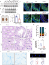
Fig. 1
Fig. 1. Loss of UBL7 leads to sperm malformation and male sterility in mice.
a WB analysis of UBL7 protein in multiple mouse tissues. b WB analysis of UBL7 protein in mouse testes at indicated ages. c Immunofluorescent staining of UBL7 in mouse seminiferous tubules at different stages. PNA labels acrosomes, DDX4 labels germ cells, DAPI labels nucleus. Scale bar, 20 μm. d Gross morphology of testes from Ubl7+/- and Ubl7−/− mice. Scale bar, 1 cm. e Ratios of testis weight to body weight of Ubl7+/- and Ubl7−/− mice. Unpaired Student’s t-test (two-sided). Error bar, n = 6 mice (4-month-old), mean ± SEM. ***p = 0.0003. Source data are provided as a Source Data file. f Hematoxylin and eosin staining of testis sections of Ubl7+/- and Ubl7−/− mice. Left panels show overall morphology of seminiferous tubules. Scale bar, 100 μm. Mid panels show seminiferous tubules at stage IX-X. Scale bar, 50 μm. Right panels show enlarged images of area in black boxes of mid panels. White triangles indicate spermatids at step 9-10. The black triangle indicates elongated spermatids at step 16. g Stacked percentage plot shows distributions of germ cells isolated from Ubl7+/- and Ubl7−/− testes. RS, round spermatids; MII, meiosis II spermatocytes; P + D, Pachytene and Diplotene spermatocytes; L + Z, Leptotene and Zygotene spermatocytes; Spg, spermatogonium. Error bar, for Ubl7+/- n = 5 mice, for Ubl7−/− n = 7 mice, mean ± SEM. Source data are provided as a Source Data file. h Count of sperm isolated from cauda epididymis of Ubl7+/- and Ubl7−/− mice. Unpaired Student’s t-test (two-sided). Error bar, n = 6 mice (4-month-old), mean ± SEM. **p = 0.0044. Source data are provided as a Source Data file. i Coomassie Blue staining of sperm isolated from cauda epididymis of Ubl7+/- and Ubl7−/− mice. Asterisks indicate abnormal bend or boundary between midpiece and principal piece. Black triangles indicate abnormal bend between or separation of sperm head and tail. Scale bar, 20 μm. j Upper panels show Hoechst 33342 staining of nucleus of Ubl7+/- and Ubl7−/− sperm. Scale bar, 5 μm. Lower panels show head morphology of Ubl7+/- and Ubl7−/− sperm. The arrows indicate the diameter of sperm around the equator region.
Fig. 2
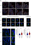
Fig. 2. Loss of UBL7 leads to manchette defects.
a Immunofluorescent staining of α-tubulin in seminiferous tubules at indicated stages. PNA labels acrosomes, DDX4 labels germ cells and DAPI labels nucleus. Scale bar, 20 μm. b Immunofluorescent staining of α-tubulin in elongating spermatids isolated from Ubl7+/- and Ubl7−/− testes. Elongating spermatids are shown in progressive steps of manchette development from left to right. Hoechst 33342 labels nucleus. Right panels show spermatids with abnormally elongated manchette and impaired head shape from Ubl7−/− testes. Scale bar, 10 μm. c Immunofluorescent staining of α-tubulin in isolated spermatids around step 14. Scale bar, 10 μm. Right panels show enlarged pictures of regions in dashed boxes of left panels. Polylines with arrows indicate the length and width of the manchettes. d, e Quantification of length and width of manchettes in spermatids around step 14. About 400 spermatids (n) were counted isolated from four Ubl7+/- mice and six Ubl7−/− mice (m) respectively. Unpaired Student’s t-test (two-sided). Violin plot, median and interquartile range (IQR). ****p < 0.0001. Source data are provided as a Source Data file.
Fig. 3
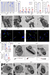
Fig. 3. Defects of HTCA, annulus and CDs in Ubl7−/− sperm.
a Morphologically classified abnormalities of Ubl7−/− sperm. Hless, headless; HNB, head-neck bending; MPB, midpiece-principal-piece bending; MPG, midpiece-principal-piece gap. Scale bar, 5 μm. b Quantitative analysis of sperm with four abnormalities. Unpaired Student’s t-test (two-sided). Error bar, n = 5 mice, mean ± SEM. Normal, MPB and MPG ****p < 0.0001, Hless **p = 0.0018, HNB ***p = 0.0003. Source data are provided as a Source Data file. c Relative ATP levels in Ubl7+/- and Ubl7−/− cauda sperm. Unpaired Student’s t-test (two-sided). Error bar, n = 4 mice, mean ± SEM. ****p < 0.0001. Source data are provided as a Source Data file. d Relative ROS levels in Ubl7+/- and Ubl7−/− cauda sperm. Unpaired Student’s t-test (two-sided). Error bar, n = 4 mice, mean ± SEM. **p = 0.0088. Source data are provided as a Source Data file. e TEM analysis of HTCA structures in Ubl7+/- and Ubl7−/− sperm. HTCA, head-tail connecting apparatus. Sc, segmented column. Cp, capitulum. Bp, basal plate. Nu, nuclei. Scale bar, 500 nm. f Immunofluorescent staining of CDs in Ubl7+/- and Ubl7−/− sperm. Ubiquitin (Ub) labels CDs; Mito tracker (Mito) labels midpieces; Hoechst 33342 (Hoechst) labels nucleus. Asterisks indicate intact CD structures. Arrows indicate CD-like vesicles detached from sperm tail. Scale bar, 20 μm. g Quantification of sperm with intact CD structures. Unpaired Student’s t-test (two-sided). Error bar, n = 4 mice, mean ± SEM. Caput sperm ***p = 0.0005, Cauda sperm ****p < 0.0001. Source data are provided as a Source Data file. h Quantification of detached CDs in sperm smear from cauda epididymis. Unpaired Student’s t-test (two-sided). Error bar, n = 4 mice, mean ± SEM. *p = 0.0222. Source data are provided as a Source Data file. i TEM analysis of shrunken CDs at bent regions of Ubl7−/− sperm. The schematic below shows cross-sections of indicated sperm. Scale bar, 1 μm. j TEM analysis of CD structures of Ubl7+/- and Ubl7−/− sperm. Abundant saccular elements and vesicles were present in Ubl7+/- sperm, while there were less (mid panel) or absence (right panel) of saccular elements and vesicles in a portion of Ubl7−/− sperm. Ratios of sperm with indicated CDs morphology were labeled at top right conner. Scale bar, 500 nm.
Fig. 4
Fig. 4. UBL7 binds to VCP complex through direct interaction with UBE4B.
a GO enrichment analysis of UBL7 binding proteins identified by FLAG-IP and mass spectrum in Ubl7flag/flag testis and spermatids. BP, biological processes. Source data are provided as a Source Data file. b WB validation of proteins identified by FLAG-IP in Ubl7flag/flag testis. Endogenous protein co-precipitated with FLAG-UBL7 were detected by indicated antibodies. c Schematic of functional domains and truncated forms of UBL7 protein. d IP of truncated forms of UBL7 and UBE4B. Flag-tagged whole-length or truncated forms of UBL7 were co-transfected with Myc-tagged UBE4B in 293 T cells. 48 h after transfection, cell lysates were subjected to IP with FLAG antibody. UBE4B co-precipitated with FLAG-UBL7 was detected by anti-Myc antibody. Cells transfected with FLAG-tag vector serve as a negative control. e IP of UBE4B and VCP. FLAG-UBE4B was co-transfected with HA-VCP or empty HA-tag vector in 293 T cells and subjected to IP with anti-HA antibody. UBE4B co-precipitated with HA-VCP was detected by FLAG antibody. f IP of UBL7 and VCP in presence of UBE4B in 293 T cells. FLAG-UBL7 was co-transfected with HA-VCP in presence or absence of Myc-UBE4B in 293 T cells. UBE4B and VCP co-precipitated with UBL7 was detected by Myc and HA antibody, respectively. g IP of UBL7 and its truncated forms with Ub-conjugates. FLAG-tagged full-length or truncated UBL7 were transfected in 293 T cells. 48 h after transfection, cells transfected with full length UBL7 were treated with MG132 (10 μM) or TAK243 (10 μM) for 1 h. Then cells were lysed for FLAG-IP. Ub-conjugates co-precipitated with FLAG-UBL7 was detected with anti-ubiquitin antibody. FLAG-GFP serves as a negative control. h A model shows that UBL7 interacts with UBE4B and indirectly binds to VCP complex. In the meantime, UBL7 binds to Ub-conjugates extracted and processed by the VCP complex.
Fig. 5
Fig. 5. UBL7 interacts with 26S proteasomes and prevents degradation of substrates.
a WB analysis of Ub receptors co-precipitated with UBL7 in 293 T cells. Ub receptors co-precipitated with FLAG-UBL7 were detected with anti-Myc antibody. FLAG-tag vector serves as a negative control. b WB analysis of PSMD2 interacting with whole-length or truncated UBL7. PSMD2 proteins pulled down by FLAG-UBL7 were detected by anti-Myc antibody. FLAG-GFP serves as a negative control. c IP of testis 26S proteasomes with UBL7 in vitro. Subunit PSMB5 contents were used as a measure of proteasomes co-precipitated with UBL7. d Activity of testis 26S proteasomes in presence of indicated concentration of UBL7 protein measured by the hydrolysis of Boc-Leu-Gly-Arg-AMC. n = 4 biological repeats for 0, 100, 200, 500 nM UBL7; n = 2 biological repeats for 1000, 1500 nM UBL7; n = 3 biological repeats for 2000 nM UBL7. Error bar, mean ± SEM. Source data are provided as a Source Data file. e Activity of testis 26S proteasomes in presence of indicated concentrations of 6Ub or UBL7 measured by the hydrolysis of Boc-Leu-Gly-Arg-AMC. n = 3 biological repeats for 0.2 μM 6Ub, 1 μM 6Ub, 0.2 μM 6Ub + 1 μM UBL7 and 1 μM 6Ub + 1 μM UBL7, n = 4 biological repeats for 0.2 μM UBL7, n = 2 biological repeats for 1 μM UBL7. Unpaired Student’s t-test (two-sided). Error bar, mean ± SEM. *p = 0.03, ***p = 0.0001. Source data are provided as a Source Data file. f, g, h WB analysis of CD3δ, TYR-C89R and UbV76-V-GFP degradation in 293 T cells expressing UBL7 or GFP control. Protein remained in cells were detected using anti-Myc antibody. The asterisks indicate unmodified forms of CD3δ and TYR-C89R, while the upper bands represent the glycosylated forms of these proteins. Quantitative analyses are shown below. CD3δ and TYR-C89R, n = 3 biological repeats. UbV76-V-GFP, n = 5 biological repeats. Unpaired Student’s t-test (two-sided). Error bar, mean ± SEM. For CD3δ, ns p = 0.0876, **p = 0.0089. For TYR-C89R, *p = 0.0362, **p = 0.0039. For UbV76-V-GFP 4 h, ns p = 0.9271, 8 h, ns p = 0.1501. Source data are provided as a Source Data file.
Fig. 6
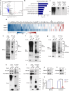
Fig. 6. UBL7 prevents target substrates degradation in testis.
a Volcano plots of proteins with differential abundance in Ubl7+/- and Ubl7−/− sperm (n = 3 mice). p-values were calculated using Unpaired Student’s t-test (two-sided). Blue dots indicate proteins significantly decreased in Ubl7−/− sperm (Fold change (FC) > 1.5, p < 0.05), red dots indicate proteins significantly increased in Ubl7−/− sperm. b GO analysis of proteins with reduced abundance in Ubl7−/− sperm. c WB analysis of Ub-conjugates that are enriched by DSP-crosslinked FLAG IP followed by TUBE IP in Ubl7flag/flag testes. d Heat map of levels of the UBL7 targeting substrates enriched by two-step IP in Ubl7−/− sperm compared to that in Ubl7+/- sperm. e WB analysis of BSCL2 immunoprecipitated by FLAG-UBL7 in Ubl7flag/flag and WT testes. f, g IP of BSCL2 or SLC2a3 with UBL7 in 293 T cells. Myc-BSCL2 and Myc-SLC2a3 were co-transfected with FLAG-UBL7 in 293 T cells. IP was performed using anti-FLAG antibody 48 h after transfection. BSCL2 and SLC2a3 pulled down by UBL7 were detected by anti-Myc antibody. FLAG-tag vector serves as a negative control. The asterisks indicate the IgG bands. h, i IP of BSCL2 or SLC2a3 with VCP and PSMD2 in 293 T cells. Myc-PSMD2 and HA-VCP were co-transfected with FLAG-BSCL2 or SLC2a3. FLAG IP was performed 48 h after transfection. PSMD2 and VCP pulled down by BSCL2 or SLC2a3 were detected by anti-Myc and anti-HA antibodies, respectively. FLAG-GFP serves as a negative control. j, k WB analysis of BSCL2 and SLC2a3 in Ubl7+/- and Ubl7−/− sperm. l quantification of WB results in j and k. Unpaired Student’s t-test (two-sided). Error bar, n = 3 mice, mean ± SEM. ***p = 0.0002. Source data are provided as a Source Data file.
Fig. 7
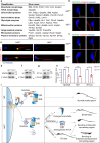
Fig. 7. Loss of substrates protected by UBL7 causes defects in Ubl7−/− sperm structures.
a List of UBL7 targeting substrates identified by two-step IP. The targeting proteins were classified by their functions or subcellular localizations. b, c Immunofluorescent staining of SPATA20 and SPATA6 in HTCA of Ubl7+/- and Ubl7−/− sperm. α-tubulin labels sperm flagella; Hoechst (Hoechst 33342) labels nucleus. Scale bar, 5 μm. d WB analysis of SPATA6 in Ubl7+/- and Ubl7−/− sperm. e Immunofluorescent staining of HK1 and SLC2a3 in Ubl7+/- and Ubl7−/− sperm. Ub labels CDs; Hoechst (Hoechst 33342) labels nucleus. Scale bar, 10 μm. f WB analysis of HK1 in Ubl7+/- and Ubl7−/− sperm. g WB analysis of IFT140 in isolated Ubl7+/- and Ubl7−/− elongating spermatids. h Quantification of WB results in d, f and g. n = 4 mice for SPATA6, n = 3 mice for the others. Unpaired Student’s t-test (two-sided). Error bar, mean ± SEM. SPATA6, **p = 0.0022, HK1, ****p < 0.0001, IFT140, **p = 0.0078. Source data are provided as a Source Data file. i A schematic diagram depicting the mechanism that loss of UBL7 causes defects in sperm.

References

    1. Meistrich, M. L. & Hess, R. A. Assessment of spermatogenesis through staging of seminiferous tubules. Spermatogenesis: Methods and Protocols, 299–307 (2013). - PubMed
    1. Ernst, C., Eling, N., Martinez-Jimenez, C. P., Marioni, J. C. & Odom, D. T. Staged developmental mapping and X chromosome transcriptional dynamics during mouse spermatogenesis. Nat. Commun.10, 1251 (2019). - PMC - PubMed
    1. Azhar, M. et al. Towards post-meiotic sperm production: genetic insight into human infertility from mouse models. Int. J. Biol. Sci.17, 2487 (2021). - PMC - PubMed
    1. Dunleavy, J. E., O’Bryan, M. K., Stanton, P. G. & O’Donnell, L. The cytoskeleton in spermatogenesis. Reproduction157, R53–R72 (2019). - PubMed
    1. Chen, Y. et al. Single-cell RNA-seq uncovers dynamic processes and critical regulators in mouse spermatogenesis. Cell Res.28, 879–896 (2018). - PMC - PubMed

Substances

LinkOut - more resources