. 2025 Oct 14;27(9):2355-2369.
doi: 10.1093/neuonc/noaf106.

Dual aVß8 Integrin and PD-1 Blockade Overcomes TGFβ-Mediated B-Cell Suppression to Enhance Anti-Tumor Immunity

Affiliations

Dual aVß8 Integrin and PD-1 Blockade Overcomes TGFβ-Mediated B-Cell Suppression to Enhance Anti-Tumor Immunity

David Hou et al. Neuro Oncol. .

Abstract

Background: Immunotherapy has revolutionized cancer treatment but has yet to be translated into brain tumors. Studies in other solid tumors suggest a central role of B-cell immunity in driving immune checkpoint blockade efficacy. In glioblastoma (GBM), tumor B cells are driven into a regulatory B-cell state that suppresses immune activation and T-cell function.

Methods: We used spatially resolved transcriptomics and multiplex immunofluorescence to characterize B-cell neighborhoods within GBM and identify enhanced TGFβ-signaling between myeloid and B cells. We generated conditional knockouts to investigate the effects of TGFβ signaling on B-cell function and survival in vivo. Additionally, we combined TGFβ blockade with PD-1 inhibition to evaluate their combined anti-glioma efficacy.

Results: Our findings reveal that myeloid cells are the primary interactors with B cells in GBM through the TGFβ pathway. Pharmacological or genetic TGFβ blockade expanded intratumoral B cells and synergized with PD-1 inhibition to enhance survival (60% tumor eradication in dual-treated mice). Therapeutic efficacy critically depended on B cells, as their depletion abolished survival benefits. Dual αVβ8/PD-1 blockade reduced B-cell-mediated suppression of CD8⁺ T-cell cytotoxicity and increased plasmablast differentiation, while partial efficacy in RagKO mice implicated ancillary roles for innate immunity.

Conclusion: Targeting TGFβ signaling using an anti-αVβ8 blocker can impact anti-tumor immunity through different possible mechanisms, of which we highlight the rescuing of B-cell function through synergy with PD-1 checkpoint blockade therapy. Our work underscores the critical role of intratumoral B-cell immunity in enhancing immunotherapy against brain tumors.

Keywords: B cells; TGFβ; checkpoint-blockade; glioblastoma; tumor microenvironment.


Conflict of interest statement

The authors declare no competing interests.

Figures
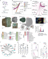
Figure 1.
Figure 1.
B-cell spatial architecture in GBM and immunosuppressive role of TGFβ (A) Illustration of computational workflow for spatial educated proximity analysis. After single-cell deconvolution using CytoSpace, a cell-cell distance graph was estimated and fit to scale-free distribution. (B) Circular plot of cell-cell proximity to B cells in GBM. Edges were weighted by power-distributed spatial correlations. (C) More granular view on the distribution of myeloid subtypes in the B cells neighborhood. (D) Example of a GBM spatial resolved transcriptomic sample after single-cell deconvolution with representative B-cell niche (B cells in red) and the close neighborhood. Two spatial transcriptomic datasets are shown representative for a primary tumor (left) and recurrent tumor (right). Color code of the cell types is indicated below. (E) Representative image of multiplex immunofluorescence staining on a GBM sample showing different cellular neighborhoods. Cells were characterized as tumor cells (SOX2+), TAMs (CD163+TMEM119), microglia (CD163TMEM119+), B cells (CD20+), and T cells (CD8+). For each sample, B-cell neighborhoods were analyzed for different cell types within 15 microns of a B cell. (F) Spatial educated receptor-ligand interaction by cell-type-specific expression inference from TAMs as sender toward B cell as receiver. R-L strength was measured by cosine similarity integrating expression and spatial information. (G) depicting the effects of TGFβ-1 on B-cell expansion. Human B cells were isolated from healthy PBMCs, activated with an expansion cocktail, and cultured with exogenous TGFβ-1, TGFβ-2, or TGFβ-3. The inhibitory effects of TGFβ-1 on B-cell expansion were mitigated by adding the TGFβ receptor inhibitor SB431542, administered daily 30 minutes before TGFβ-1. All experiments were repeated in triplicates. (H) Histogram showing the cellular expansion of activated B cells cultured with exogenous TGFβ-1 or mock treatment, as measured by eFluor-450 proliferation dye. B cells were activated with a B-cell expansion cocktail, and TGFβ1 was added daily starting from day 1, inhibiting B-cell proliferation. (I) Bar plot with experimental triplicates showing the reduction in B-cell expansion index after 7 days of activation in the presence of TGFβ1, illustrating the inhibitory effects of TGFβ1 on B-cell proliferation. BDM: bone-derived macrophages; MHC: major histocompatibility complex; Hypox: hypoxia; Perivas. CAF: perivascular cancer-associated fibroblast, SMC: smooth muscle cell; cDC: conventional dendritic cells; pDC: plasmacytoid dendritic cells; CD4 IFN: interferon gamma-producing CD4 T cells; CD8 cyto: cytotoxic CD8 T cell; TAM anti_inf.: anti-inflammatory tumor-associated macrophages; TGF: tumor growth factor; BMP: bone morphogenetic protein; ACVR: activin A receptor. ns = P > .05, *P < .05, **P < .01, ***P < .001, ****P < .0001.
Figure 2.
Figure 2.
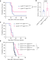
Conditional Tgfb1 knockouts promote B-cell infiltration and animal survival and synergize with PD-1 blockade (A) Lyz2-Tgfb1 transgenic mice with Tgfb1 knocked out of myeloid cells (LyzMcre/WT-Tgfb1flox/flox) were challenged with CT2A glioma cells. Animal survival was assessed in these experimental mice compared to wild-type controls (LyzMcre/WT-Tgfb1flox/WT). Knockout of Tgfb1 trended toward improved animal survival. (B) Lyz2-Tgfb1 transgenic mice with Tgfb1 knocked out of myeloid cells (LyzMcre/WT-Tgfb1flox/flox) were challenged with CT2A glioma cells. After 14 days, the tumors were analyzed for B-cell infiltration via flow cytometry. Knockout of Tgfb1 trended toward increased B-cell counts in the tumor. (C) Survival curve after CT2A tumor challenge for CD19cre/WT-Tgfb1flox/flox vs CD19cre/WT-Tgfb1flox/WT mice where Tgfb1 was knocked out of B cells. (D) A transgenic mouse model with an inducible knockout of TGFb-R2 on B cells (Cd19ert-cre/WT-Tgfbr2flox/flox) was combined with or without PD-1 blockade and assessed for animal survival compared to Cd19ert-cre/WT-Tgfbr2flox/WT controls with or without PD-1 blockade. Cd19ert-cre/WT-Tgfbr2flox/flox + PD-1 had significantly improved survival compared to both Cd19ert-cre/WT-Tgfbr2flox/flox without PD-1 and Cd19ert-cre/WT-Tgfbr2flox/WT + PD-1. ns = P > .05, *P < .05, **P < .01, ***P < .001, ****P < .0001.
Figure 3.
Figure 3.
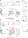
Inhibiting TGFβ by blocking αVβ8 integrin synergizes with PD-1 blockade to eradicate tumors in a B-cell-dependent manner (A) Bulk RNA sequencing of murine B cells from CT2A tumors and spleen for various integrin chains. αV and β8 integrin chains are upregulated in tumor B cells compared to splenic B cells. (B) Experimental schema for animal survival experiments for Figure 3C-E. (C) Survival curves of WT mice challenged with CT2A tumors receiving a combination of αVβ8 integrin blockade and PD-1 checkpoint blockade. Dual therapy with both αVβ8 integrin blockade and PD-1 checkpoint blockade led to tumor eradication in nearly 60% of mice, significantly outperforming single-agent therapy and untreated controls. (D) Survival curves of long-term survivors from (C) that were rechallenged with CT2A tumors on the opposite hemisphere to evaluate immune memory response. Long-term survivors in each treatment group were divided into two, with half receiving intracranial CD20 B-cell depleting antibodies before tumor rechallenge. Only animals who maintained an intact B-cell compartment and originally received dual αVβ8 integrin blockade and PD-1 checkpoint blockade could prevent tumor re-engraftment. (E) Bar plots from flow cytometry analysis of B-cell infiltration and proliferation in tumor-bearing mice after no treatment, mono treatment with αVβ8 blockade or PD-1 blockade, or dual αVβ8 and PD-1 blockade. (F) Survival curves of B-cell knockout mice challenged with CT2A tumors receiving dual αVβ8 integrin blockade compared to untreated controls. While the median survival was improved with dual therapy, there was no tumor eradication in any of the groups. (G) Survival curves of RagKO mice, which lack T and B cells, challenged with CT2A tumors receiving dual αVβ8 integrin blockade compared to untreated controls. (H) Bar plot showing B-cell expansion in a murine model with αVβ8 integrin knocked out specifically in B cells. PD-1 therapy enhanced B-cell proliferation in tumors in these mice compared to controls.
Figure 4.
Figure 4.
αVβ8 integrin blockade + PD-1 blockade reverses B-cell immunosuppression and improves T-cell immunity (A) CD4+ T-cell suppression assay with B cells isolated from tumors that underwent no treatment, monotherapy with PD-1 or αVβ8 blockade, or dual PD-1 and αVβ8 blockade. T cells were isolated from the spleen of tumor-bearing mice that did not receive any treatment. T-cell proliferation was measured via dilution of proliferation dye eFluor 450, and effector function was measured by IL17 and IFN-γ expression. (B) CD8+ T-cell suppression assay with B cells isolated from tumors that underwent no treatment, monotherapy with PD-1 or αVβ8 blockade, or dual PD-1 and αVβ8 blockade. T-cell proliferation was measured via dilution of proliferation dye eFluor 450, and effector function was measured by GzmB and IFN-γ expression. (C) Intratumoral CD4+ and CD8+ T-cell phenotypes were analyzed after treatment with isotype control, anti-αVβ8 Fab, and PD-1, or dual anti-αVβ8 + PD1 blockade. CD4+ T cells demonstrated increased proliferation as measured by Ki67 staining after treatment with anti-αVβ8 Fab or dual treatment. Dual anti-αVβ8 + PD1 blockade significantly improved CD8+ T-cell proliferation and GzmB expression in the brain. ns = P > .05, *P < .05, **P < .01, ***P < .001, ****P < .0001.
Figure 5.
Figure 5.
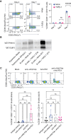
αVβ8 integrin blockade + PD-1 blockade promote B-cell plasmablast differentiation (A) Representative dot plots and bar graph demonstrating CD38 and CD20 expression in human B cells after in-vitro culture with TGFβ-1 vs mock dimethyl sulfoxide (DMFO). Plasmablast phenotype (CD38+CD20) was evaluated at days 4 and 7 of culture. All experiments were repeated in triplicates. (B) Western blots for IgG heavy and light chains in B-cell culture media. The same number of B cells (0.5 × 106) were cultured in antibody-free media and activated with B-cell expansion cocktail. TGFβ-1 was added to the experimental group. The supernatant was sampled at days 4 and 7 for analysis of IgG antibodies. (C) Representative flow cytometry plots and cumulative bar graphs of intratumoral B-cell plasmablast differentiation (CD38+CD20) in tumor-bearing mice after no treatment, mono treatment with αVβ8 blockade or PD-1 blockade, or dual αVβ8 and PD-1 blockade. B cells in both the tumor and draining deep cervical lymph nodes (dcLN) were evaluated. ns = P > .05, **P < .01, ****P < .0001.

References

    1. Hodges TR, Ott M, Xiu J, et al. Mutational burden, immune checkpoint expression, and mismatch repair in glioma: implications for immune checkpoint immunotherapy. Neuro Oncol. 2017;19(8):1047–1057. - PMC - PubMed
    1. Thorsson V, Gibbs DL, Brown SD, et al. ; Cancer Genome Atlas Research Network. The immune landscape of cancer. Immunity. 2018;48(4):812–830.e14. - PMC - PubMed
    1. Nduom EK, Weller M, Heimberger AB.. Immunosuppressive mechanisms in glioblastoma. Neuro Oncol. 2015;17(Suppl 7):vii9–vii14. - PMC - PubMed
    1. Dapash M, Hou D, Castro B, Lee-Chang C, Lesniak MS.. The interplay between glioblastoma and its microenvironment. Cells. 2021;10(9):2257. - PMC - PubMed
    1. Antonios JP, Soto H, Everson RG, et al. Immunosuppressive tumor-infiltrating myeloid cells mediate adaptive immune resistance via a PD-1/PD-L1 mechanism in glioblastoma. Neuro Oncol. 2017;19(6):796–807. - PMC - PubMed

MeSH terms