. 2025 Apr 9:19:1454924.
doi: 10.3389/fnint.2025.1454924. eCollection 2025.

Novel cVEMP procedure reveals sexual dimorphism in peak to trough latency

Affiliations

Novel cVEMP procedure reveals sexual dimorphism in peak to trough latency

Max Gattie et al. Front Integr Neurosci. .

Abstract

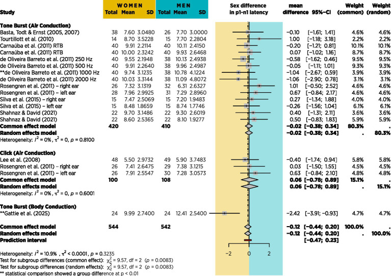
Introduction: Sex difference in latency for cervical vestibular-evoked myogenic potential (VEMP) has been reported in Brown Norway rats. Human investigations of sex difference in VEMP latency have shown inconsistent results, although there are indicators of sexual dimorphism in vestibular function and a higher reporting rate for vestibular disorder in women than in men.

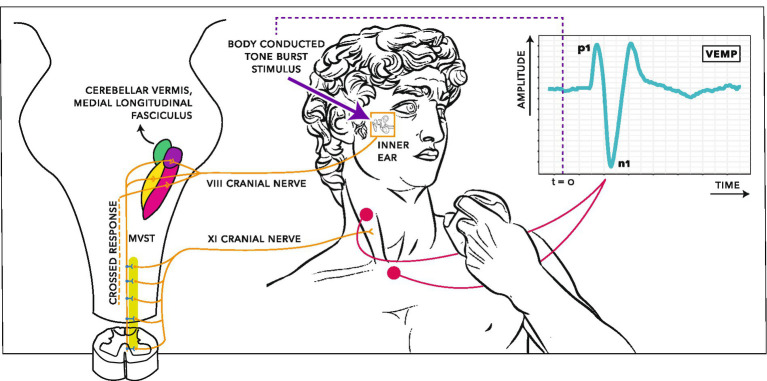
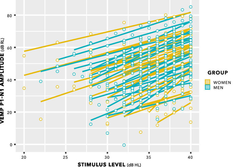
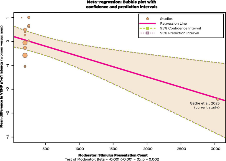
Methods: Sex effects in human VEMP were re-evaluated here using a procedure adapting clinical protocols for higher sensitivity. VEMP was compared between 24 women and 24 men using a novel procedure that (1) controlled neck tension with biofeedback and a padded head bar; (2) used body-conducted stimuli to eliminate sound exposure concerns and collect appreciably more data than is feasible with air-conducted stimuli; which in turn (3) increased statistical power because there were sufficient data for a linear mixed effects regression modelling analysis.

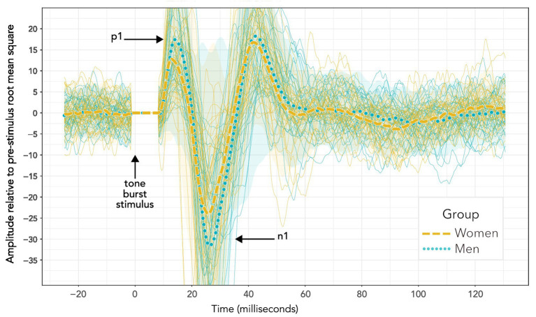
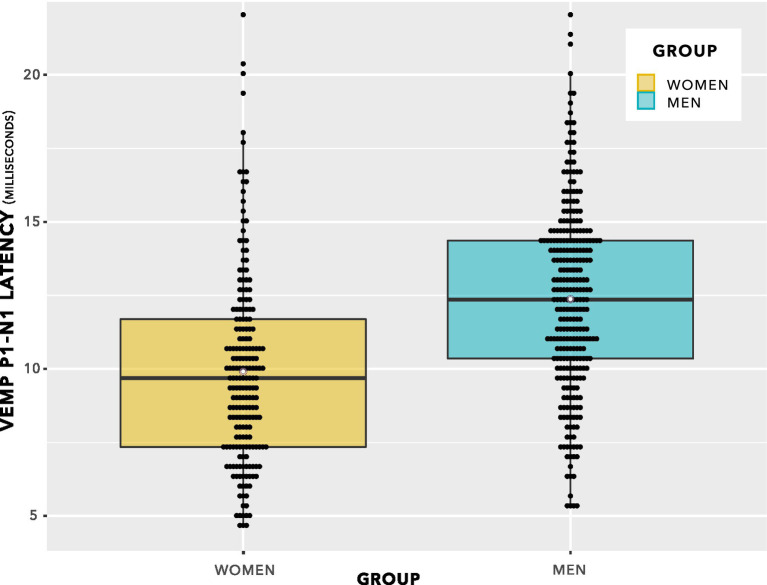
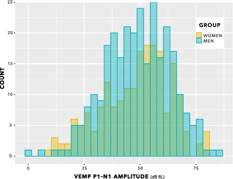
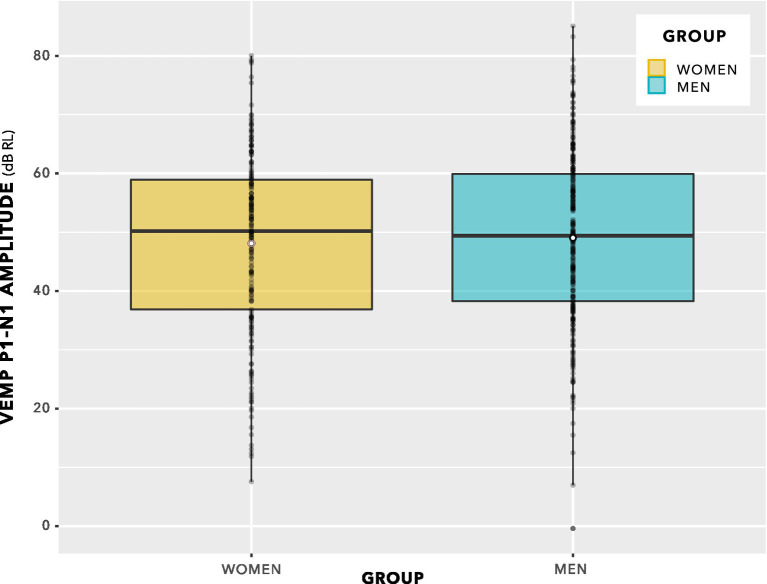
Results: Women had significantly shorter VEMP peak to trough latency than men. The sex difference of 2.4 ms (95% CI [-0.9, -3.9], p = 0.0020) was 21% of the mean 11.4 ms VEMP peak to trough latency measured across women and men. There was no significant sex difference in VEMP peak to trough amplitude. These findings are a reversal of several prior studies in humans, reviewed here with a simulation indicating the studies may have been underpowered.

Discussion: Findings are consistent with those in Brown Norway Rats, for which a study design featuring a custom rodent holder to control neck tension, extension of test sequences in comparison to those typically used in VEMP protocols for humans, and insertion of electrodes subcutaneously will have increased sensitivity compared to that achievable with clinical VEMP protocols for humans. Findings are interpreted as sex hormones affecting myelination or synaptic response; sexual dimorphism in neck/head size may also have contributed. The vestibular periphery and brainstem are highly conserved across vertebrates with similar findings in rat and human supporting use of VEMP as a reliable, non-invasive indicator of vestibular function. VEMP measures in humans may require higher sensitivity than is achievable using current clinical protocols in order to produce consistent results.

Keywords: cVEMP; dimorphism; sex; vestibular; vestibular-evoked myogenic potential (VEMP).


Conflict of interest statement

The authors declare that the research was conducted in the absence of any commercial or financial relationships that could be construed as a potential conflict of interest.

Figures

Figure 1
Figure 1
Electrophysiological recording arrangement for cervical vestibular-evoked myogenic potentials (VEMPs). The stimulus is a vibratory tone burst. Energy from the tone burst deflects hair cells in the vestibular system, creating an electrical impulse along the VIII cranial nerve. These vestibular afferents synapse in the vestibular nucleus then descend along the medial vestibulospinal tract (MVST). The MVST connects to the XI cranial nerve, which branches to synapse on motoneurons innervating the ipsilateral sternocleidomastoid. The brief activity from a tone burst results in a brief inhibition of tonic activity in the muscle fibres of the sternocleidomastoid. This inhibitory potential can be recorded between electrodes on the sternum and the belly of the sternocleidomastoid, creating a wave form with a characteristic peak (p1) and trough (n1) at approximately 13 ms and 23 ms after stimulus onset. The vestibulo-collic reflex also includes crossed and ascending components, not shown in detail. Based on Kim et al. (2010), Oh et al. (2016), and Colebatch et al. (2016). Creative Commons CC BY-NC-SA 4.0.
Figure 2
Figure 2
Participants were asked to push against a padded bar with their foreheads, maintaining tension in the sternocleidomastoid tension as close as possible to 50 µV root mean square throughout testing. The Eclipse clinical software provided biofeedback helping participants to monitor sternocleidomastoid tension.
Figure 3
Figure 3
VEMP grand averages recorded following 500 Hz tone bursts presented using bone conduction at 40 dB HL. Distinct sequences for individuals within each group are also shown (more lightly drawn traces than the grand averages) along with 95% confidence intervals (shaded areas). Visual inspection of the waveforms suggests a difference in p1–n1 amplitude, but not p1–n1 latency. However, such an interpretation is misleading. Firstly, these data do not enable a valid statistical comparison, since there were uneven presentation counts across women and men. Secondly, averaging the data to enable statistical comparison shows a significant group difference in p1–n1 latency but not p1–n1 amplitude. Statistical analyses are reported in section 3, and discussed in section 4.
Figure 4
Figure 4
Initial model for statistical analysis of VEMP p1–n1 amplitude measurements. Potential confounders were reviewed in section 2.5, and were assessed as part of the model where possible.
Figure 5
Figure 5
VEMP p1–n1 latency histogram. Indication is of approximately normal distributions for the female and male groups, with some suggestion of a difference in means of the distributions.
Figure 6
Figure 6
VEMP p1–n1 latencies. The even spacing on the ordinate was due to the 3 kHz sampling rate. VEMP p1–n1 latencies are grouped in steps of 0.33 ms, which was the maximum resolution of the Eclipse when using VEMP protocols. To provide an indication of when several data points were recorded at the same VEMP p1–n1 latency, data points have been plotted using the beeswarm feature in ggplot (R Core Team, 2020). This feature has introduced variation along the abscissa corresponding to the quantity of data present. Mean values for each group are shown as white data points. The box plot suggests there will be a group difference in VEMP p1–n1 latency, however it contains repeated measurements per participant and as such does not entirely reflect data appropriate for use in statistical analysis. Statistical analyses are reported in section 3, and discussed in section 4.
Figure 7
Figure 7
VEMP p1–n1 latencies versus stimulus level for women and men. There was no interaction. Sampling resolution of the Eclipse meant that data points were grouped in steps of 0.33 ms (see note at Figure 5). To provide an indication of when data from both women and men were recorded at the same stimulus level and VEMP p1–n1 latency, data points have been plotted using the beeswarm feature in ggplot (R Core Team, 2020). This feature introduced variation along the abscissa corresponding to the quantity of data present. The variation does not reflect stimulus level used. Stimulus level was always an integer multiple of 1 dB.
Figure 8
Figure 8
VEMP p1–n1 amplitude histogram. Indication is of approximately normal distributions for the female and male groups. There is no suggestion of a difference in means of the distributions.
Figure 9
Figure 9
VEMP p1–n1 amplitudes. Since there are repeated measures per participant, this is not a valid statistical comparison. However, there is no indication of a statistically significant group difference. Statistical analysis was via linear mixed-effects regression modelling, and is described in the main text.
Figure 10
Figure 10
VEMP p1–n1 amplitude versus stimulus level. Individual lines of best fit are per participant. The slopes have a mean value of 1.89 with SD 0.25, supporting use of a fixed slope linear mixed effects model.
Figure 11
Figure 11
The model for VEMP p1–n1 amplitude described in figure 4, updated following data collection. Only stimulus level predicted VEMP p1–n1 amplitude.
Figure 12
Figure 12
Flow diagram showing literature review process.
Figure 13
Figure 13
Meta-analysis based on 9 of the 20 studies from table 1 for which mean p1-n1 latency difference with standard deviations could be obtained. No account is taken of stimulus presentation count, however the effect is shown in Figure 14.
Figure 14
Figure 14
Meta-regression with studies from Figure 13 that also reported stimulus presentation count.
Figure 15
Figure 15
95% confidence intervals for the analyses shown in Table 2. Participant counts were balanced (e.g., 48 participants are 24 women, 24 men). Group differences are shown as prolongation in men’s VEMP p1–n1 latencies compared to those of women. The mean latency difference is shown for each condition as a vertical orange line, with horizontal bars either side showing the 95% confidence interval. When the 95% confidence interval crosses zero, the data analyses in the simulations did not establish a statistically significant group difference. Such analyses are depicted in a lighter colour (yellow), whereas analyses for which a statistically significant group difference could be established are depicted in a darker colour (green).

Similar articles

References

    1. Ahmed S. B., Qamar A., Imram M., Fahim M. F. (2020). Comparison of neck length, relative neck length and height with incidence of cervical spondylosis. Pak. J. Med. Sci. 36, 219–223. doi: 10.12669/pjms.36.2.832, PMID: - DOI - PMC - PubMed
    1. Akin F. W., Murnane O. D., Proffitt T. M. (2003). The effects of click and tone-burst stimulus parameters on the vestibular evoked myogenic potential (VEMP). J. Am. Acad. Audiol. 14, 500–509. doi: 10.3766/jaaa.14.9.5 - DOI - PubMed
    1. Arshad M., Stanley J. A., Raz N. (2016). Adult age differences in subcortical myelin content are consistent with protracted myelination and unrelated to diffusion tensor imaging indices. NeuroImage 143, 26–39. doi: 10.1016/j.neuroimage.2016.08.047, PMID: - DOI - PMC - PubMed
    1. Asakura S., Kamogashira T. (2021). Sudden bilateral hearing loss after vestibular-evoked myogenic potentials. Clin. Case Rep. 9:e05025. doi: 10.1002/ccr3.5025, PMID: - DOI - PMC - PubMed
    1. Ashford A., Huang J., Zhang C., Wei W., Mustain W., Eby T., et al. . (2016). The cervical vestibular-evoked myogenic potentials (cVEMPs) recorded along the sternocleidomastoid muscles during Head rotation and flexion in Normal human subjects. J. Assoc. Res. Otolaryngol. 17, 303–311. doi: 10.1007/s10162-016-0566-8, PMID: - DOI - PMC - PubMed

LinkOut - more resources