. 2025 Apr 23:29:0194.
doi: 10.34133/bmr.0194. eCollection 2025.

Multiaction Antimicrobial, Anti-inflammatory, and Prohealing Hydrogel as a Novel Strategy for Preventing Postoperative Pancreatic Fistula

Affiliations

Multiaction Antimicrobial, Anti-inflammatory, and Prohealing Hydrogel as a Novel Strategy for Preventing Postoperative Pancreatic Fistula

Yuan Zhou et al. Biomater Res. .

Abstract

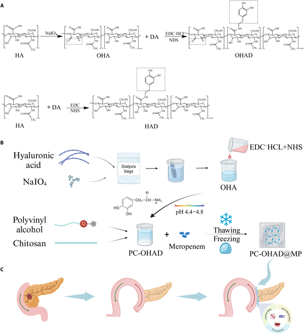
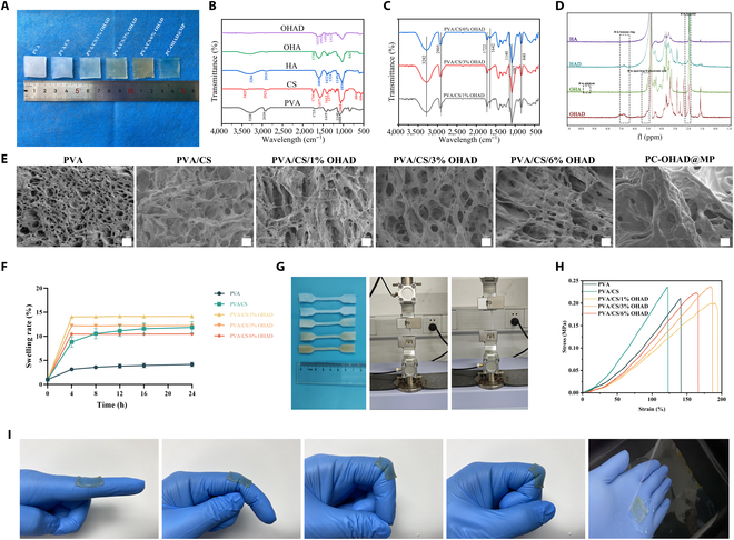
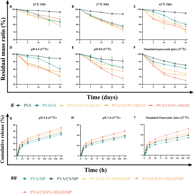
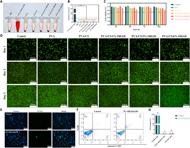
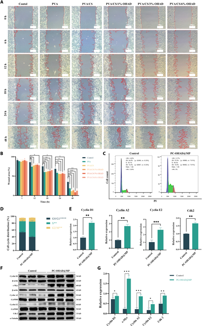
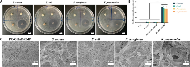
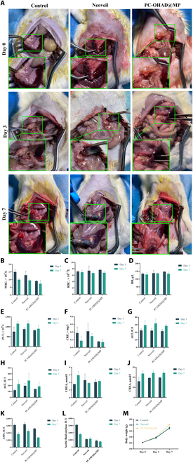
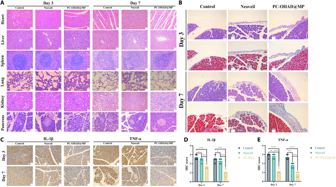
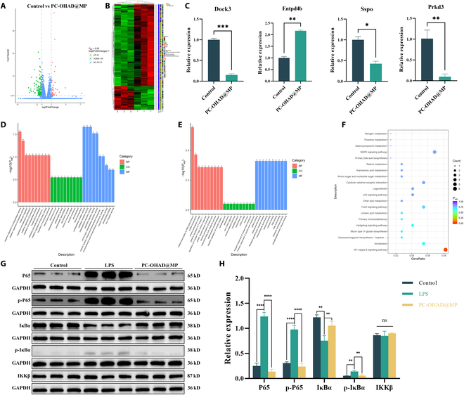
Postoperative pancreatic fistula remains a challenging complication after pancreaticoduodenectomy. Addressing this issue requires effective strategies to promote anastomotic healing. In this study, we developed a novel hydrogel designed to close pancreaticoenteric anastomosis after pancreaticoduodenectomy. The hydrogel-composed of polyvinyl alcohol, chitosan, and dopamine-modified oxidized hyaluronic acid-exhibited excellent antibacterial, anti-inflammatory, and wound healing properties. It was designed to conform well to the anastomotic site for clinical application. The hydrogel demonstrated good biocompatibility, appropriate mechanical strength, low swelling, and strong adhesive properties, meeting specific requirements for pancreaticoenteric anastomosis environments. Moreover, by activating the cell cycle, it promoted cell proliferation and migration, thereby accelerating anastomotic closure. Addition of the potent broad-spectrum antibiotic meropenem further enhanced its antibacterial efficacy, targeting common microbial species involved in delayed healing and fistula formation after pancreatic surgery. In a rat model of pancreatic fistula, the hydrogel effectively sealed the anastomosis, filled potential suture gaps, and exerted antibacterial, anti-inflammatory, and tissue regeneration-promoting effects around the anastomotic site. Therefore, this hydrogel, with its ideal degradation properties, shows promising application prospects in closing pancreaticoenteric anastomosis following pancreaticoduodenectomy, thereby offering an effective solution to reduce complications such as pancreatic fistula after pancreatic surgery.


Conflict of interest statement

Competing interests: The authors declare that they have no competing interests.

Figures

Fig. 1.
Fig. 1.
Preparation process and applications of hydrogels. (A) Modification and modification process of hyaluronic acid. (B) Schematic diagram of the hydrogel preparation process. (C) Main application scenarios and methods of hydrogels.
Fig. 2.
Fig. 2.
Appearance and characterization of hydrogels. (A) Appearance of the hydrogel (digital camera view). (B) FTIR spectra of hydrogels with single different components. (C) FTIR spectra of synthesized hydrogels with different OHAD concentrations. (D) 1H NMR spectra of HA, HAD, OHA, and OHAD. (E) Electron microscopy images of hydrogels with different compositions. (F) Swelling curves of hydrogels with different compositions (n = 3). (G) Preparation of tensile samples of hydrogels with different compositions and experimental process (n = 5). (H) Stress–strain curves of hydrogel samples with different compositions. (I) Adhesive properties of hydrogels in different scenarios.
Fig. 3.
Fig. 3.
Degradation and loaded drug release profiles of hydrogels under different conditions (# indicates panels A to F; ## indicates panels G to I) (n = 3).
Fig. 4.
Fig. 4.
In vitro compatibility of hydrogels. (A) Appearance of hydrogel in vitro hemolysis test (digital camera view) (n = 3). (B) Statistical results of hemolysis rate of hydrogels in vitro. (C) Statistical results of cell viability of hydrogel samples with different compositions. (D) Dual staining of live and dead cells under the microscope for hydrogel samples with different composition (n = 3). (E) TUNEL staining under the microscope for PC-OHAD@MP hydrogel (n = 3). (F) Apoptosis flow cytometry results for PC-OHAD@MP hydrogel (n = 3). (G) Statistical results of apoptosis flow cytometry for PC-OHAD@MP hydrogel.
Fig. 5.
Fig. 5.
Hydrogel enhances cell proliferation and migration. (A) Scratch assay under the microscope for hydrogel samples with different compositions (n = 3). (B) Statistical results of scratch assay for hydrogel samples with different compositions. (C) Cell cycle flow cytometry results for PC-OHAD@MP hydrogel (n = 3). (D) Statistical results of cell cycle apoptosis for PC-OHAD@MP hydrogel. (E) Relative mRNA expression levels of key regulatory genes in the cell cycle for PC-OHAD@MP hydrogel (n = 3). (F) Quantitative analysis results of protein expression levels of key regulatory genes in the cell cycle for PC-OHAD@MP hydrogel (n = 3). (G) Statistical results of protein expression levels of key regulatory genes in the cell cycle for PC-OHAD@MP hydrogel.
Fig. 6.
Fig. 6.
Hydrogel exhibits strong antibacterial activity. (A) Appearance of inhibition zones in agar diffusion assay under a digital camera for PVA, PVA/CS, PVA/CS/3% OHAD, and PC-OHAD@MP hydrogels against Staphylococcus aureus (S. aureus), Escherichia coli (E. coli), Pseudomonas aeruginosa (P. aeruginosa), and Klebsiella pneumoniae (K. pneumoniae) (n = 3). (B) Statistical results of inhibition zones in agar diffusion assay. (C) Coculture of PC-OHAD@MP and PVA/CS/3% OHAD hydrogels with S. aureus, E. coli, P. aeruginosa, and K. pneumoniae under an electron microscope.
Fig. 7.
Fig. 7.
In vivo biocompatibility of hydrogel and reduction of postoperative pancreatic fistula. (A) Appearance of surgical sites at postoperative days 0, 3, and 7 under a digital camera for the control group, Neoveil, and PC-OHAD@MP (n = 5). (B to L) Blood routine tests, CRP, ALT, AST, urea, CREA, blood amylase, and abdominal amylase levels at postoperative days 0, 3, and 7 for the control group, Neoveil, and PC-OHAD@MP (n = 5). (M) Preoperative weight gain curve at postoperative days 0, 3, and 7 for the control group, Neoveil, and PC-OHAD@MP (n = 5).
Fig. 8.
Fig. 8.
In vivo biocompatibility, promotion of collagen fiber deposition, and anti-inflammatory capability of PC-OHAD@MP. (A) Histological examination of heart, liver, spleen, lung, kidney, and pancreatic tissues stained with H&E at postoperative days 3 and 7 for the control group, Neoveil, and PC-OHAD@MP (n = 5). (B) Masson’s trichrome staining of pancreatic tissues at suture sites at postoperative days 3 and 7 for the control group, Neoveil, and PC-OHAD@MP (n = 5). (C) Immunohistochemical staining of IL-1β and TNF-α levels in pancreatic tissues at suture sites at postoperative days 3 and 7 for the control group, Neoveil, and PC-OHAD@MP (n = 5). (D) Statistical results of immunohistochemical staining for IL-1β levels in pancreatic tissues at suture sites at postoperative days 3 and 7 for the control group, Neoveil, and PC-OHAD@MP. (E) Statistical results of immunohistochemical staining for TNF-α levels in pancreatic tissues at suture sites at postoperative days 3 and 7 for the control group, Neoveil, and PC-OHAD@MP.
Fig. 9.
Fig. 9.
Hydrogel promotes cell proliferation, migration, and anti-inflammatory mechanisms. (A) Volcano plot showing differentially expressed genes between the control group and the PC-OHAD@MP group (n = 3). (B) Heatmap displaying differentially expressed genes between the control group and the PC-OHAD@MP group (n = 3). (C) Relative mRNA expression levels of Dock3, Entpd4b, Sspo, and Prkd3 between the control group and the PC-OHAD@MP group (n = 3). (D) Up-regulated Gene Ontology (GO) enrichment analysis results between the control group and the PC-OHAD@MP group (n = 3). (E) Down-regulated GO enrichment analysis results between the control group and the PC-OHAD@MP group (n = 3). (F) Kyoto Encyclopedia of Genes and Genomes (KEGG) enrichment analysis results between the control group and the PC-OHAD@MP group (n = 3). (G) Protein expression levels of key genes in the NF-κB pathway (P65, p-P65, p-IκBα, IκBα, and IKKβ) between the control group and the PC-OHAD@MP group (n = 3). (H) Quantitative analysis of protein expression levels of key genes in the NF-κB pathway (P65, p-P65, p-IκBα, IκBα, and IKKβ) between the control group and the PC-OHAD@MP group.

Similar articles

References

    1. Wang X, Bai Y, Cui M, Zhang Q, Zhang W, Fang F, Song T. Modified Blumgart anastomosis without pancreatic duct-to-jejunum mucosa anastomosis for pancreatoduodenectomy: A feasible and safe novel technique. Cancer Biol Med. 2018;15(1):79–87. - PMC - PubMed
    1. Simon R. Complications after pancreaticoduodenectomy. Surg Clin North Am. 2021;101(5):865–874. - PubMed
    1. Malgras B, Dokmak S, Aussilhou B, Pocard M, Sauvanet A. Management of postoperative pancreatic fistula after pancreaticoduodenectomy. J Visc Surg. 2023;160(1):39–51. - PubMed
    1. Bassi C, Dervenis C, Butturini G, Fingerhut A, Yeo C, Izbicki J, Neoptolemos J, Sarr M, Traverso W, Buchler M, et al. . International study group on pancreatic fistula, postoperative pancreatic fistula: An international study group (ISGPF) definition. Surgery. 2005;138(1):8–13. - PubMed
    1. Li Z, Wei A, Xia N, Zheng L, Yang D, Ye J, Xiong J, Hu W. Blumgart anastomosis reduces the incidence of pancreatic fistula after pancreaticoduodenectomy: A systematic review and meta-analysis. Sci Rep. 2020;10(1):17896. - PMC - PubMed

LinkOut - more resources