. 2025 Apr 9:12:1564648.
doi: 10.3389/fvets.2025.1564648. eCollection 2025.

ORNIPURAL® as conventional therapy versus mixture of Curcuma longa extract and pomegranate peel extract as homeotherapy in dogs with dexamethasone-induced hepatopathy: clinicolaboratory, ultrasonographic, and histopathological monitoring

Affiliations

ORNIPURAL® as conventional therapy versus mixture of Curcuma longa extract and pomegranate peel extract as homeotherapy in dogs with dexamethasone-induced hepatopathy: clinicolaboratory, ultrasonographic, and histopathological monitoring

Arafat Khalphallah et al. Front Vet Sci. .

Abstract

Introduction: Curcuma longa extract and pomegranate peel extract as homeotherapy have numerous therapeutic uses, mainly for anti-inflammatory, immunomodulatory, and hepatoprotective efficacy. The current study compared ORNIPURAL® (as a commercial hepatoprotective drug) and a herbal mixture of Curcuma longa extract and pomegranate peel extract [as homeotherapy] in dogs with dexamethasone-induced hepatopathy throughout a 42-day long-term study.

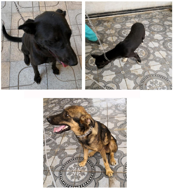
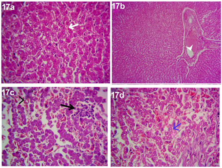
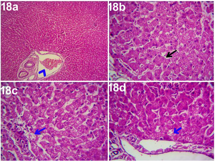
Methods: The study was conducted on mongrel dogs (n = 30) throughout three phases of the experiment: an acclimatization phase (14 days), a steroidal-induced hepatopathy phase (14 days), and a treatment phase (14 days, i.e., either with ORNIPURAL® or with herbal mixtures). The investigated dogs undergoing complete clinical and ultrasonographic examinations as well as hematological analysis and serum hepatorenal biomarkers that were estimated in days 0 (control group), 7 (hepatopathy group), 14 (hepatopathy group), 21 (treatment group), and 28 (treatment group). Histopathology of the liver was conducted for some dogs on days 0, 14, and 28 after the euthanization of these animals.

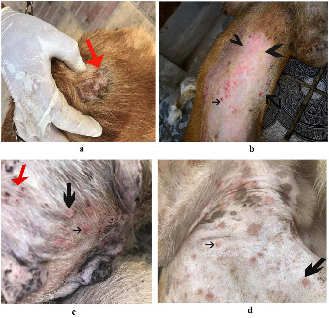
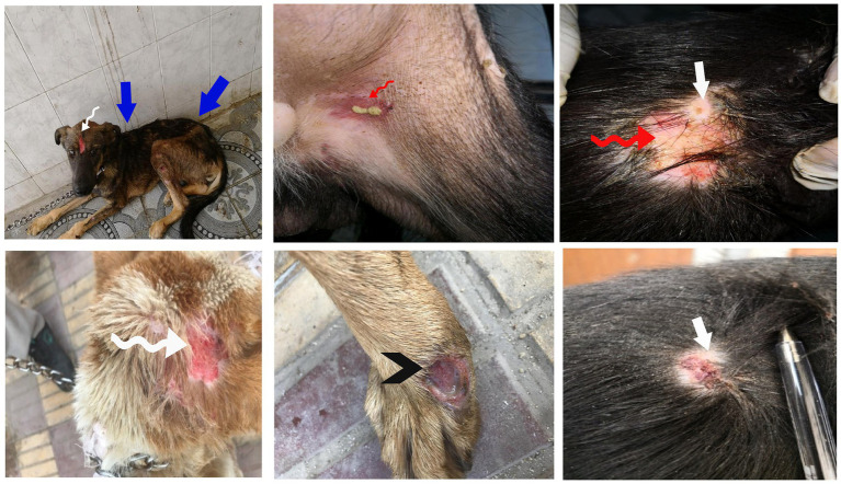
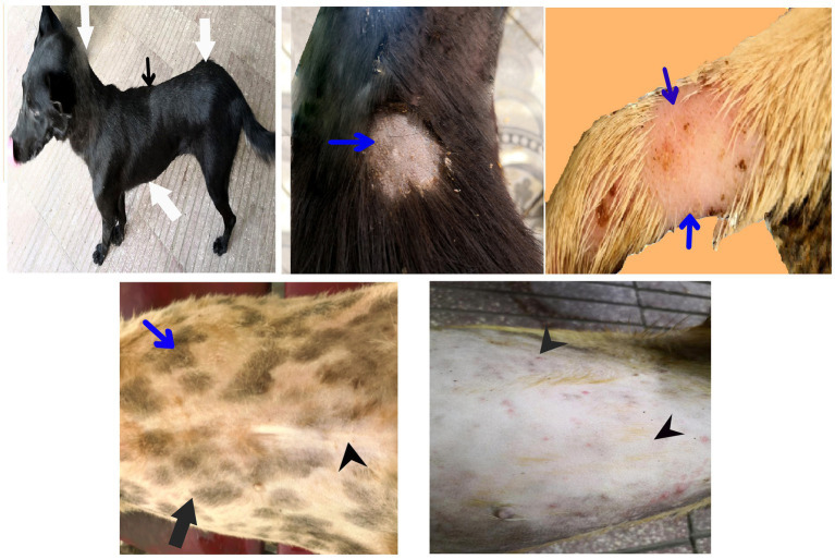
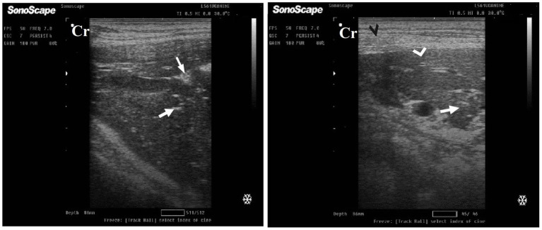
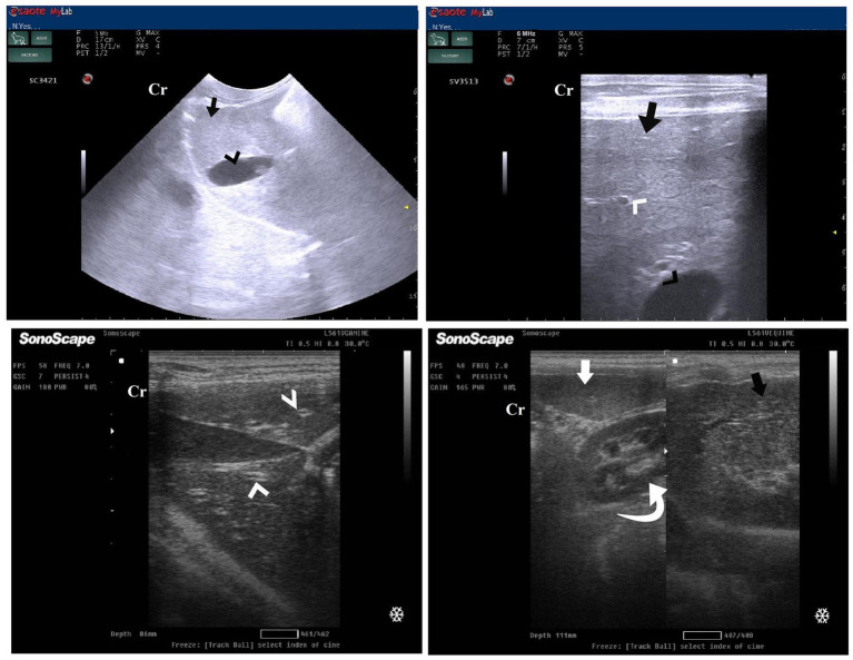
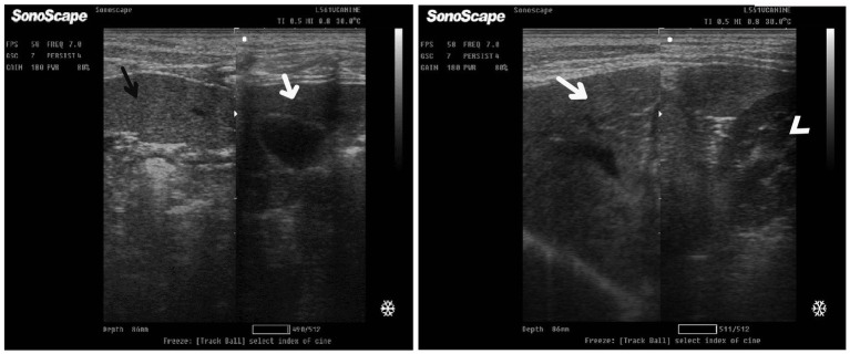
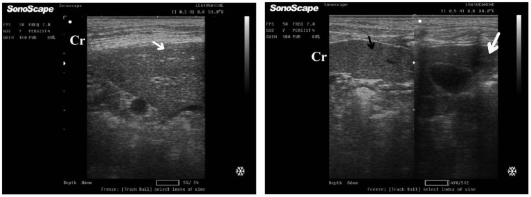
Results and conclusion: The present study reported the most remarkable efficacy of both ORNIPURAL® and a herbal mixture of Curcuma longa extract and pomegranate peel extract as hepatoprotective medicaments in the therapy of dexamethasone-induced fatty liver in dogs. Therefore, a 14-day treatment with either a herbal mixture or ORNIPURAL® in treated dogs (treatment groups) induced an unmistakable improvement in their clinical status, blood pictures, and serum hepatorenal parameters as well as characteristic sonographic and histopathological findings compared with those in dexamethasone-induced hepatic lipidosis (hepatopathy groups). Compared to dogs treated with ORNIPURAL®, this clinical improvement was more evident in dogs treated with an herbal mixture. Moreover, no significant alterations in blood pictures and serum hepatorenal indices were demonstrated between ORNIPURAL® and herbal-treated dogs. Overall, the herbal mix of Curcuma longa extract and pomegranate peel extract had higher efficacy and greater potency than conventional therapy that uses ORNIPURAL® in treating dogs with hepatopathy. The study also recommended the parallel use of this herbal mixture as well as ORNIPURAL® in long-term therapeutic strategies in dogs with dexamethasone-induced fatty liver as both minimized dexamethasone side effects. Ultrasonography alone was not enough to evaluate hepatobiliary disorders in canines.

Keywords: Curcuma longa extract; ORNIPURAL®; dogs; liver histopathology; pomegranate peel extract; steroidal hepatopathy; ultrasonography.


Conflict of interest statement

The authors declare that the research was conducted in the absence of any commercial or financial relationships that could be construed as a potential conflict of interest.

Figures

Figure 1
Figure 1
The image shows heathy control dogs in the control group (day 0). They were active and alert with healthy skin, no signs of dehydration, and suitable body weights.
Figure 2
Figure 2
The image shows dogs in the steroidal-induced hepatopathy group after 7 days following dexamethasone injection (DexaHepatogr; day 7). The hepatopathy-affected dogs showed the development of skin abscesses (black arrows), skin rashes (blackhead arrows), and alopecia (red arrows).
Figure 3
Figure 3
The image shows dogs in the steroidal-induced hepatopathy group after 14 days following dexamethasone injection (DexaHepatogr; day 14). The severity of these clinical findings was more evident on day 14 than on day 7 in DexaHepatogr. The hepatopathy-affected dogs showed skin lacerations (red curved arrows), ulcers (white curved arrows), and ruptured dermal abscesses (blackhead arrows). Lethargy and loss of body conditions (blue arrows) and areas of alopecia (white arrows) were observed more than on day 14 of DexaHepatogr.
Figure 4
Figure 4
The image shows dogs in the herbal mixture-treated group (Herbmix-gr) after 7 days of treatment with an herbal mixture of pomegranate peel extract and C. longa extract (Herbmix-gr; day 21). The affected dogs expressed clear improvement in their body conditions (white arrows) and no lethargy with almost complete disappearance of alopecia (black arrows), skin rashes, skin lacerations (blue arrows), and dehydration signs as well as complete healing of ruptured skin abscess (black head arrows) were observed.
Figure 5
Figure 5
The image shows dogs in ORNIPURAL® treated group (Ornipgr) after 7 days following ORNIPURAL® (Ornipgr; day 21) for five-time treatment/10 days with ORNIPURAL® at one-day intervals. The affected dogs were still suffering from poor health conditions, emaciation, and loss of body weight (white arrows). Areas of alopecia (black arrows), skin rashes (blue arrows), skin lacerations (blue head arrows), and ruptured skin abscesses (black arrows), as well as skin abscesses (red arrows), still appeared.
Figure 6
Figure 6
The image shows dogs in the herbal mixture-treated group (Herbmix-gr) after 14 days following treatment with a herbal mixture of pomegranate peel extract and C. longa extract (Herbmix-gr; day 28). The examined dogs on day 28, after treatment with the herbal mixture (Herbmix-gr) for 14 days, restored their body conditions and body weights (white arrows) with the healthy appearance of their skin. Complete healing of skin lacerations (black head arrows) and disappearance of skin abscesses (black arrows) and alopecia (curved white arrows) were observed.
Figure 7
Figure 7
The image shows dogs in ORNIPURAL® treated group (Ornipgr) after 14 days following treatment with ORNIPURAL® (Ornipgr; day 28). Complete healing and disappearance of skin lesions (abscess, lacerations, and erosions) were observed (white arrows). The affected dogs were still suffering from poor health conditions, emaciation, and loss of body weight (curved red arrows). Areas of alopecia (Black arrows) were still appeared.
Figure 8
Figure 8
Ultrasonogram in a 3-year-old male dog in the control group (day 0) with healthy liver imaged through the left lateral recumbency, that is, intercostal approach (right 11th ICSs) (A) or dorsal recumbency (subcostal approach) (B) using 7 MHz linear ultrasound transducer. It showed a normal ultrasonographic image of the hepatobiliary system. The normal hepatic parenchyma (A) has a uniform medium level of echogenicity, with interruption caused by the hepatic and portal veins (white arrows). The echotexture was coarser and more hypoechoic compared with the spleen (white head arrows) (B). Compared with the kidney, the liver had an increased echogenicity (curved white arrows) (B). The gall bladder (A) was anechoic and ovoid with a tapered neck (white asterisk). The wall of the gall bladder was not very clear at all. Cr: Cranial.
Figure 9
Figure 9
Ultrasonogram of longitudinal views of the liver in a 2-year-old male dog in a steroidal-induced hepatopathy group (DexaHepatogr) imaged through the dorsal recumbency (subcostal approach) using a 7 MHz linear ultrasound transducer. DexaHepatogr (day 7) showed a slight increase in echogenicity of the hepatic parenchyma (white head arrows), but it was still less than that of the spleen (black head arrows). The liver showed a focal hyperechoic area (white arrows) within the parenchyma. The hepatic parenchyma had a heterogeneous nature. Cr: Cranial.
Figure 10
Figure 10
Ultrasonogram of transverse views (A,B) and longitudinal views (C,D) and of the liver in a 2-year-old female dog in a steroidal-induced hepatopathy group (DexaHepatogr) imaged through the dorsal recumbency (subcostal approach) using 6 and 7 MHz linear ultrasound transducer or 5 MHz micro convex ultrasound. DexaHepatogr on day 14 showed a diffuse increase in hepatic parenchymal echogenicity (black arrows) as it was hyperechoic in comparison to the renal cortex (white curved arrows) and was like echogenicity of splenic parenchyma (white arrows). The diffuse and homogenous hyperechogenicity of the hepatic parenchyma caused indistinct visualization of the hepatic vessel wall. The hepatic parenchyma sometimes appeared heterogeneous and contained hypoechoic foci, most likely due to concurrent nodular hyperplasia (white head arrows). The gall bladder (black head arrows) was anechoic and ovoid with a tapered neck. The wall of the gall bladder was a thin echogenic line. Cr: Cranial.
Figure 11
Figure 11
Ultrasonogram of longitudinal views of the liver in a 2-year-old female dog in a herbal mixture-treated group (Herbmix-gr; day 21) imaged through the dorsal recumbency (subcostal approach) using 7 MHz linear ultrasound transducer. It showed normal hepatic parenchyma as a uniform medium level of echogenicity (white arrows). Compared with the spleen, the liver should have a reduced echogenicity (black arrows). Compared with the kidney, the liver had an increased echogenicity (white head arrows). Cr: Cranial.
Figure 12
Figure 12
Ultrasonogram of longitudinal views of the liver in a 4-year-old male dog in an herbal mixture-treated group (Herbmix-gr; day 28) imaged through the dorsal recumbency (subcostal approach) using 7 MHz linear ultrasound transducer. It showed normal hepatic parenchyma as a uniform medium level of echogenicity (white arrows). The liver parenchyma had reduced echogenicity compared to the spleen (black arrows). Cr: Cranial.
Figure 13
Figure 13
Ultrasonogram of longitudinal views of the liver in a 3-year-old male dog in an ORNIPURAL®-treated group (Ornipgr; day 21) imaged through the dorsal recumbency (subcostal approach) using a 9 MHz linear ultrasound transducer. It showed a diffuse increase in hepatic parenchymal echogenicity (black arrows). The diffuse and homogenous hyperechogenicity of the hepatic parenchyma caused indistinct visualization of the hepatic vessel wall. The hepatic parenchyma sometimes appeared heterogeneous and contained hypoechoic foci, most likely due to concurrent nodular hyperplasia (white head arrows). The gall bladder (white asterisk) was anechoic and ovoid with a tapered neck. The wall of the gall bladder was fragile and was not clear. Cr: Cranial.
Figure 14
Figure 14
Ultrasonogram of longitudinal views of the liver in a 3-year old s-male dog in an ORNIPURAL® treated group (Ornipgr; day 28) imaged through the dorsal recumbency (subcostal approach) using a 7 MHz linear ultrasound transducer. The liver parenchyma became slightly normal as it had uniform homogenous numerous echoes (white arrows), with interruption caused by the hepatic and portal veins. The hepatic vasculatures (white asterisk) were relatively imaged. However, the hepatic parenchyma echogenicity was like the echogenicity of splenic parenchyma (white head arrows). Cr: Cranial.
Figure 15
Figure 15
Photomicrographs of the liver in a healthy dog (Day 0) showing normal of most hepatic parenchyma.
Figure 16
Figure 16
Photomicrograph of the liver in a dog at steroidal-induced hepatopathy group (DexaHepatogr day 14) showing perivascular hydropic degenerations within hepatic parenchyma (black arrows) and thrombus formation (curved black arrows) within hepatic blood vessels beside necrotic tunica intema (head black arrows). [H&E X 400].
Figure 17
Figure 17
Photomicrograph of the liver in a dog in an herbal mixture-treated group (Herbmix-gr; day 28) showing normal of most hepatic parenchyma (White arrows), focal necrotic area infiltrated with mononuclear cells (Black arrows), microsteatosis in some hepatocytes (blue arrows) and dilated hepatic blood vessels (white head arrows) and sinusoids (black head arrows) [H&E X100 for (a); H&E X400 for (b–d)].
Figure 18
Figure 18
Photomicrograph of the liver in a dog at ORNIPURAL® treated group (Ornipgr; day 28) showing normal hepatic parenchyma (black arrows), unicellular necrotic changes of some hepatocytes (blue arrows) and congestion of portal vein (Blue head arrows) [H&E X100 for (a); H&E X400 for (b–d)].

Similar articles

References

    1. Rothuizen J. Hepatitis in dogs. In: Weber O, Protzer U, (editors). Comparative hepatitis. Birkhäuser Advances in Infectious Diseases. Switzerland: Basel, Birkhäuser Basel (2008).
    1. Dirksen K, Verzijl T, Grinwis GC, Favier RP, Penning LC, Burgener IA, et al. . Use of serum MicroRNAs as biomarker for hepatobiliary diseases in dogs. J Vet Intern Med. (2016) 30:1816–23. doi: 10.1111/jvim.14602, PMID: - DOI - PMC - PubMed
    1. Fieten H, Hooijer-Nouwens BD, Biourge VC, Leegwater PA, Watson AL, van den Ingh TSGAM, et al. . Association of dietary copper and zinc levels with hepatic copper and zinc concentration in Labrador retrievers. J Vet Intern Med. (2012) 26:1274–80. doi: 10.1111/j.1939-1676.2012.01001.x, PMID: - DOI - PubMed
    1. Eman SR, Kubesy AA, Baraka TA, Torad FA, Shaymaa IS, Mohammed FF. Evaluation of hepatocyte-derived microRNA-122 for diagnosis of acute and chronic hepatitis of dogs. Vet World. (2018) 11:667–73. doi: 10.14202/vetworld.2018.667-673, PMID: - DOI - PMC - PubMed
    1. Poldervaart JH, Favier RP, Penning LC, van den Ingh TS, Rothuizen J. Primary hepatitis in dogs: a retrospective review (2002-2006). J Vet Intern Med. (2009) 23:72–80. doi: 10.1111/j.1939-1676.2008.0215.x, PMID: - DOI - PubMed

LinkOut - more resources