

Comparative Study of Acid Etching and SLA Surface Modification for Titanium Implants
- PMID: 40271856
- PMCID: PMC11990087
- DOI: 10.3390/ma18071632
Comparative Study of Acid Etching and SLA Surface Modification for Titanium Implants
Abstract
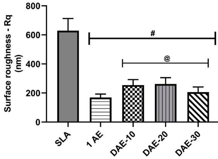
The dust generated during the sandblasting process of the sandblasted and acid-etched (SLA) method, commonly used to treat the surface of Ti dental implants, poses significant challenges in maintaining a clean manufacturing environment and ensuring safe working conditions. Nevertheless, surface modification remains crucial for improved performance of Ti dental implants. To address this problem and propose a clean and simple surface modification process to potentially replace SLA modification, this study aimed to characterize the surfaces of commercially pure Ti (cp-Ti) samples treated by acid etching and compare them with SLA-treated samples in terms of surface roughness (Rq), wettability (assessed through contact angle measurements), mineralized matrix deposition (evaluated through simulated body fluid [SBF] soaking), cell viability, cell differentiation (assessed based on alkaline phosphatase activity), and mineralization (assessed using MTT assay). Acid-etched surfaces exhibited nano- and micro-roughness and higher hydrophilicity than SLA surfaces, which is conducive to forming a highly bioactive TiO2 surface. Moreover, acid-etched samples exhibited earlier hydroxyapatite deposition after SBF soaking than SLA samples. Furthermore, the acid-etched surfaces were nontoxic and displayed significantly higher cell viability and differentiation after seven days than SLA surfaces. These findings suggest that acid etching is a viable alternative to the SLA method, likely offering superior surface bioactivity and biocompatibility.
Keywords: cell adhesion; dental implants; osseointegration; surface-active agents; titanium.
Conflict of interest statement
The funders had no role in the design of the study; in the collection, analyses, or interpretation of data; in the writing of the manuscript, or in the decision to publish the results.
Figures


Similar articles
-
Comparison of Roughness, Wettability, and SEM Features between Sandblasted Acid-Etched and Oxidized Titanium Dental Implants.J Long Term Eff Med Implants. 2024;34(4):57-63. doi: 10.1615/JLongTermEffMedImplants.2023049632. J Long Term Eff Med Implants. 2024. PMID: 38842233
-
Micro/nano topography of selective laser melting titanium inhibits osteoclastogenesis via mediation of macrophage polarization.Biochem Biophys Res Commun. 2021 Dec 3;581:53-59. doi: 10.1016/j.bbrc.2021.09.021. Epub 2021 Sep 15. Biochem Biophys Res Commun. 2021. PMID: 34655976
-
Surface Characteristics and Biocompatibility of Ultrafine-Grain Ti after Sandblasting and Acid Etching for Dental Implants.ACS Biomater Sci Eng. 2019 Oct 14;5(10):5107-5115. doi: 10.1021/acsbiomaterials.9b00579. Epub 2019 Sep 27. ACS Biomater Sci Eng. 2019. PMID: 33455258
-
Effects of different acid etching protocols on the enhanced osteogenic potential of titanium surfaces: An in vitro study.Dent Mater J. 2025 May 29;44(3):241-249. doi: 10.4012/dmj.2024-115. Epub 2025 Apr 29. Dent Mater J. 2025. PMID: 40301062
-
[The role of surface roughness in promoting osteointegration].Refuat Hapeh Vehashinayim (1993). 2003 Jul;20(3):8-19, 98. Refuat Hapeh Vehashinayim (1993). 2003. PMID: 14515625 Review. Hebrew.
Cited by
-
In Vivo Evaluation of Laser-Textured Air Plasma in Osseointegration of Dental Implants.Materials (Basel). 2025 Aug 14;18(16):3810. doi: 10.3390/ma18163810. Materials (Basel). 2025. PMID: 40870128 Free PMC article.
-
Residual-Free Micro-Nano Titanium Surfaces via Titanium Blasting and Single Acid-Etching: A Cleaner Alternative.Bioengineering (Basel). 2025 Jul 5;12(7):735. doi: 10.3390/bioengineering12070735. Bioengineering (Basel). 2025. PMID: 40722427 Free PMC article.
References
-
- Davenport R. The Routledge History of Death Since 1800. Taylor & Francis; London, UK: New York, NY, USA: 2021. Patterns of death, 1800–2020: Global rates and causes; pp. 21–44.
-
- van Noort R. Titanium: The implant material of today. J. Mater. Sci. 1987;22:3801–3811. doi: 10.1007/BF01133326. - DOI
-
- Singh A.G., Saini P., Pabla B.S. In-vitro mineralization of bio-coated 3D printed metallic implant under simulated body conditions. Mater. Today Proc. 2022;50:1946–1952. doi: 10.1016/j.matpr.2021.09.321. - DOI
-
- Eliades T. Passive film growth on titanium alloys: Physicochemical and biologic considerations. Int. J. Oral Maxillofac. Implants. 1997;12:621–627. - PubMed
Grants and funding
LinkOut - more resources
Full Text Sources
Miscellaneous
