. 2025 May 20;99(5):e0187824.
doi: 10.1128/jvi.01878-24. Epub 2025 Apr 24.

Structural and functional significance of Aedes aegypti AgBR1 flavivirus immunomodulator

Affiliations

Structural and functional significance of Aedes aegypti AgBR1 flavivirus immunomodulator

Ane Martinez-Castillo et al. J Virol. .

Abstract

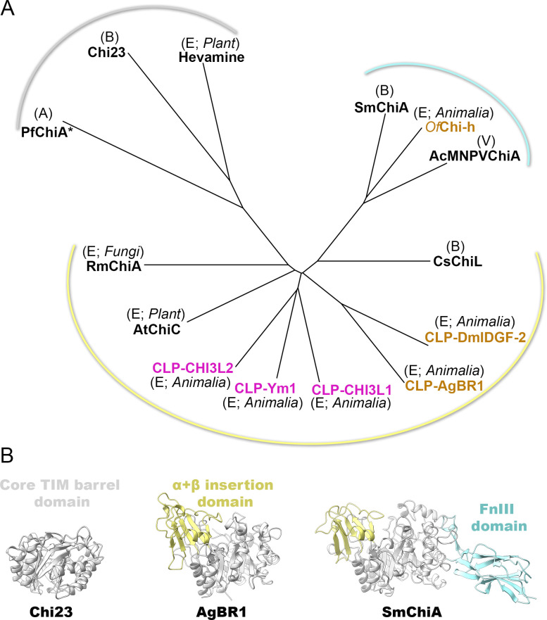
Zika virus (ZIKV), an arbovirus, relies on mosquitoes as vectors for its transmission. During blood feeding, mosquitoes inoculate saliva containing various proteins. Recently, AgBR1, an Aedes aegypti salivary gland protein, has gained attention for its immunomodulatory potential, along with another protein, called NeSt1. We have determined the crystal structure of AgBR1 at 1.2 Å resolution. Despite its chitinase-like fold, we demonstrated that AgBR1 does not bind to chitobiose or chitinhexaose, while a key mutation in the catalytic site abrogates enzymatic activity, suggesting that the protein's function has been repurposed. Our study also shows that AgBR1 and NeSt1, when presented to murine primary macrophages, alter cellular pathways related to virus entry by endocytosis, immune response, and cell proliferation. AgBR1 (and NeSt1) do not directly bind to the Zika virus or modulate its replication. We propose that their immunomodulatory effects on Zika virus transmission are through regulation of host-cell response, a consequence of evolutionary cross talk and virus opportunism. These structural and functional insights are prerequisites for developing strategies to halt the spread of mosquito-borne disease.IMPORTANCEOur study informs on the structural and functional significance of a mosquito salivary gland protein, AgBR1 (along with another protein called NeSt1), in the transmission of the Zika virus (ZIKV), a mosquito-borne virus that has caused global health concerns. By analyzing AgBR1's three-dimensional structure in combination with cellular and interaction studies, we discovered that AgBR1 does not function like typical proteins in its family-it does not degrade sugars. However, we show that it primes immune cells in a way that could help the virus enter cells more easily but not by interacting with the virus or altering viral replication. This finding is significant because it reveals how mosquito proteins, repurposed by evolution, can influence virus transmission without the virus's direct presence. Understanding how proteins like AgBR1 work could guide the development of new strategies to prevent Zika virus spread, with potential relevance for other mosquito-borne viruses.

Keywords: Zika virus; cellular pathways; crystal structure; immunomodulators; mosquito salivary proteins; mosquito-borne disease.


Conflict of interest statement

The authors declare no conflict of interest.

Figures

Fig 1
Fig 1
AgBR1 crystal structure and comparison with human and insect chitinase-like proteins. (A) AgBR1 crystal structure represented in cartoon containing β-sheets numbered from 1 to 8, surrounded by α-helices numbered from 1 to 8, colored in blue; in yellow, the α+β insertion domain between 285 and 375 residues. The black dashed oval indicates the putative catalytic site located on strand β4 (gray), with the residue leading to the abrogation of enzymatic activity in red. Right: the consensus sequence for catalytic activity with the sequence in AgBR1 on top. Left inset: the 2Fo–Fc density map contoured at the 1σ level of the two NAG sugar molecules attached to N225. (B) Structural superimposition of AgBR1 (light blue) onto insect DmIDGF-2 (orange) and human CHI3L2 (light magenta) represented as cartoon tubes. Cyan spheres in the left image indicate the last (R165) and first (V186) visible residues of the disordered loop, whereas the red and blue spheres in the right image mark the N- and C-terminal ends, respectively. Black numbered dashed circles mark some of the loops with different lengths and conformations (loop 1: L82-K86; loop 2: L109-P118; disordered loop: F158-E190; loop 3: V210-F214; loop 4: V220-V224; loop 5: Y266-N276; loop 6: P347-P360; loop 7: I378-G386).
Fig 2
Fig 2
Composite of AgBR1 and sugar molecules. Surface representation of AgBR1 protein (semitransparent white) with the model depicted in light blue cartoon tube. Superimposed are GlcNAc6 (represented as a stick model, with oxygen and carbon atoms colored red and green, respectively) and GlcNAc2 (represented as a stick model, with oxygen and carbon atoms colored red and light magenta, respectively), derived from superimposition onto chitin-bound CHI3L2 structures (PDB ids 4p8x, 4p8v). Insets in the center and on the right show clashes at sugar positions -2 and -1 with loop 2, and at positions +1, +2, and +3 with loop 4 and residues preceding the disordered loop (R165-V186).
Fig 3
Fig 3
AgBR1 and NeSt1 effect on BMDM cells. BMDM cells incubated with 10 µg of Alexa-488-labeled AgBR1 (A) or NeSt1 (B) during 2 h at 37°C. The protein is internalized in both cases after incubation. (C) Volcano plot representing the proteins differentially expressed in BMDM cells in the presence or absence of AgBR1 and NeSt1. Blue and red dots represent upregulated and downregulated proteins above the significance threshold, respectively. (D and E) Innate immune response network extracted from STRING based on the BMDM over-/sub-expressed proteins in the presence of AgBR1 or NeSt1. The colored lines indicate the associations with different levels of certainty; green activation, red inhibition, blue binding, pink post-translation modified, black reaction, yellow expression, and gray evidence view. (F and G) Top five canonical pathways predicted by IPA for AgBR1 and NeSt1.
Fig 4
Fig 4
Structure-based superimposition of GH18 chitinase family proteins. (A) Structure-based phylogenetic tree of selected chitinases and chitinases-like of the GH18 family (Drosophila melanogaster Imaginal Disc Growth Factor-2 [DmIDGF-2], PDB id 1jnd; YKL-40-like protein 1 [Ym1], PDB id 1vf8; human chitinase 3-like protein 2 [CHI3L2], PDB id 4p8u; human chitinase 3-like protein 1 [CHI3L1], PDB id 1hjx; Arabidopsis thaliana Chitinase C [AtChiC], PDB id 3aqu; Serratia marcescens Chitinase A [SmChiA], PDB id 1ctn; Autographa californica Multiple Nucleopolyhedrovirus Chitinase A [AcMNPVChiA], PDB id 5dez; hevamine [from tree Hevea brasiliensis], PDB id 2hvm; Pyrococcus furiosus Chitinase A [PfChiA*, where the asterisk indicates the absence of the CBM domain in the structure, despite its presence in the sequence], PDB id 2dsk; Pseudoalteromonas aurantia Chi23, PDB id 6k7z; Ostrinia furnacalis [OfChi-h], PDB id 5gpr; Rhizomucor miehei Chitinase 1 [RmChi1], PDB id 5xwf; Chitiniphilus shinanonensis [CsChiL], PDB id 6kst). The labels (A), (B), and (E) within the tree denote the kingdoms Archaea, Bacteria, Eukarya, respectively, and (V) stands for viruses, to which the chitinase and chitinase-like proteins (CLPs) belong. CLPs from mammals and insects are colored in magenta and gold, respectively. The tree displays three major clusters, each delineated by a curved line colored in gray, yellow, and cyan, representing the shared structural module depicted in (B). (B) Cartoon representation of the conserved structural fold (gray, left) and add-on motifs (yellow, center, and cyan, right) in chitinase and chitinase-like proteins.
Fig 5
Fig 5
Putative adaptive function of AgBR1. Schematic illustrating the repurposing of a GH18-type chitinase with a common TIM barrel fold, known for binding and degrading chitin polymers (light on), into a structurally homologous protein that lacks both enzymatic and chitin-binding activity (light off); inactive but chitin binding (light half on). The repurposed mosquito salivary gland protein AgBR1 does not physically interact with the Zika virus or alter its replication, but it primes the immune response in murine macrophages by regulating viral entry through endocytotic pathways and influencing cytokine responses.

Similar articles

References

    1. Gavor E, Choong YK, Liu Y, Pompon J, Ooi EE, Mok YK, Liu H, Kini RM, Sivaraman J. 2022. Identification of Aedes aegypti salivary gland proteins interacting with human immune receptor proteins. PLoS Negl Trop Dis 16:e0010743. doi:10.1371/journal.pntd.0010743 - DOI - PMC - PubMed
    1. Arcà B, Ribeiro JM. 2018. Saliva of hematophagous insects: a multifaceted toolkit. Curr Opin Insect Sci 29:102–109. doi:10.1016/j.cois.2018.07.012 - DOI - PubMed
    1. Conway MJ, Londono-Renteria B, Troupin A, Watson AM, Klimstra WB, Fikrig E, Colpitts TM. 2016. Aedes aegypti D7 saliva protein inhibits dengue virus infection. PLoS Negl Trop Dis 10:e0004941. doi:10.1371/journal.pntd.0004941 - DOI - PMC - PubMed
    1. Jin L, Guo X, Shen C, Hao X, Sun P, Li P, Xu T, Hu C, Rose O, Zhou H, Yang M, Qin CF, Guo J, Peng H, Zhu M, Cheng G, Qi X, Lai R. 2018. Salivary factor LTRIN from Aedes aegypti facilitates the transmission of Zika virus by interfering with the lymphotoxin-β receptor. Nat Immunol 19:342–353. doi:10.1038/s41590-018-0063-9 - DOI - PubMed
    1. Fontaine A, Diouf I, Bakkali N, Missé D, Pagès F, Fusai T, Rogier C, Almeras L. 2011. Implication of haematophagous arthropod salivary proteins in host-vector interactions. Parasit Vectors 4:187. doi:10.1186/1756-3305-4-187 - DOI - PMC - PubMed

MeSH terms

LinkOut - more resources