Comparative Study
. 2025 Jul 31;121(8):1215-1227.
doi: 10.1093/cvr/cvaf074.

Sex and sex hormonal regulation of the atrial inward rectifier potassium current (IK1): insights into potential pro-arrhythmic mechanisms

Affiliations
Comparative Study

Sex and sex hormonal regulation of the atrial inward rectifier potassium current (IK1): insights into potential pro-arrhythmic mechanisms

Lucilla Giammarino et al. Cardiovasc Res. .

Abstract

Aims: Pronounced sex-differences are known in the incidence of atrial fibrillation (AF). In this study, we aimed to investigate the atrial electrophysiological properties that may underlie sex-differences in AF incidence in the younger population, focusing on IK1, a cardiac ion current important for action potential (AP) stability and triggered activity.

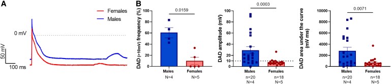
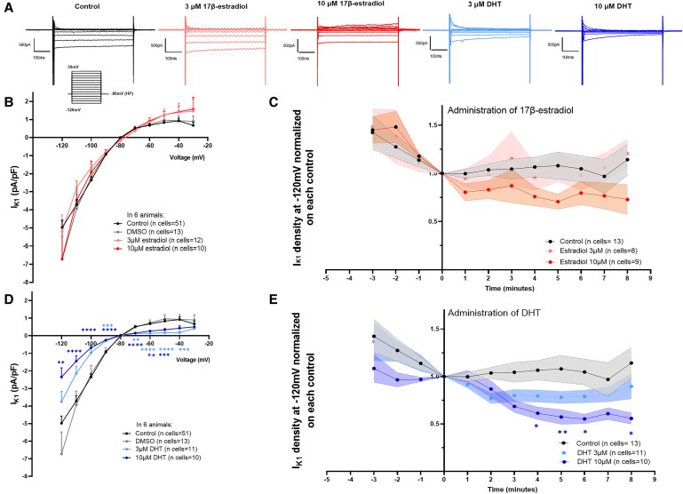
Methods and results: We assessed sex-differences in P-wave morphology in 12-lead ECG in healthy young New Zealand White rabbits. Males presented longer PWD and larger P-wave area compared to females. Patch-clamp experiments were performed in isolated rabbit atrial cardiomyocytes (CMs). Male atrial CMs presented higher delayed after depolarizations (DAD) incidence, amplitude, and area under the curve (AUC) than females, potentially facilitating the presence of atrial triggered activity in males. Male atrial CMs showed a less hyperpolarized resting membrane potential (RMP), a 50% smaller IK1, and a 26% reduction in Kir2.1 protein expression, a pore forming subunit of IK1, than females. Dihydrotestosterone (DHT) effects were investigated acutely and semi-chronically ex vivo. Experiments showed that the sex-difference in IK1 could be mimicked by DHT. In female atrial CMs, acute and semi-chronic (24 h) DHT administration reduced IK1. In the presence of a PKC-inhibitor, DHT-mediated IK1 reduction was not observed in atrial female CMs, suggesting it to be PKC-mediated. Chronic DHT-effects were investigated in vivo in female rabbits after hormone-releasing pellet implantation. After 2 weeks, animals showed a significantly prolonged and larger P-wave, a smaller atrial IK1 and a trend towards an increased DAD amplitude and AUC.

Conclusion: Sex impacts on atrial electrophysiology, leading to sex-differences in P-wave morphology, triggered activity, RMP, and IK1. These sex-differences can be mimicked by sex hormone-treatment, suggesting that sex hormones-particularly DHT-play a pivotal role in mediating sex-differences in atrial electrophysiology. Such sex-differences might impact on the propensity to develop AF, particularly in the younger population.

Keywords: Atrial fibrillation; IK1; Sex differences; Sex hormones.


Conflict of interest statement

Conflict of interest: U.S. received consultancy fees or honoraria from Università della Svizzera Italiana (USI, Switzerland) and Roche Diagnostics (Switzerland). U.S. received research grants from EP Solutions Inc. (Switzerland), Roche Diagnostics (Switzerland) and Bayer Healthcare (Germany). U.S. is co-founder and shareholder of YourRhythmics BV, a spin-off company of the University Maastricht.

Figures

Graphical Abstract
Graphical Abstract
Figure 1
Figure 1
Sex differences in atrial ECG parameters in wild-type rabbits and healthy young volunteers. (A) Representative ECG traces from lead II recorded in surface ECGs in rabbits; top panel male animal, lower panel female animal. (B) Representative surface ECG traces from lead II recorded in humans, top panel men, lower panel women. Dashed lines: black represents the start of the P-wave in both sexes; red represents the end of the P-wave in female sex; blue represents the end of the P-wave in male sex. (C) Sex differences in P-wave parameters (duration, amplitude, and area) in rabbits. Biological replicates (N, animals): females = 15 and males = 12. (D) Sex differences in P-wave parameters in humans. Biological replicates (N, individuals): females = 10 and males = 10. Results are expressed as mean ± SEM. Unpaired t-test or Mann–Whitney test.
Figure 2
Figure 2
Sex DAD in atrial AP in isolated atrial rabbit CMs. (A) Representative AP traces in female and male atrial CMs with spontaneous DAD in control condition. (B) Sex differences in DAD frequency (DAD defined as delayed membrane depolarization higher than 10 mV from RMP expressed in % per animal), and DAD amplitude and DAD area under the curve. N indicates the number of biological replicates (N, animals): males = 4 and females = 5; n indicates the total number of individual cells recorded from males = 20, from females = 18. Results are expressed as mean ± SEM. Unpaired t-test or Mann–Whitney test.
Figure 3
Figure 3
Sex differences on RMP, IK1 density, KCNJ2 mRNA expression in isolated rabbit atrial CMs and Kir2.1 protein expression in atrial tissues. (A) Sex differences in RMP recorded in isolated atrial CMs using whole-cell patch-clamp in current-clamp condition. Biological replicates (N, animals): males = 4, females = 3, with ‘n’ individual cells recorded in males = 43, in females = 31. Mann–Whitney test was performed. (B) Representative IK1 traces from male and female atrial CMs recorded using protocol in (C). (C) Sex differences in IK1 I-V relationship in atrial CMs from both sexes. Biological replicates (N, animals): males = 6, females = 6, with ‘n’ individual cells recorded in males = 59, in females = 51. Two-way ANOVA and Sidak post hoc analyses were performed. (D) Sex differences in KCNJ2 mRNA expression. GAPDH mRNA was used as housekeeping gene to normalize values. Biological replicates (N, animals): males = 4 and females = 3. (E) Representative western blot showing sex differences Kir2.1 protein expression (∼45 kDa) in rabbit atria. Total protein content was used to normalize protein content. (F) Bar graphs represent western blot quantification of Kir2.1 on the total protein content in the left atrium (left), in the right atrium (centre) and overall, in the atria (right). Biological replicates (N, animals): males = 4, females = 5, number of individual atria investigated (n) in males = 8, in females = 10 (two atria per rabbit). Results are expressed as mean ± SEM. Unpaired t-test in panel (D) and (F). *P ≤ 0.05, and ****P ≤ 0.0001.
Figure 4
Figure 4
Acute sex hormones effects on IK1 density in female rabbit atrial CMs. (A) Representative IK1 traces of whole-cell patch-clamp recordings in atrial CMs isolated from females. (B, D) Acute effect (5 min administration) of 17β-estradiol (B) and DHT (D) on atrial IK1 density, in female atrial CMs from six animals (N = 6 biological replicates, animals). (C, E) Time course of sex hormones effects on atrial IK1 density in females (C, estradiol; E, DHT); plots represent current values recorded at −120 mV, normalized for the last value obtained before sex hormone administration. Results are expressed as mean ± SEM. The total number of individual cells (n) measured per group is indicated in parentheses. Two-way ANOVA, Dunnett in panel (B) and (D) and Sidak post hoc analysis in panel (C) and (E). **P ≤ 0.01, ***P ≤ 0.001, and ****P ≤ 0.0001.
Figure 5
Figure 5
Semi-chronic (24 h) sex hormones effects on IK1 density in rabbit atrial CMs from both sexes and acute effect of physiological DHT concentration in female atrial CMs. (A, B) IK1 I-V relationship showing the semi-chronic effects of 17β-estradiol (left panels) and DHT (right panels) on IK1 in atrial CMs isolated from females in (A) (N = 4 biological replicates, animals) and from males in (B) (N = 3 biological replicates, animals). (C) (Left) Representative IK1 traces in a control CM and in a CM acutely (∼2 h) treated with 10 nM DHT concentration. (Right) IK1 I-V relationship showing the acute effects of a physiological (10 nM) concentration of DHT on IK1 in atrial CMs isolated from females (N = 5 biological replicates, animals). Results are expressed as mean ± SEM, with the number of individual cells (n) measured indicated in parentheses. Two-way ANOVA was performed. *P ≤ 0.05, **P ≤ 0.01, ***P ≤ 0.001, and ****P ≤ 0.0001.
Figure 6
Figure 6
Chronic (2 weeks) DHT effects in atrial ECG parameters in rabbits. (A, D) Representative ECG traces from lead II recorded in rabbits; top panel pre-pellet implantation, lower panel same animal 2 weeks post-pellet implantation; (A) DHT pellet, (D) placebo pellet. (B, E) animals before pellet implantation, respectively. (C, F) P-wave parameters (duration, amplitude, area) in pre- and post-pellet implanted rabbits. Data represent N = 9 biological replicates/animals per group. Results are expressed as mean ± SEM. Paired t-test. (A, B, C) DHT pellet; (D, E, F) placebo pellet.
Figure 7
Figure 7
Chronic (2 weeks) DHT effects on DAD and IK1 density in atrial CMs isolated from pellet-implanted female rabbits. (A) Representative AP traces in DHT and Placebo implanted female rabbits with spontaneous DAD in control condition. (B) Chronic DHT effects in DAD frequency (DAD defined as delayed membrane depolarization higher than 10 mV from RMP expressed in % per animal), DAD amplitude and DAD area under the curve. Biological replicates (N, animals): DHT = 4, Placebo = 4; with total individual cells measured (n) in DHT = 20, in Placebo = 12. (C) Representative IK1 traces of whole-cell patch-clamp recordings in atrial CMs isolated from DHT and Placebo implanted female rabbits. (D) IK1 I-V relationship showing the DHT chronic effects on IK1 in atrial CMs isolated from implanted female rabbits. Biological replicates (N, animals): DHT = 4, Placebo = 4; with total individual cells measured (n) in DHT = 38, in Placebo = 36. Results are expressed as mean ± SEM. Mann–Whitney test in (A) and (B), Two-way ANOVA and Sidak post hoc analysis in (D). *P ≤ 0.05, **P ≤ 0.01, and ***P ≤ 0.001.

Comment in

References

    1. Andrade JG, Deyell MW, Lee AYK, Macle L. Sex differences in atrial fibrillation. Can J Cardiol 2018;34:429–436. - PubMed
    1. Benjamin EJ, Levy D, Vaziri SM, D'Agostino RB, Belanger AJ, Wolf PA. Independent risk factors for atrial fibrillation in a population-based cohort. The framingham heart study. JAMA 1994;271:840–844. - PubMed
    1. Friberg J, Scharling H, Gadsbøll N, Truelsen T, Jensen GB; Copenhagen City Heart Study . Comparison of the impact of atrial fibrillation on the risk of stroke and cardiovascular death in women versus men (The Copenhagen City Heart Study). Am J Cardiol 2004;94:889–894. - PubMed
    1. Westerman S, Wenger N. Gender differences in atrial fibrillation: a review of epidemiology, management, and outcomes. Curr Cardiol Rev 2019;15:136–144. - PMC - PubMed
    1. Odening KE, Deiß S, Dilling-Boer D, Didenko M, Eriksson U, Nedios S, Ng FS, Roca Luque I, Sanchez Borque P, Vernooy K, Wijnmaalen AP, Yorgun H. Mechanisms of sex differences in atrial fibrillation: role of hormones and differences in electrophysiology, structure, function, and remodelling. Europace 2019;21:366–376. - PubMed

Publication types

Substances

LinkOut - more resources