. 2025 Jun 26;188(13):3603-3622.e27.
doi: 10.1016/j.cell.2025.03.046. Epub 2025 Apr 23.

Encoding and decoding selectivity and promiscuity in the human chemokine-GPCR interaction network

Affiliations

Encoding and decoding selectivity and promiscuity in the human chemokine-GPCR interaction network

Andrew B Kleist et al. Cell. .

Abstract

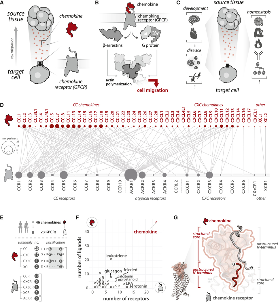
In humans, selective and promiscuous interactions between 46 secreted chemokine ligands and 23 cell surface chemokine receptors of the G-protein-coupled receptor (GPCR) family form a complex network to coordinate cell migration. While chemokines and their GPCRs each share common structural scaffolds, the molecular principles driving selectivity and promiscuity remain elusive. Here, we identify conserved, semi-conserved, and variable determinants (i.e., recognition elements) that are encoded and decoded by chemokines and their receptors to mediate interactions. Selectivity and promiscuity emerge from an ensemble of generalized ("public/conserved") and specific ("private/variable") determinants distributed among structured and unstructured protein regions, with ligands and receptors recognizing these determinants combinatorially. We employ these principles to engineer a viral chemokine with altered GPCR coupling preferences and provide a web resource to facilitate sequence-structure-function studies and protein design efforts for developing immuno-therapeutics and cell therapies.

Keywords: GPCR; chemokine; chemotaxis; data science; machine learning; polymorphism; protein-protein interaction; selectivity determinants; short linear motif; unstructured protein.


Conflict of interest statement

Declaration of interests B.F.V and F.C.P. have ownership interests in Protein Foundry, LLC and XLock Biosciences, Inc.

Figures

Figure 1.
Figure 1.. Structural and functional organization of the chemokine-GPCR system
(A) Secreted chemokines are sensed by receptors on migrating cells. (B) Chemokine binding to GPCRs activates G protein and β-arrestin signaling/recruitment, resulting in cell migration. (C) The chemokine-GPCR system underlies development and immune regulation and is exploited in disease. (D) Interaction network between chemokine ligands (top) and receptors (bottom) (compiled from Table S1; interaction strength ≥ 2 represented, see STAR Methods). Node size scaled to number of binding partners. (E) Chemokine ligand (red) and receptor (gray) subfamilies. (F) The number of GPCR ligands and receptors grouped by family (STAR Methods). (G) Reciprocal structured-to-unstructured binding mode (PDB: 7F1T). The receptor’s unstructured N terminus interacts with the chemokine’s structured core, and vice versa. See also Figure S1.
Figure 2.
Figure 2.. Minimal encoding of generalized chemokine-GPCR recognition
(A) Residue-residue contacts for 16 chemokine-GPCR complexes. (B) Contact fingerprint representing all unique contacts (rows) and complexes (columns). Example fingerprint shown with black/white denoting presence/absence of contact between equivalent residues. * denotes two models used. (C) Human paralog alignments were used to calculate paralog conservation scores, with CCN example given for position B2.3. (D) Residue-residue contacts among 16 chemokine-GPCR complexes (points), and human paralog conservation scores of chemokine (y axis) and receptor (x axis) residues comprising each contact. Receptor paralog scores are calculated among non-atypical receptors (STAR Methods). (E) 12/16 complexes have ≥1 contact between conserved chemokine/receptor residues (human paralogs) involving disulfide regions. Paralog conservation scores shown as pie charts. (F) Interactions between paralog conserved residues by analogy to Lego bricks. (G) Contacts between conserved (human paralogs) chemokine and GPCR residues (dark gray) are 3% of overall contacts. Contacts involving ≥1 variable residue are 97% of contacts (light blue). (H) Effects of CXCR4 mutagenesis on CXCL12 binding. Log2 enrichment scores reflect CXCL12-GFP binding to cells harboring WT versus mutated CXCR4 (STAR Methods). Statistical testing by Kruskal-Wallis test with p values determined by post hoc Dunn test with Bonferroni correction for multiple comparisons. *p value < 0.05, ***p value < 0.001, ****p value < 0.0001. p values for all other pairwise comparisons > 0.05. Boxplot boxes reflect the median (central line), first and third quartiles (box boundaries), and largest/smallest values no further than 1.5x the interquartile range (whiskers). Raw data from Heredia et al. See also Figures S2 and S3.
Figure 3.
Figure 3.. Subfamily-specific sensors encode distinct chemokine-GPCR binding modes
(A) CC and CXC chemokines differ by the presence (CXC) or absence (CC) of a residue (“X”) between conserved N-terminal cysteines. (B) Predominance of interactions among like subfamily chemokines/GPCRs (chi-squared test p = 6.76e-11). (C) Top: logistic regression models were trained to classify a sequence as CC or CXC by residue identity at each sequence position. Positions ranked according to accuracy in subfamily prediction (STAR Methods). Bottom: CC and CXC complexes used to identify consensus subfamily contacts. Contacts in CCL5:CCR5 complexes were considered degenerate (STAR Methods). (D) Residue-residue contacts among CC and CXC complexes (points), with subfamily-predictive scores of chemokine (y axis) and receptor (x axis) positions comprising each contact. (E) Consensus, CC- and CXC-specific contacts involving subfamily-predictive positions. (F) CC- and CXC-specific consensus contacts from (E), with residue positions (chemokine: red, receptor: gray) represented as pie charts depicting position-specific subfamily scores. (G) CC- and CXC-specific consensus contact (top) with subfamily scores and residue sequence logos (middle). CC and CXC complexes (bottom) with puzzle pieces depicting how the same chemokine position (NTc.Cm3) uniquely accommodates CC versus CXC receptor residues (bottom). (H) β-arrestin recruitment by NanoLuc Binary Technology (NanoBiT) with CCR5 (top 3 panels; versus CCL3, CCL4, and CCL5) and CXCR4 (bottom panel; versus CXCL12) receptor mutants at positions 1×24 and 6×58. All experiments n = 3. Error bars reflect SEM. See Table S2. See also Figure S4.
Figure 4.
Figure 4.. Customization of chemokine-GPCR interactions through structure- and sequence-level changes
(A) Customization through structure-level (contact differences) and sequence-level (residue differences among structurally preserved contacts) changes. (B) Comparison of the percent identical/equivalent contacts at the interface among all chemokine-GPCR complexes (y axis, related to structure-level changes) and mean pairwise percent identities of interface residues among paired chemokines and GPCRs (x axis, related to sequence-level changes). Points denote a comparison of a pair of chemokine-GPCR complexes; examples given for the three groups of selectivity-network relatedness. (C) Generalized (i.e., distinguish chemokines/receptors from other molecules/GPCRs), subfamily (i.e., distinguish between chemokines/receptors of different subfamilies, CC or CXC), and network (i.e., found only within specific chemokine-receptor pairs that share interactions) recognition features. (D) Positive selectivity features facilitate interactions, and negative selectivity features disfavor interactions at generalized, subfamily, and network “layers” of encoding. (E) Positive selectivity example. 11 shared contacts among identical residues encode CXCL8 recognition by CXCR1 and CXCR2 (STAR Methods). (F) Selectivity preferences are encoded using compact, discrete interface regions. (G) Negative selectivity example. The contact cxb1.1–7×27 is preserved among 9/16 chemokine-GPCR complexes but has low paralog conservation (left). Unfavorable chemokine-GPCR interactions are likely to prevent noncognate chemokine-GPCR pairs. See also Figure S5.
Figure 5.
Figure 5.. Encoding interactions via SLiMs in unstructured regions
(A) Enumeration of all 2-, 3-, and 4-mer fragments for unstructured regions (chemokine/GPCR N termini; GPCR ECL2) using a sliding window approach (STAR Methods). (B) Inferring fragment functional roles based on ortholog/paralog conservation. Fragments conserved in ≥50% of orthologs are called putative SLiMs. (C) CCL28 structure (PDB: 6CWS) depicting N-terminal residues 1–5 and fragment ortholog conservation. (D) LogEC50 of CCL28 N-terminal truncation variants values from calcium flux experiments on CCR3 (left) and CCR10 (right) -expressing cells. Error reflects SEM of nonlinear fit of logEC50 value. All conditions n = 3. See Table S2. (E) Most frequent variants in chemokines (left) and receptors (right) among interface positions from gnomAD. (F) Allele frequency of ACKR1 Gly42 and Asp42. Gly42 allele frequency inferred as 1 Asp42 allele frequency. (G) Log2-fold ratio of chemokine EC50 values for ACKR1 Asp42 versus Gly42 in bioluminescence resonance energy transfer (BRET)-based binding assay (STAR Methods). See Table S2. (H) ITC performed by injecting 200 μM ACKR1(1–60) Gly42 (left) and Asp42 (right) into WT-CXCL12. Thermograms representative of n = 2 replicates. (I) Immune, functional, and phenotypic trade-offs between ACKR1 Gly42 and Asp42 alleles. See also Figures S6 and S7.
Figure 6.
Figure 6.. Rational design of altered selectivity using a promiscuous viral chemokine.
(A) HHV-8-mediated expression of vMIP-II, which binds CC and CXC receptors to modulate host immunity. (B) Distribution of “prediction probability scores” among chemokine interface residues from chemokine-GPCR complexes by histogram (STAR Methods; Figure S7F). Scores assess likelihood that a queried residue belongs to a CC (i.e., closer to 0) or CXC (i.e., closer to 1) chemokine. Interface residues from Zheng et al. model were used for CCL5. (C) Percentage interface residues from (B) comprising CC- versus CXC-like residues and mapping onto CCL5/vMIP-II structures. (D) Positions of vMIP-II mutants tested. (E) Log fold change of IC50 (or EC50 for CCR3) of vMIP-II mutants versus WT for vMIP-II “reversion” mutants, tested at CCR3, CCR5, and CXCR4 in β-arrestin recruitment assays (Figure S7I; STAR Methods). All data n = 3. WT vMIP-II and mutants tested as agonists (CCR3) or antagonists (CCR5: in competition with CCL5; CXCR4: in competition with CXCL12). See Table S2. See also Figure S7.
Figure 7.
Figure 7.. Encoding and decoding chemokine-GPCR selectivity and promiscuity
(A) Encryption model for chemokine-GPCR selectivity encoding. (B) Chemokine-GPCR network editing applications. (C) Chemokine regulation of complex multicellular circuits for therapeutic applications.

References

    1. McCormick B, Chu JY, and Vermeren S. (2017). Cross-talk between Rho GTPases and PI3K in the neutrophil. Small GTPases 10, 187–195. 10.1080/21541248.2017.1304855. - DOI - PMC - PubMed
    1. Jin T, Xu X, and Hereld D. (2008). Chemotaxis, chemokine receptors and human disease. Cytokine 44, 1–8. 10.1016/j.cyto.2008.06.017. - DOI - PMC - PubMed
    1. DeFea KA (2007). Stop that cell! Beta-arrestin-dependent chemotaxis: a tale of localized actin assembly and receptor desensitization. Annu. Rev. Physiol 69, 535–560. 10.1146/annurev.physiol.69.022405.154804. - DOI - PubMed
    1. Zou YR, Kottmann AH, Kuroda M, Taniuchi I, and Littman DR (1998). Function of the chemokine receptor CXCR4 in haematopoiesis and in cerebellar development. Nature 393, 595–599. 10.1038/31269. - DOI - PubMed
    1. Ma Q, Jones D, Borghesani PR, Segal RA, Nagasawa T, Kishimoto T, Bronson RT, and Springer TA (1998). Impaired B-lymphopoiesis, myelopoiesis, and derailed cerebellar neuron migration in CXCR4- and SDF-1-deficient mice. Proc. Natl. Acad. Sci. USA 95, 9448–9453. 10.1073/pnas.95.16.9448. - DOI - PMC - PubMed

LinkOut - more resources