. 2025 Apr 23;26(4):353-370.
doi: 10.1631/jzus.B2300678.

Roles of PANoptosis and related genes in acute liver failure: neoteric insight from bioinformatics analysis and animal experiment verification

[Article in English, Chinese]
Affiliations

Roles of PANoptosis and related genes in acute liver failure: neoteric insight from bioinformatics analysis and animal experiment verification

[Article in English, Chinese]
Tiantian Ge et al. J Zhejiang Univ Sci B. .

Abstract

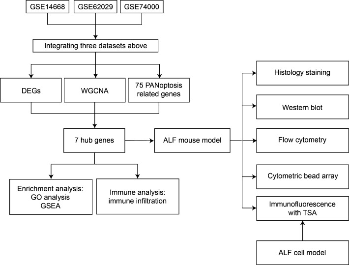
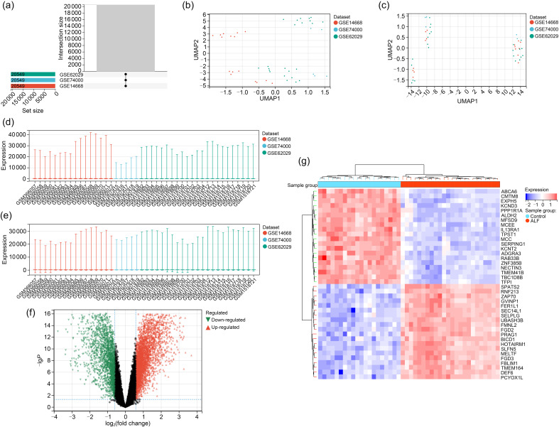
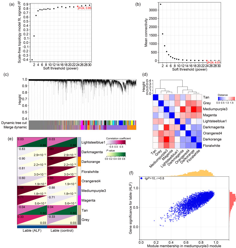
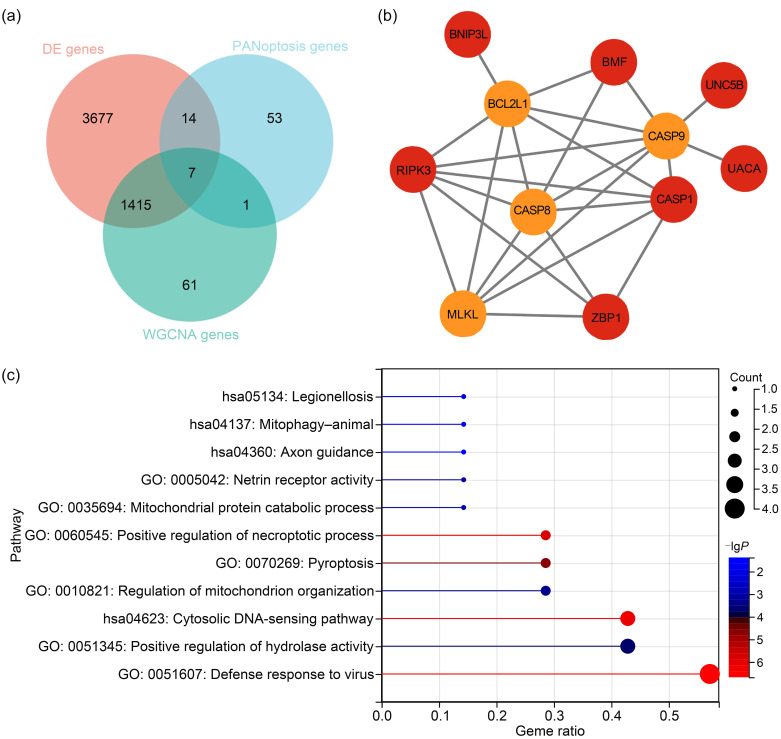
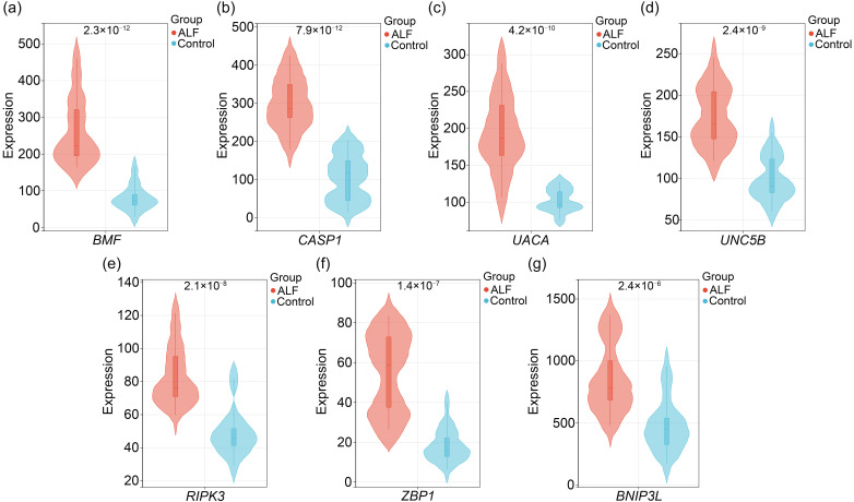
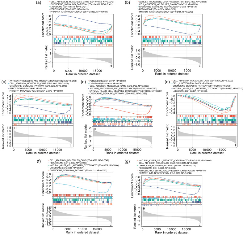
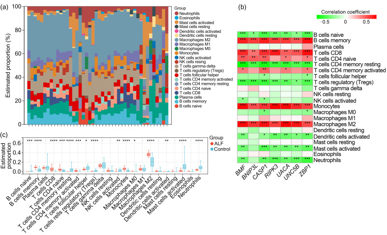
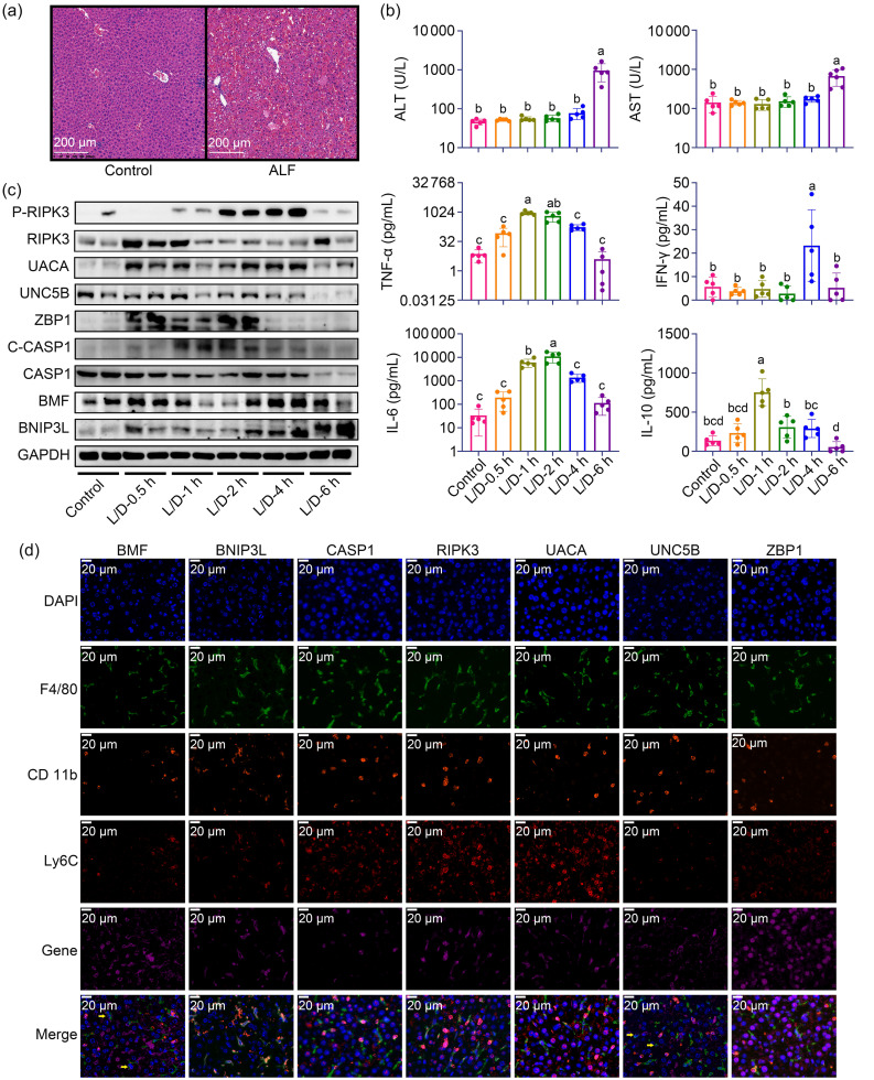
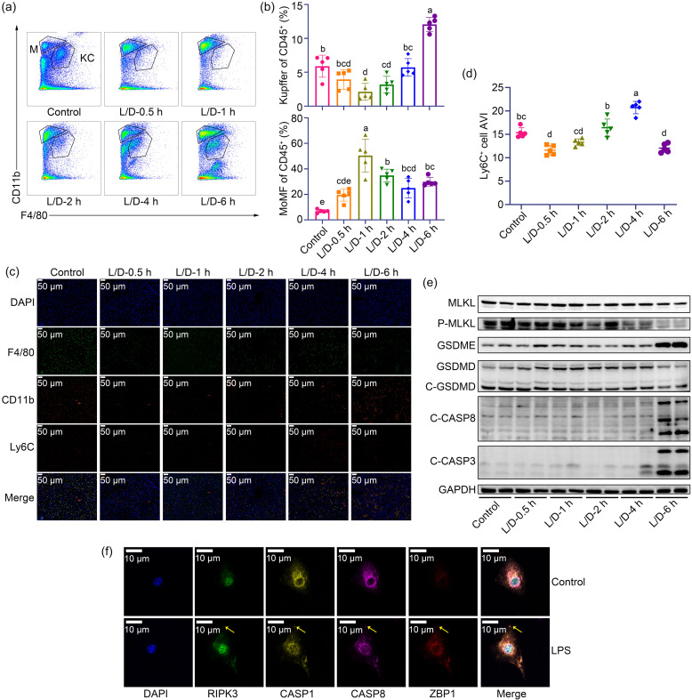
BACKGROUND: PANoptosis has the features of pyroptosis, apoptosis, and necroptosis. Numerous studies have confirmed the diverse roles of various types of cell death in acute liver failure (ALF), but limited attention has been given to the crosstalk among them. In this study, we aimed to explore the role of PANoptosis in ALF and uncover new targets for its prevention or treatment. METHODS: Three ALF-related datasets (GSE14668, GSE62029, and GSE74000) were downloaded from the Gene Expression Omnibus (GEO) database to identify differentially expressed genes (DEGs). Hub genes were identified through intersecting DEGs, genes obtained from weighted gene co-expression network analysis (WGCNA), and genes related to PANoptosis. Gene Ontology (GO), Kyoto Encyclopedia of Genes and Genomes (KEGG), protein‍‒‍protein interaction (PPI) analyses and gene set enrichment analysis (GSEA) were performed to determine functional roles. Verification was performed using an ALF mouse model. RESULTS: Our results showed that expression of seven hub genes (B-cell lymphoma-2-modifying factor (BMF), B-cell lymphoma-2-interacting protein 3-like (BNIP3L), Caspase-1 (CASP1), receptor-interacting protein kinase 3 (RIPK3), uveal autoantigen with coiled-coil domains and ankyrin repeats protein (UACA), uncoordinated-5 homolog B receptor (UNC5B), and Z-DNA-binding protein 1 (ZBP1)) was up-regulated in liver samples of patients. However, in the ALF mouse model, the expression of BNIP3L, RIPK3, phosphorylated RIPK3 (P-RIPK3), UACA, and cleaved caspase-1 was up-regulated, while the expression of CASP1 and UNC5B was down-regulated. The expression of ZBP1 and BMF increased only during the development of ALF, and there was no significant change in the end stage. Immunofluorescence of mouse liver tissue showed that macrophages expressed all seven markers. Western blot results showed that pyroptosis, apoptosis, and necroptosis were always involved in lipopolysaccharide (LPS)/ d-galactosamine (d-gal)‍-induced ALF mice. The ALF cell model showed that bone marrow-derived macrophages (BMDMs) form PANoptosomes after LPS stimulation. CONCLUSIONS: Our results suggest that PANoptosis of macrophages promotes the development of ALF. The seven new ALF biomarkers identified and validated in this study may contribute to further investigation of diagnostic markers or novel therapeutic targets of ALF.

泛凋亡具有焦亡、凋亡和坏死性凋亡的特征。尽管大量研究已证实各类型细胞的死亡在急性肝衰竭(ALF)中发挥着不同作用,但对它们间的互作关注较少。本研究旨在探讨泛凋亡在ALF中的作用,并挖掘可作为预防或治疗ALF的新靶点。本研究首先从基因表达综合数据库(GEO)中下载三个与ALF相关的数据集(GSE14668、GSE62029和GSE74000),从中筛选差异表达基因(DEGs),并通过加权基因共表达网络分析(WGCNA)对上述基因和泛凋亡相关基因集取交集得到枢纽基因;通过基因本体论(GO)、京都基因与基因组百科全书(KEGG)、蛋白质-蛋白质互作(PPI)和基因集富集分析(GSEA)确定枢纽基因功能;最后利用ALF小鼠和细胞模型进行验证。结果表明,在ALF患者肝脏样本中,7个枢纽基因(BMFBNIP3LCASP1RIPK3UACAUNC5BZBP1)表达水平均上调;在ALF小鼠模型中,BNIP3L、RIPK3、P-RIPK3、UACA和cleaved caspase-1表达上调,而CASP1和UNC5B的表达下调;ZBP-1和BMF的表达仅在造模过程中升高,终末期则无明显变化。小鼠肝组织免疫荧光显示,这7个枢纽基因在巨噬细胞中均有表达;western blot结果表明,在脂多糖(LPS)/ d-氨基半乳糖( d-gal)引起的ALF小鼠模型中,焦亡、凋亡和坏死性凋亡均有发生;ALF细胞模型实验显示,骨髓源巨噬细胞在LPS刺激后形成泛凋亡小体。综上,巨噬细胞的泛凋亡有可能加速ALF的进展,此外本研究鉴定并验证的7个ALF枢纽基因将有希望作为未来深入研究ALF的诊断标志物或新的治疗靶点。.

泛凋亡具有焦亡、凋亡和坏死性凋亡的特征。尽管大量研究已证实各类型细胞的死亡在急性肝衰竭(ALF)中发挥着不同作用,但对它们间的互作关注较少。本研究旨在探讨泛凋亡在ALF中的作用,并挖掘可作为预防或治疗ALF的新靶点。本研究首先从基因表达综合数据库(GEO)中下载三个与ALF相关的数据集(GSE14668、GSE62029和GSE74000),从中筛选差异表达基因(DEGs),并通过加权基因共表达网络分析(WGCNA)对上述基因和泛凋亡相关基因集取交集得到枢纽基因;通过基因本体论(GO)、京都基因与基因组百科全书(KEGG)、蛋白质-蛋白质互作(PPI)和基因集富集分析(GSEA)确定枢纽基因功能;最后利用ALF小鼠和细胞模型进行验证。结果表明,在ALF患者肝脏样本中,7个枢纽基因(BMFBNIP3LCASP1RIPK3UACAUNC5BZBP1)表达水平均上调;在ALF小鼠模型中,BNIP3L、RIPK3、P-RIPK3、UACA和cleaved caspase-1表达上调,而CASP1和UNC5B的表达下调;ZBP-1和BMF的表达仅在造模过程中升高,终末期则无明显变化。小鼠肝组织免疫荧光显示,这7个枢纽基因在巨噬细胞中均有表达;western blot结果表明,在脂多糖(LPS)/d-氨基半乳糖(d-gal)引起的ALF小鼠模型中,焦亡、凋亡和坏死性凋亡均有发生;ALF细胞模型实验显示,骨髓源巨噬细胞在LPS刺激后形成泛凋亡小体。综上,巨噬细胞的泛凋亡有可能加速ALF的进展,此外本研究鉴定并验证的7个ALF枢纽基因将有希望作为未来深入研究ALF的诊断标志物或新的治疗靶点。

Keywords: Acute liver failure; Biomarker; Gene Expression Omnibus (GEO); PANoptosis; Therapeutic target.


Figures

Fig. 1
Fig. 1. Flow diagram of the study process. DEGs: differentially expressed genes; WGCNA: weighted gene co-expression network analysis; ALF: acute liver failure; GO: Gene Ontology; GSEA: gene set enrichment analysis; TSA: tyramide signal amplification.
Fig. 2
Fig. 2. Screening for differential genes. (a) Intersection sizes of the three datasets. (b, c) Gene expression level statistics of the datasets before and after de-batching. (d, e) Principal component analysis (PCA) of datasets before and after de-batching. (f) Volcano plot of differentially expressed genes (DEGs): up-regulated DEGs are represented by red nodes, down-regulated DEGs by green nodes, and genes with no significant differential expression by black nodes. (g) Heatmap of DEG expression levels: azure indicates control samples, orange indicates acute liver failure (ALF) samples, red indicates high gene expression, and purple indicates low gene expression.
Fig. 3
Fig. 3. Weighted gene co-expression network analysis (WGCNA). (a, b) Analysis of the scale-free fit index for various soft-thresholding powers. (c) Gene trees of co-expression gene modules are shown in various colors. (d) Heatmap of eigengene adjacency. (e) Heatmap of the association between acute liver failure (ALF) and modules. The mediumpurple3 module is shown to be significantly correlated with the ALF. The numbers in the top and bottom triangles represent the correlation coefficient and P value, respectively. (f) Correlation plot between gene significance and module membership of genes included in the mediumpurple3 module. r: correlation coefficient.
Fig. 4
Fig. 4. Selection of the PANoptosis-related genes. (a) Venn diagram showing the seven PANoptosis-related genes from the intersection of DEGs, mediumpurple3 module via weighted gene co-expression network analysis (WGCNA), and 75 genes that refer to PANoptosis. (b) The proteinprotein interaction (PPI) network revealed that the seven hub genes were linked directly or indirectly. The genes in the orange circle are the hub genes, and those in the yellow circle are the bridge genes needed to connect the hub genes. (c) Enrichment analyses of Kyoto Encyclopedia of Genes and Genomes (KEGG) and Gene Ontology (GO) pathways related to the seven hub genes.
Fig. 5
Fig. 5. Violin plots of the expression of seven PANoptosis-related genes in the acute liver failure (ALF) and control groups. (a) Expression of B-cell lymphoma-2-modifying factor (BMF). (b) Expression of Caspase-1 (CASP1). (c) Expression of uveal autoantigen with coiled-coil domains and ankyrin repeats protein (UACA). (d) Expression of uncoordinated-5 homolog B receptor (UNC5B). (e) Expression of receptor-interacting protein kinase 3 (RIPK3). (f) Expression of Z-DNA-binding protein 1 (ZBP1). (g) Expression of B-cell lymphoma-2-interacting protein 3-like (BNIP3L). The number at the top of the violin diagram for each gene represents a P-value of <0.0001.
Fig. 6
Fig. 6. Gene set enrichment analysis (GSEA) of seven PANoptosis-related genes. (a) GSEA of Caspase-1 (CASP1). (b) GSEA of receptor-interacting protein kinase 3 (RIPK3). (c) GSEA of Z-DNA-binding protein 1 (ZBP1). (d) GSEA of B-cell lymphoma-2-modifying factor (BMF). (e) GSEA of B-cell lymphoma-2-interacting protein 3-like (BNIP3L). (f) GSEA of uncoordinated-5 homolog B receptor (UNC5B). (g) GSEA of uveal autoantigen with coiled-coil domains and ankyrin repeats protein (UACA). H represents high-expression group; L represents low-expression group.
Fig. 7
Fig. 7. Immune cell infiltration in acute liver failure (ALF). (a) Proportion of 22 types of immune cells in the ALF group. (b) Correlations among seven PANoptosis-related genes and 22 immune cell types. (c) Comparison of the proportions of 22 kinds of immune cells between the ALF and control groups. * P<0.05; ** P<0.01; *** P<0.001; **** P<0.0001.
Fig. 8
Fig. 8. Verification of the animal model. (a) Hematoxylin and eosin (H&E) staining of liver sections. (b) Levels of serum alanine aminotransferase (ALT), aspartate aminotransferase (AST), tumor necrosis factor-α (TNF-α), interferon-γ (IFN-γ), interleukin (IL)-6, and IL-10. (c) Changes of the expression levels of nine kinds of proteins with the time of modeling by western blot. (d) Cellular localization analyses of the seven genes through immunofluorescence with tyramide signal amplification (TSA). Data are expressed as mean±standard deviation (n=5). The presence of the same letter between two groups indicates no significant difference between them, and the absence of the same letter indicates a significant difference between them (P<0.05). The golden arrows indicate macrophages expressing BMF or UNC5B. BMF: B-cell lymphoma-2-modifying factor; BNIP3L: B-cell lymphoma-2-interacting protein 3-like; CASP1: Caspase-1; C-CASP1: cleaved CASP1; DAPI: 4',6-diamidino-2-phenylindole; GAPDH: glyceraldehyde-3-phosphate dehydrogenase; L/D: lipopolysaccharide/d-galactosamine; RIPK3: receptor-interacting protein kinase 3; P-RIPK3: phosphorylated RIPK3; UACA: uveal autoantigen with coiled-coil domains and ankyrin repeats protein; UNC5B: uncoordinated-5 homolog B receptor; ZBP1: Z-DNA-binding protein 1.
Fig. 9
Fig. 9. Changes in macrophages, pyroptosis, apoptosis, and necroptosis in the liver after LPS/d-gal stimulation (labeled L/D). (a) Representative flow cytometry plots of Kupffer cells (labeled KC) and MoMFs (labeled M) of each group. (b) Percentages of Kupffer cells and MoMFs in hepatic CD45+ cells. (c) Immunofluorescence results of macrophages in liver tissue at different time points after LPS/d-gal stimulation. (d) Average fluorescence intensity (AVI) of Ly6C+ cells. (e) Trends of biomarkers of pyroptosis, apoptosis, and necroptosis. (f) Immunofluorescence results of PANoptosomes in bone marrow-derived macrophages (BMDMs) after LPS stimulation for 6 h (63× oil immersion objective). The golden arrows show the PANoptosome protein complex. Data are expressed as mean±standard deviation (n=5). The presence of the same letter between two groups indicates no significant difference between them, and the absence of the same letter indicates a significant difference between them (P<0.05). C-GSDMD: cleaved gasdermin D; C-CASP8: cleaved Caspase-8; C-CASP3: cleaved Caspase-3; DAPI: 4',6-diamidino-2-phenylindole; d-gal: d-galactosamine; LPS: lipopolysaccharide; MoMFs: monocyte-derived macrophages.

Similar articles

References

    1. Bernal W, Wendon J, 2013. Acute liver failure. N Engl J Med, 369(26): 2525-2534. 10.1056/NEJMra1208937 - DOI - PubMed
    1. Chen YJ, Jiao DY, Liu Y, et al. , 2023. FBXL4 mutations cause excessive mitophagy via BNIP3/BNIP3L accumulation leading to mitochondrial DNA depletion syndrome. Cell Death Differ, 30(10): 2351-2363. 10.1038/s41418-023-01205-1 - DOI - PMC - PubMed
    1. Choi SW, Oh H, Park SY, et al. , 2022. Netrin-1 attenuates hepatic steatosis via UNC5b/PPARγ‍-‍mediated suppression of inflammation and ER stress. Life Sci, 311: 121149. 10.1016/j.lfs.2022.121149 - DOI - PubMed
    1. Christgen S, Zheng M, Kesavardhana S, et al. , 2020. Identification of the panoptosome: a molecular platform triggering pyroptosis, apoptosis, and necroptosis (panoptosis). Front Cell Infect Microbiol, 10: 237. 10.3389/fcimb.2020.00237 - DOI - PMC - PubMed
    1. Duan YW, Chen SX, Li QY, et al. , 2022. Neuroimmune mechanisms underlying neuropathic pain: the potential role of TNF-α-necroptosis pathway. Int J Mol Sci, 23(13): 7191. 10.3390/ijms23137191 - DOI - PMC - PubMed