

[18F]NaF PET/CT Imaging of Iliac Bones to Assess Bone Turnover
- PMID: 40274673
- PMCID: PMC12162772
- DOI: 10.1007/s11307-025-02003-6
[18F]NaF PET/CT Imaging of Iliac Bones to Assess Bone Turnover
Abstract
Purpose: This study investigated the effects of laterality, age, gender, BMI, and physical activity level on iliac bone turnover using [18F]NaF PET/CT.
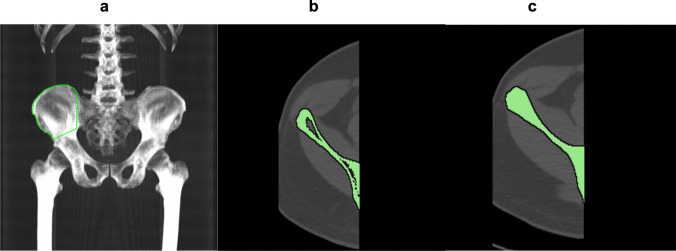
Procedures: Fifty-nine males and 44 females from the CAMONA study were analyzed. A region of interest (ROI) was drawn to segment the iliac bone using Hounsfield unit thresholds and morphological closing algorithm. [18F]NaF SUVmean was compared between the left and right iliac bones using a paired t-test, while Pearson correlation coefficient assessed changes with age, BMI, and physical activity level.
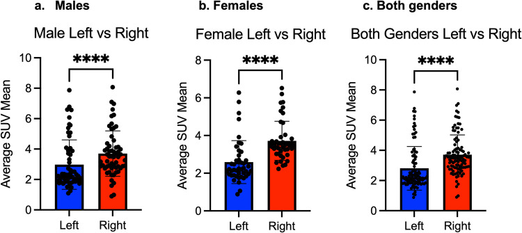
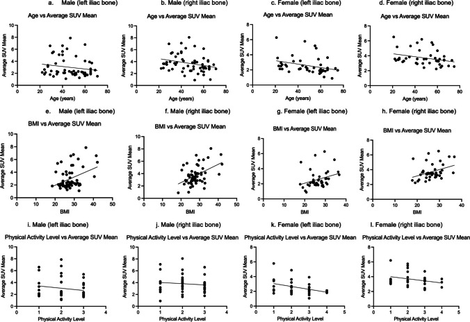
Results: [18F]NaF uptake was higher in right iliac bone than left in males, females, and the combined-group. In males, SUVmean was 2.98 ± 1.63 (1.1-7.87) on left and 3.71 ± 1.49 (1.49-3.7) on right. In females, SUVmean was 2.59 ± 1.14 (0.88-6.27) on left and 3.72 ± 1.04 (2.22-6.51) on right. Combined, SUVmean was 2.81 ± 1.44 (0.88-7.87) on left and 3.71 ± 1.31 (0.89-8.07) on right. [18F]NaF uptake negatively correlated with age (right: r = - 0.27, P = 0.006; left: r = - 0.22, P = 0.02), stronger in females (right: r = - 0.30, P = 0.04; left: r = - 0.31, P = 0.04) than males (right: r = - 0.26, P = 0.04; left: r = - 0.18, P = 0.18). SUVmean correlated positively with BMI in males (right: r = 0.47, P = 0.0002; left: r = 0.38, P = 0.0027), females (right: r = 0.36, P = 0.0168; left: r = 0.30, P = 0.0505), and combined-group (right: r = 0.43, P < 0.0001; left: r = 0.37, P = 0.0001). No significant correlation was found between SUVmean and physical activity in males, while in females, a negative correlation was observed on left (r = - 0.37, P = 0.0390) but not on right (r = - 0.27, P = 0.1302), and when combined, the correlation remained significant on left (r = - 0.24, P = 0.0372) but not on right (r = - 0.16, P = 0.1541).
Conclusions: [18F]NaF uptake was higher in the right iliac bone and declined with age, particularly in females. The positive correlation between SUVmean and BMI; and the negative correlation between SUVmean and physical activity suggest metabolic influences on bone turnover. [18F]NaF PET/CT may serve as a tool for assessing bone metabolism and turnover in asymptomatic individuals.
Keywords: Bone; Iliac bone; Metabolism; Osteoporosis; Positron emission tomography (PET); Positron emission tomography/computed tomography (PET/CT); [18F]-sodium fluoride ([18F]NaF).
© 2025. The Author(s).
Conflict of interest statement
Declarations. Ethical Approval: All procedures performed in studies involving human participants were in accordance with the ethical standards of the institutional and/or national research committee and with the 1964 Helsinki declaration and its later amendments or comparable ethical standards. Conflicts of Interest: The authors declare that they have no conflicts of interest in regard to this study.
Figures
References
-
- Sarafrazi N, Wambogo EA, Shepherd JA (2021) Osteoporosis or low bone mass in older adults: United States, 2017–2018. NCHS Data Brief 405:1–8 - PubMed
MeSH terms
Substances
LinkOut - more resources
Full Text Sources
