

Multimodal imaging analysis of autosomal recessive Parkinson's disease
- PMID: 40274726
- PMCID: PMC12289758
- DOI: 10.1007/s12149-025-02053-4
Multimodal imaging analysis of autosomal recessive Parkinson's disease
Abstract
Objective: Pathophysiological backgrounds of idiopathic Parkinson's disease (IPD) and autosomal recessive monogenic Parkinson's disease (AR-PD) have common features that can be assessed through multimodal imaging. In this study, the striatal and myocardial dopaminergic innervation, brain 18F-FDG metabolism, resting-state functional activity of basal ganglia network (BGN) and white-matter (WM) microstructure were evaluated in AR-PD with respect to IPD, to investigate whether AR-PD can be subtyped as "brain-first" parkinsonism according to recent etiopathogenetic classification effort.
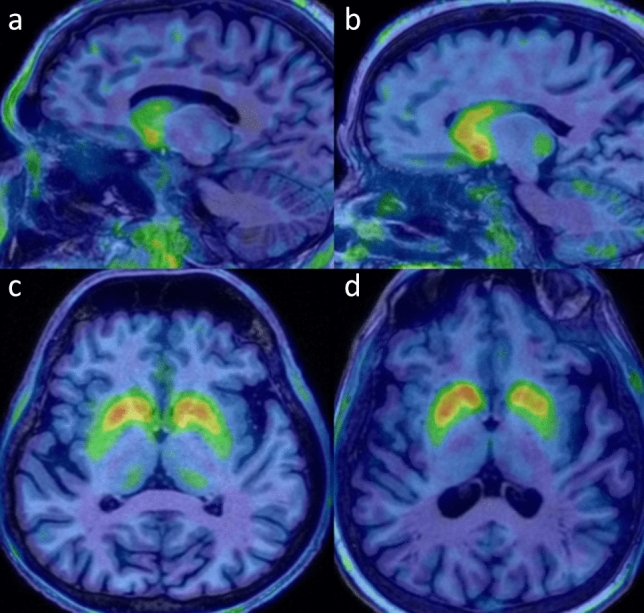
Methods: Forty patients (17 with Parkin, 3 with DJ-1 mutations and 20 with IPD) were included. Striatal dopaminergic innervation was assessed semi-quantitatively by 18F-DOPA PET, and cardiac 18F-DOPA uptake was also evaluated. Brain 18F-FDG PET images were evaluated visually. Resting-state functional MRI and diffusion tensor imaging (DTI) were used to assess the BGN activity and WM microstructural alterations.
Results: AR-PD patients showed significantly decreased 18F-DOPA uptake in caudate corpus compared to both IPD and controls, with a more symmetrical striatal dopaminergic denervation. Myocardial 18F-DOPA uptake in AR-PD was similar to controls, while it was significantly reduced in IPD. There was no significant difference in cortical 18F-FDG metabolism and functional activity of BGN between PD groups. The DTI data revealed more extensive WM microstructural damage in AR-PD compared to IPD.
Conclusions: AR-PD group showed additional significant decreased 18F-DOPA uptake in caudate corpus and more symmetrical striatal denervation. Additionally, relatively preserved myocardial innervation, cortical metabolic and WM microstructural changes suggest the possibility of "brain-first" type progression in AR-PD. Also, 18F-DOPA PET/CT may be a practical tool for evaluating dopaminergic innervation of striatum and heart together, but further evaluation is needed in this area.
Keywords: 18F-DOPA; 18F-FDG; Monogenic; Multimodal imaging; Parkinson’s disease.
© 2025. The Author(s).
Figures

References
-
- Balestrino R, Schapira A. Parkinson disease. Eur J Neurol. 2020;27:27–42. - PubMed
-
- Horsager J, Andersen KB, Knudsen K, Skjærbæk C, Fedorova TD, Okkels N, et al. Brain-first versus body-first Parkinson’s disease: a multimodal imaging case-control study. Brain. 2020;143:3077–88. - PubMed
-
- Horsager J, Knudsen K, Sommerauer M. Clinical and imaging evidence of brain-first and body-first Parkinson’s disease. Neurobiol Dis. 2022;164: 105626. - PubMed
-
- Ribeiro M-J, Thobois S, Lohmann E, Du Montcel ST, Lesage S, Pelissolo A, et al. A multitracer dopaminergic PET study of young-onset parkinsonian patients with and without parkin gene mutations. J Nucl Med. 2009;50:1244–50. - PubMed
MeSH terms
Substances
LinkOut - more resources
Full Text Sources
Medical
Research Materials
Miscellaneous
