. 2025 Apr 24;20(1):195.
doi: 10.1186/s13023-025-03711-6.

Using chanarin-dorfman syndrome patient fibroblasts to explore disease mechanisms and new treatment avenues

Affiliations

Using chanarin-dorfman syndrome patient fibroblasts to explore disease mechanisms and new treatment avenues

Mor Angel et al. Orphanet J Rare Dis. .

Abstract

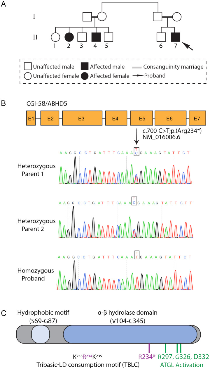
Background: Chanarin-Dorfman syndrome (CDS) is a multisystemic autosomal recessive rare disorder. CDS is caused by variants in the abhydrolase domain containing 5 (ABHD5) encoding gene (CGI-58), which ultimately leads to excessive lipid storage, and therefore a high abundance of cellular lipid droplets (LDs). Although the molecular etiology of the disease was described many years ago, no treatment for CDS is currently available.

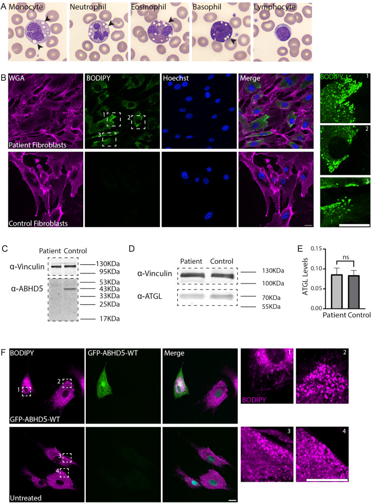
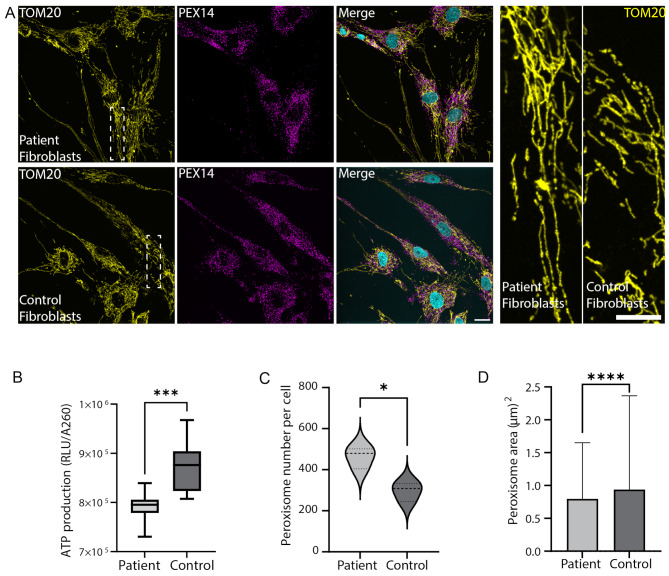
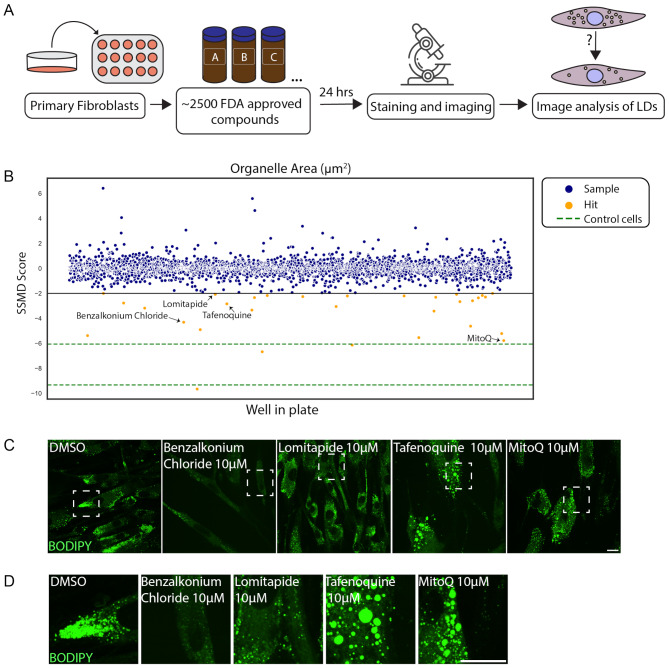
Results: To further characterize the molecular basis of the disease and to uncover new treatment avenues, we used skin fibroblasts originating from a young patient diagnosed with CDS due to a homozygous nonsense mutation. We show that dysfunctional ABHD5 does not only affect LDs, but also influences other metabolic-related organelles; the mitochondria and peroxisomes. Additionally, we found that expressing functional ABHD5 in CDS patient cells reduced LD number. Finally, we developed and applied a high content-based drug repurposing screen based on a collection of ∼2500 FDA approved compounds, yielding several compounds that affected LD total area and size.

Conclusions: Our findings enhance the understanding of the dysfunction underlying CDS and propose new avenues for the treatment of CDS patients.

Keywords: Chanarin-Dorfman syndrome; Drug repurposing; Lipid droplets; Mitochondria; Neutral lipid storage; Peroxisomes.


Conflict of interest statement

Declarations. Ethics approval and consent to participate: Family consented to this study according to an approved institutional review board (IRB) protocol (0038 − 14). Consent for publication: The patients’ parents signed on a consent for publication. Competing interests: The authors have no conflict of interest.

Figures

Fig. 1
Fig. 1
Characterization and diagnosis of a CDS patient. (A) Patient pedigree. The diagnosed patient (i.e., proband) is marked with an arrow. (B) Illustration of the CGI-58 gene, encoding for the ABHD5 protein, as sequenced by Sanger sequencing. Exons are marked with E. Arrow marks the location of the proband mutation. (C) Domain organization of the ABHD5 protein. The human ABHD5 contains 349 amino acids, comprising a hydrophobic domain (LD-binding site) and an α/β hydrolase domain; the three residues involved in ATGL activation (R297, G326 and D332 are highlighted in green). The R234* ABHD5 mutation reported here (red) is localized to the tribasic-LD consumption motif (TBLC)
Fig. 2
Fig. 2
LDs accumulate in the patient fibroblasts and can be reversed by overexpressing ABHD5-WT. (A) Images of representative cells detected in the CDS patient. Lipid vacuoles are marked by black arrowheads. (B) Primary skin fibroblasts derived from the proband and from a healthy individual (GM00498 cells) were stained with WGA for membrane segmentation (magenta), and with BODIPY to mark LDs (green). DNA was stained with Hoechst H33342 (blue). Enlargements of designated areas are in the boxed regions at right as numbered. Scale bars = 20 μm. (C-D) Western blot analysis of proteins extracts from skin fibroblasts derived from CDS patient cells, and from control skin fibroblasts cells (GM40098). Anti-Vinculin was used for loading control. Blots were incubated with (C) anti-ABHD5 and (D) anti-ATGL. (E) Quantifications of ATGL signal levels as seen in D. Data were analyzed using Fiji, and statistical analysis was carried out by two-tailed T-test (n = 3, ns = non-significant). Bar graph illustrates the mean ± SD. (F) Immortalized skin fibroblasts originating from the proband were electroporated with GFP-ABHDT-WT plasmid (green), followed by fixation after 24 h, and BODIPY staining to mark LDs (magenta). The experiment was performed three times (n = 12 positive cells). Enlargements of designated areas are in the boxed regions at right as numbered. Scale bar = 20 μm
Fig. 3
Fig. 3
Mitochondria and peroxisomes are affected in CDS patient fibroblasts. (A) Primary skin fibroblasts originating from the proband, and control skin fibroblasts (GM00498) were immuno-stained with anti-TOM20 to mark mitochondria (yellow), and with anti-PEX14 to mark peroxisomes (magenta). DNA staining is in cyan. Scale bar = 20 μm. Enlargements of designated areas are in the boxed regions at right. Scale bar = 10 μm. (B) Box plot represents ATP production in the patient and control (GM00498) skin fibroblasts expressed as relative fluorescence units (RFU) per cell content measured by methylene blue absorbance 620 nm (A620) (***p < 0.001, n = 10), (C) Violin plot quantifying number of peroxisomes per cell as seen in A. Data were analyzed using two-tailed T-test (*p < 0.05, more than 100 cells were quantified per patient or control cells, n = 3 independent biological replicates). (D) Bar graph quantifying area of peroxisomes as seen in A. Data were analyzed using two-tailed Mann-Whitney test (****p < 0.0001, n > 33,000 peroxisomes per patient or control cells, more than 150 cells were quantified). Bar graph illustrates the mean ± SD
Fig. 4
Fig. 4
Drug repurposing screen on CDS patient cells reveals drugs that reduce LD total area in the patient fibroblasts (A) Illustration of high content drug repurposing screen. (B) Scatter plot representing all compounds screened and analyzed for total LD area. Compounds below − 2 (line marked in black) are determined as hits (yellow), that found significant in all four categories (i.e., compactness, form factor, area and total area). Other samples that were tested are marked in blue, and the score for control cells (GM05400) as a reference is marked in green. Black arrows point at the four compounds shown in C. (C) Primary skin fibroblasts obtained from the proband were treated with 10µM Benzalkonium Chloride, Lomitapide, Tafenoquine or MitoQ for 24 h. Cells were fixed and stained with BODIPY to mark LDs (green). (D) Enlargements of the marked boxes in C. Scale bar = 20 μm

References

    1. Schweiger M, Lass A, Zimmermann R, Eichmann TO, Zechner R. Neutral lipid storage disease: genetic disorders caused by mutations in adipose triglyceride lipase/PNPLA2 or CGI-58/ABHD5. Am J Physiol Endocrinol Metab. 2009;297(2):E289–96. - PubMed
    1. Nur BG, Gencpinar P, Yuzbasioglu A, Emre SD, Mihci E. Chanarin-Dorfman syndrome: Genotype-Phenotype correlation. Eur J Med Genet. 2015;58(4):238–42. - PubMed
    1. Akiyama M, Sawamura D, Nomura Y, Sugawara M, Shimizu H. Truncation of CGI-58 protein causes malformation of lamellar granules resulting in ichthyosis in Dorfman-Chanarin syndrome. J Invest Dermatol. 2003;121(5):1029–34. - PubMed
    1. Bruno C, Bertini E, Di Rocco M, Cassandrini D, Ruffa G, De Toni T, et al. Clinical and genetic characterization of Chanarin-Dorfman syndrome. Biochem Biophys Res Commun. 2008;369(4):1125–8. - PubMed
    1. Missaglia S, Coleman RA, Mordente A, Tavian D. Neutral lipid storage diseases as cellular model to study lipid droplet function. Cells. 2019;8(2). - PMC - PubMed

MeSH terms

Substances

Supplementary concepts