. 2025 Apr 9:32:101751.
doi: 10.1016/j.mtbio.2025.101751. eCollection 2025 Jun.

An NIR-responsive "4A hydrogel" encapsulating wormwood essential oil: through antibacterial, antioxidant, anti-inflammation, and angiogenic to promote diabetic wound healing

Affiliations

An NIR-responsive "4A hydrogel" encapsulating wormwood essential oil: through antibacterial, antioxidant, anti-inflammation, and angiogenic to promote diabetic wound healing

Mengjuan Tao et al. Mater Today Bio. .

Abstract

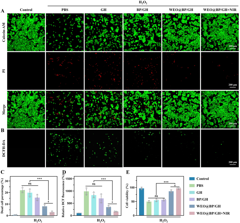
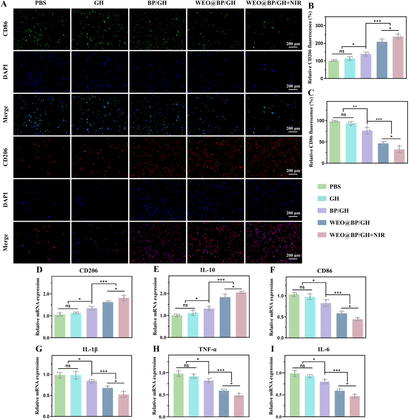
The incorporation of hydrogels with biocompatible functional components to develop wound dressings exhibiting potent antibacterial, antioxidant, anti-inflammatory, and angiogenic properties to promote diabetic wound healing is highly desirable yet continues to pose a significant challenge. In this study, wormwood essential oil (WEO) is successfully encapsulated within black phosphorus (BP) using a physical extrusion technique. Subsequently, this composite is encapsulated within biocompatible gelatin methacrylate (GelMA) and hyaluronic acid methacrylate (HAMA) hydrogels to create multifunctional hydrogel dressing (WEO@BP/GH). In comparison to traditional hydrogels, BP enhances the encapsulation stability of WEO and improves the microenvironmental regulation capabilities through NIR-triggered release of WEO. Systemic in vitro experiments demonstrate that synergistic interaction between the diverse bioactive components of WEO and photothermal effects of BP results in highly effective antibacterial activities against S. aureus and E. coli, antioxidant of scavenging ROS, anti-inflammation of downregulating M1/M2 macrophages ratio, and angiogenic properties. Moreover, the in vivo tests demonstrate that WEO@BP/GH hydrogel significantly enhances high-performance diabetic wound repair through the acceleration of hemostasis, promotion of collagen deposition, regulation of inflammatory responses, and facilitation of vascularization. The findings indicate that WEO@BP/GH hydrogel holds considerable promise as a candidate for microenvironment regulation and effective diabetic wound healing across various clinical applications.

Keywords: Black phosphorus; Microenvironment; Multifunctional hydrogel; Wormwood essential oil.


Conflict of interest statement

The authors declare no conflict of interest.

Figures

Image 1
Graphical abstract
Fig. 1
Fig. 1
Schematic illustration of the preparation of the WEO@BP/GH hydrogel and its mechanism to promote diabetic wound healing through antibacterial, antiinflammation, antioxidant, and angiogenic properties.
Fig. 2
Fig. 2
Fabrication and characterization of the WEO@BP nanoparticles. A) Schematic illustration of the fabrication and NIR-trigged release of the WEO@BP nanoparticles. B, C) SEM images of WEO and WEO@BP nanoparticles, respectively. Scale bar: 200 nm. D, E) TEM images of WEO and WEO@BP nanoparticles, respectively. Scale bar: 200 nm. F) DLS evaluation of WEO@BP nanoparticles. G) Gas chromatography analysis of WEO nanoparticles. H, I) Cumulative release of WEO@BP/GH hydrogels with and without NIR irradiation within 30 min (H) and 120 h (I).
Fig. 3
Fig. 3
Physiochemical characterization of the hydrogels. A) Optical images of the fabricated GH, BP/GH, and WEO@BP/GH hydrogels. B) SEM images of the hydrogels at different magnifications. The red arrow refers to WEO@BP nanoparticles. Scale bar in low magnification: 200 μm. Scale bar in high magnification: 20 μm. C) Raman spectra of the hydrogels. D) Dynamic rheological evaluation of the UV-crosslinking processes of GH and WEO@BP/GH hydrogels. E-H) Rheological analysis of the strain sweep (E), dynamic step-strain (F), frequency sweep (G), and viscosity (H) of GH and WEO@BP/GH hydrogels. (For interpretation of the references to color in this figure legend, the reader is referred to the Web version of this article.)
Fig. 4
Fig. 4
Antibacterial abilities of the hydrogels in vitro. A) Infrared thermal images of the WEO@BP/GH hydrogel. B) Temperature rise curves of the WEO@BP/GH hydrogel under various NIR power intensities (0.5, 1.0, and 1.5 W/cm2). C) Photothermal stabilities of the WEO@BP/GH hydrogel under 1.0 W/cm2 NIR power intensity within four heating and cooling cycles. D) Digital images of the viable S. aureus and E. coli bacterial clones on agar plates treated with PBS and different hydrogels. E) Live/dead staining of the S. aureus and E. coli bacteria. Scale bar: 100 μm. F, G) The corresponding bacterial survival ratios of S. aureus (F) and E. coli (G) were calculated in agar plates. H, I) Quantitative analysis of the relative PI fluorescence ratio of S. aureus (H) and E. coli (I) in live/dead staining. ns: not significant, ∗∗p < 0.01, and ∗∗∗p < 0.001.
Fig. 5
Fig. 5
Antioxidant abilities of the hydrogels in vitro. A, B) Calcein-AM/PI staining (A) and ROS staining (B) of HaCaT cells treated with 500 μM H2O2 and different hydrogels. Scale bar: 200 μm. C, D) Quantitative analysis of dead cell percentage (C) and relative DCF fluorescence (D) after treatments with 500 μM H2O2 and different hydrogels. E) CCK-8 analysis of the HaCaT cells viability after co-culturing with 500 μM H2O2 and different hydrogels. ns: not significant, ∗p < 0.05, and ∗∗∗p < 0.001.
Fig. 6
Fig. 6
Anti-inflammation capacities of the hydrogels in vitro. A) Fluorescence microscopic images of CD206 (green) and CD86 (red) in THP-1 cells treated with various samples. B, C) Quantitative analysis of relative fluorescence of CD206 (B) and CD86 (C) in THP-1 cells. D-I) qRT-PCR analysis of the relative mRNA expression of CD206 (D), IL-10 (E), CD86 (F), IL-1β (G), TNF-α (H), and IL-6 (I) in THP-1 cells treated with various samples. ns: not significant, ∗p < 0.05, ∗∗p < 0.01, and ∗∗∗p < 0.001. (For interpretation of the references to color in this figure legend, the reader is referred to the Web version of this article.)
Fig. 7
Fig. 7
Cytocompatibility and angiogenic properties of the hydrogels in vitro. A, B) Microscopic images of calcein-AM/PI staining (A) and the quantitative analysis of calcein-AM ration (B) of HaCaT cells treated with PBS and different hydrogels for 1 d, 3 d, and 5 d. Scale bar: 200 μm. C) CCK-8 results of HaCaT cells after co-culturing with PBS and different hydrogels. D) Microscopic images of wound healing using HaCaT cells treated with different hydrogels at 0 and 24 h. Scale bar: 500 μm. E) Quantitative analysis of the wound healing ratios. F) Microscopic images of tube formation using HUVECs treated with different hydrogels at 2 h and 4 h. Scale bar: 200 μm. G, H) Quantitative analysis of the branch points (G) and tube length (H). ns: not significant, ∗p < 0.05, ∗∗p < 0.01, and ∗∗∗p < 0.001.
Fig. 8
Fig. 8
Diabetic wound healing performance of the hydrogels in vivo. A) Schematic illustration of the diabetic wound modeling, treatment, and evaluation. B) Optical images of the wounds under various treatments on days 0, 3, 7, 10, and 14 (n = 4). Scale bar: 1 mm. C) Schematic diagram of wounds under various treatments on days 0, 3, 7, 10, and 14. Scale bar: 1 mm. D) Infrared thermal images of SD rats under NIR irradiation. E) Temperature rise curves of different groups under NIR irradiation. F) Wound remaining areas of different groups at various time points. ns: not significant, ∗p < 0.05, ∗∗p < 0.01, and ∗∗∗p < 0.001.
Fig. 9
Fig. 9
Histological evaluation of the wound tissues in vivo. A, B) H&E (A) and Masson's trichrome (B) staining of the wound tissues on day 14 after various treatments. Scale bars: 1 mm and 100 μm. C, D) Quantitative analysis of scar width (C) and collagen deposition (D) on day 14 after various treatments. ns: not significant, ∗p < 0.05, ∗∗p < 0.01, and ∗∗∗p < 0.001.
Fig. 10
Fig. 10
Immunohistochemical and immunofluorescence staining of the wound tissues in vivo. A, B) Immunohistochemical staining of MPO (A) and VEGF (B) in tissue sections on day 14. Scale bar: 100 μm. C-H) Immunofluorescence staining of IL-6 (C), CD31 (D), CD86 (E), CD206 (F), DHE (G), and MMP-9 (H) in tissue sections on day 14. Scale bar: 100 μm. I-P) Quantitative analysis of MPO (I), VEGF (J), IL-6 (K), CD31 (L), CD86 (M), CD206 (N), DHE (O), and MMP-9 (P) in tissue sections on day 14. ns: not significant, ∗p < 0.05, ∗∗p < 0.01, and ∗∗∗p < 0.001.
Fig. 11
Fig. 11
Diabetic wound healing mechanisms. A) Venn diagram of the differential gene counts in WEO@BP/GH + NIR group, control group, and diabetic wound healing. B, C) The volcano plot (B) and heatmap (C) of the significantly differentially expressed genes in wound sites after WEO@BP/GH + NIR treatment relative to the control group. D) Protein-protein interaction network of the significantly differentially expressed genes. E, F) KEGG pathway enrichment evaluation. G) GO enrichment analysis of the differentially expressed genes.

References

    1. Wang F., Sun Q., Li Y., Xu R., Li R., Wu D., Huang R., Yang Z., Li Y. Hydrogel encapsulating wormwood essential oil with broad‐spectrum antibacterial and immunomodulatory properties for infected diabetic wound healing. Adv. Sci. 2024;11(3) doi: 10.1002/advs.202305078. - DOI - PMC - PubMed
    1. Meng N., Zhou C., Sun Z., Chen X., Xiong S., Tao M., Qin Y., Hu K., Ma L., Tian D., Zhu F., Yang Y. Tailored gelatin methacryloyl-based hydrogel with near-infrared responsive delivery of qiai essential oils boosting reactive oxygen species scavenging, antimicrobial, and anti-inflammatory activities for diabetic wound healing. Int. J. Biol. Macromol. 2024;263(Pt 2) doi: 10.1016/j.ijbiomac.2024.130386. - DOI - PubMed
    1. Qi X., Cai E., Xiang Y., Zhang C., Ge X., Wang J., Lan Y., Xu H., Hu R., Shen J. An immunomodulatory hydrogel by hyperthermia-assisted self-cascade glucose depletion and ros scavenging for diabetic foot ulcer wound therapeutics. Adv. Mater. 2023;35(48) doi: 10.1002/adma.202306632. - DOI - PubMed
    1. Khan A.U.R., Huang K., Khalaji M.S., Yu F., Xie X., Zhu T., Morsi Y., Jinzhong Z., Mo X. Multifunctional bioactive core-shell electrospun membrane capable to terminate inflammatory cycle and promote angiogenesis in diabetic wound. Bioact. Mater. 2021;6(9):2783–2800. doi: 10.1016/j.bioactmat.2021.01.040. - DOI - PMC - PubMed
    1. Bai Q., Han K., Dong K., Zheng C., Zhang Y., Long Q., Lu T. Potential applications of nanomaterials and technology for diabetic wound healing. Int. J. Nanomed. 2020;15:9717–9743. doi: 10.2147/IJN.S276001. - DOI - PMC - PubMed

LinkOut - more resources