. 2025 Apr 22;1(3):None.
doi: 10.1016/j.celbio.2025.100051.

Desmoplastic tumor priming using clinical-stage corticosteroid liposomes

Affiliations

Desmoplastic tumor priming using clinical-stage corticosteroid liposomes

Tarun Ojha et al. Cell Biomater. .

Abstract

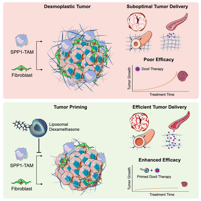
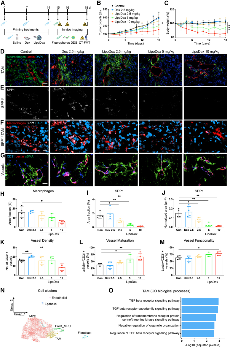
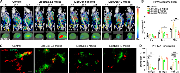
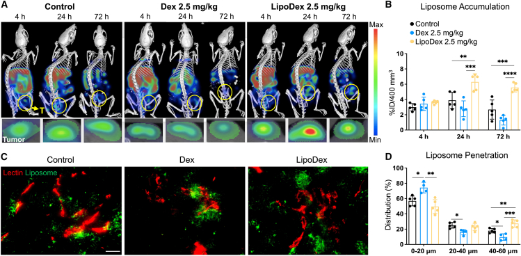
Inflammation is a hallmark of cancer. It contributes to a heterogeneous, hyperpermeable, and poorly perfused tumor vasculature, as well as to a dense and disorganized extracellular matrix, which together negatively affect drug delivery. Reasoning that glucocorticoids have pleiotropic effects, we use clinical-stage dexamethasone liposomes (LipoDex) to prime the tumor microenvironment for improved drug delivery and enhanced treatment efficacy. We show that LipoDex priming improves tumor vascular function and reduces extracellular matrix deposition. Single-cell sequencing corroborates LipoDex-mediated inhibition of pro-inflammatory, pro-angiogenic, and pro-fibrogenic gene expression in mononuclear cells, tumor-associated macrophages, and cancer-associated fibroblasts. Multimodal optical imaging illustrates that LipoDex pre-treatment increases the tumor accumulation and intratumoral distribution of subsequently administered polymeric and liposomal drug delivery systems. Using Doxil as a prototypic nanodrug, we finally show that LipoDex priming promotes antitumor treatment efficacy. Altogether, our findings demonstrate that desmoplastic tumors can be primed for improved drug targeting and therapy using clinical-stage glucocorticoid liposomes.

Keywords: cancer-associated fibroblasts; corticosteroids; drug targeting; liposomes; nanomedicine; tumor microenvironment; tumor priming; tumor-associated macrophages.


Conflict of interest statement

The authors declare no competing interests.

Figures

None
Graphical abstract
Figure 1
Figure 1
Study design The potential of LipoDex to modulate the TME and improve tumor-targeted drug delivery was assessed in a highly stromal MLS ovarian cancer mouse model. First, the effects of free Dex and LipoDex priming on cellular (TAM and fibroblast) and structural (vessels, hyaluronan, and collagen) components of the TME were studied, by performing histology and scRNA-seq. Next, the effects of free Dex and LipoDex priming on the tumor accumulation and penetration of fluorophore-labeled 10–20 nm-sized polymeric and 100 nm-sized liposomal nanocarriers were assessed, using multimodal and multiscale optical imaging. In addition, the effect of free Dex and LipoDex priming on the therapeutic efficacy of Doxil was assessed.
Figure 2
Figure 2
LipoDex treatment modulates TAM and tumor vasculature (A) Experimental setup. (B) Relative MLS tumor growth in mice treated with saline, free Dex, and three doses of LipoDex (n = 9–3). (C) Relative body weight loss (n = 9–3). (D–G) Immunofluorescence images of F4/80 as a pan-macrophage marker (D), SPP1 as a pro-fibrogenic macrophage marker (E and F), and CD31, rhodamine-lectin, and αSMA as markers for all, perfused, and pericyte-covered vessels, respectively (G). (H–J) Area fractions of F4/80 (H), SPP1 (I), and normalized area fraction of SPP1 (J) indicate dose-dependent depletion of TAM and SPP1+ TAM by LipoDex. (K–M) Quantification of tumor blood vessel density (K), vessel maturation (L), and vessel functionality (M). (N) UMAP plots show distinct clusters of cells in control and 2.5 mg/kg LipoDex-treated tumor. (O) The top five significantly downregulated biological processes in TAM by 2.5 mg/kg LipoDex treatment include pathways associated with TGF-β signaling (all p ≤ 0.05). Values represent average ± SD. In (B) and (C), statistical analysis was performed using one-way ANOVA with Tukey’s multiple comparisons test. In (H)–(M), two-tailed t test were performed. ns > 0.05, ∗p ≤ 0.05, and ∗∗p ≤ 0.01, and ∗∗∗p ≤ 0.001. Scale bars represent 50 μm (D and G) and 20 μm (E and F).
Figure 3
Figure 3
LipoDex treatment reduces ECM deposition (A and B) Immunofluorescence microscopy analysis of hyaluronan (A) and collagen I (B). (C) Two-photon microscopy images of collagen fibers, obtained via second-harmonic generation imaging. (D–F) Quantifications of hyaluronan (D), collagen I (E), and total collagen content in two-photon microscopy (F), exemplifying that even low-dose LipoDex treatment significantly reduces ECM deposition in MLS tumors. (G) Top five significantly downregulated biological processes in CAFs by 2.5 mg/kg LipoDex treatment include ECM associated pathways (all p ≤ 0.05). Values represent average ± SD. For (D)–(F), two-tailed t tests were performed; ns > 0.05, ∗p ≤ 0.05, and ∗∗p ≤ 0.01. Scale bars represent 50 μm.
Figure 4
Figure 4
LipoDex priming improves the tumor accumulation and penetration of polymeric nanocarriers (A) Top: whole-body CT-FMT images showing longitudinal PHPMA biodistribution and tumor accumulation in MLS tumor-bearing mice. Bottom panel: CT-segmented tumor slices (in green) exemplify FMT-assessed polymer accumulation (color-coded clouds). (B) Normalized PHPMA tumor accumulation quantification in control vs. LipoDex-treated mice, exemplifying that the 2.5 mg/kg dose of the liposomal GC improves polymer tumor targeting. (C) Fluorescence microscopy analysis of polymer extravasation and penetration out of lectin-stained tumor blood vessels into the interstitium. (D) Quantification of PHPMA intratumoral distribution reveals that all LipoDex doses enhance polymer penetration as compared to the saline-treated controls. Values represent average ± SD. One-way ANOVA with Tukey’s multiple comparisons test was performed for statistical analysis. ns > 0.05, ∗p ≤ 0.05, ∗∗p ≤ 0.01, ∗∗∗p ≤ 0.001, and ∗∗∗∗p ≤ 0.0001. Scale bar represents 50 μm.
Figure 5
Figure 5
LipoDex priming improves the tumor accumulation and penetration of liposomal nanocarriers (A) Top: whole-body CT-FLT imaging exemplifies PEGylated liposome biodistribution and tumor accumulation in untreated, free Dex-treated, and LipoDex-treated mice. Bottom panel: CT-segmented tumor slices (green) show liposome tumor accumulation (color-coded clouds). (B) Quantification of liposome tumor accumulation demonstrates significant enhancement in overall tumor accumulation upon LipoDex priming compared to free Dex treatment and saline controls. (C) Fluorescence microscopy images depict extravasation and penetration of liposomes out of lectin-labeled tumor blood vessels into the interstitium. (D) Quantification of intratumoral distribution reveals that LipoDex priming significantly enhances liposome penetration into deeper tumor compartments. Values represent average ± SD. One-way ANOVA with Tukey’s multiple comparisons test was performed. ns > 0.05, ∗p ≤ 0.05, ∗∗p ≤ 0.01, ∗∗∗p ≤ 0.001, and ∗∗∗∗p ≤ 0.0001. Scale bar represents 50 μm.
Figure 6
Figure 6
LipoDex priming enhances cancer nanomedicine treatment efficacy (A) Experimental setup. (B–H) Tumor growth curves showing that LipoDex boosted the antitumor activity of Doxil, while free Dex negatively affected therapeutic outcome. (I) Relative tumor volume change at day 20 post therapy start. (J–O) Fluorescence microscopy analysis of collagen I (J and M), doxorubicin tumor accumulation (K and N), and apoptosis (L and O), substantiating the added value of LipoDex priming. Values represent average ± SD. Brown-Forsythe ANOVA with Dunnett's multiple comparisons test (I) and two-tailed t test were performed (M–O). ns > 0.05, ∗p ≤ 0.05, ∗∗p ≤ 0.01, ∗∗∗p ≤ 0.001, and ∗∗∗∗p ≤ 0.0001. Scale bars represent 50 μm (J and L) and 2 mm (K).

References

    1. De Lázaro I., Mooney D.J. Obstacles and opportunities in a forward vision for cancer nanomedicine. Nat. Mater. 2021;20:1469–1479. doi: 10.1038/s41563-021-01047-7. - DOI - PubMed
    1. Nia H.T., Munn L.L., Jain R.K. Physical traits of cancer. Science. 2020;370 doi: 10.1126/science.aaz0868. - DOI - PMC - PubMed
    1. Sutherland T.E., Dyer D.P., Allen J.E. The extracellular matrix and the immune system: A mutually dependent relationship. Science. 2023;379 doi: 10.1126/science.abp8964. - DOI - PubMed
    1. Nguyen L.N.M., Ngo W., Lin Z.P., Sindhwani S., MacMillan P., Mladjenovic S.M., Chan W.C.W. The mechanisms of nanoparticle delivery to solid tumours. Nat Rev Bioeng. 2024;2:201–213. doi: 10.1038/s44222-024-00154-9. - DOI
    1. Mitchell M.J., Billingsley M.M., Haley R.M., Wechsler M.E., Peppas N.A., Langer R. Engineering precision nanoparticles for drug delivery. Nat. Rev. Drug Discov. 2021;20:101–124. doi: 10.1038/s41573-020-0090-8. - DOI - PMC - PubMed

LinkOut - more resources