Cervical Necrotizing Fasciitis in an Uncontrolled Diabetic Male Patient: A Multimodal Management Approach
- PMID: 40276410
- PMCID: PMC12021304
- DOI: 10.7759/cureus.81177
Cervical Necrotizing Fasciitis in an Uncontrolled Diabetic Male Patient: A Multimodal Management Approach
Abstract
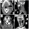
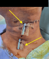
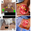
Cervical necrotizing fasciitis is a rare, life-threatening infection, often odontogenic. We report a case of idiopathic cervical necrotizing fasciitis in an uncontrolled diabetic patient, successfully managed through a multidisciplinary approach with good wound healing. A 54-year-old male with type 2 diabetes mellitus presented with a 10-day history of painful neck swelling and purulent discharge on the left side of the neck. Clinical examination revealed signs of necrotizing infection, confirmed by laboratory tests and contrast-enhanced computed tomography (CECT), which showed a large necrotic collection with gas formation extending to the supra-glottic region. Management included broad-spectrum antibiotics, insulin therapy, and urgent surgical debridement. Extensive necrosis involving the neck and laryngeal structures necessitated a second debridement on day seven, followed by negative pressure wound therapy. A split-thickness skin graft on day 14 led to complete healing, and the patient was discharged on day 21 with full recovery at the one-month follow-up. Cervical necrotizing fasciitis poses a high risk due to its proximity to vital structures and potential for mediastinal spread. Early diagnosis through imaging and clinical evaluation, along with aggressive surgical debridement and advanced wound care, is essential. This case underscores the importance of a multimodal strategy and optimal diabetes control in facilitating recovery and minimizing hospital stay. Idiopathic cervical necrotizing fasciitis, though rare, demands prompt diagnosis and intervention. An integrated approach, incorporating early imaging, repeated debridement, negative pressure therapy, and skin grafting, can significantly enhance patient outcomes. In this case, comprehensive management resulted in recovery within 21 days, shorter than the typical hospital stay for similar cases.
Keywords: cervical; debridement; idiopathic origin; multi-disciplinary; necrotizing fasciitis; skin grafting; wound negative pressure therapy.
Copyright © 2025, Bandaru et al.
Conflict of interest statement
Human subjects: Consent for treatment and open access publication was obtained or waived by all participants in this study. Ministry of Health and Prevention, Research Ethical Committee, RAK Subcommittee, United Arab Emirates issued approval MOHAP/REC/2025/11-2025-F-M. Conflicts of interest: In compliance with the ICMJE uniform disclosure form, all authors declare the following: Payment/services info: All authors have declared that no financial support was received from any organization for the submitted work. Financial relationships: All authors have declared that they have no financial relationships at present or within the previous three years with any organizations that might have an interest in the submitted work. Other relationships: All authors have declared that there are no other relationships or activities that could appear to have influenced the submitted work.
Figures


Similar articles
-
Cervical Necrotizing Fasciitis: An Institutional Experience.Cureus. 2022 Dec 10;14(12):e32382. doi: 10.7759/cureus.32382. eCollection 2022 Dec. Cureus. 2022. PMID: 36632261 Free PMC article.
-
Cervical Necrotizing Fasciitis, Diagnosis and Treatment of a Rare Life-Threatening Infection.Ear Nose Throat J. 2023 Mar;102(3):NP109-NP113. doi: 10.1177/0145561321991341. Epub 2021 Feb 11. Ear Nose Throat J. 2023. PMID: 33570428
-
Vacuum assisted closure therapy in the management of cervico-facial necrotizing fasciitis: a case report and review of the literature.Minerva Stomatol. 2014 Apr;63(4):135-44. Minerva Stomatol. 2014. PMID: 24705043 Review. English, Italian.
-
A Case Report of Necrotizing Fasciitis Managed With the Application of Negative Pressure Wound Therapy to Achieve Better Wound Closure.Cureus. 2023 Sep 28;15(9):e46140. doi: 10.7759/cureus.46140. eCollection 2023 Sep. Cureus. 2023. PMID: 37900373 Free PMC article.
-
Necrotizing fasciitis following postpartum tubal ligation. A case report and review of the literature.Arch Gynecol Obstet. 1995;256(1):35-8. doi: 10.1007/BF00634347. Arch Gynecol Obstet. 1995. PMID: 7726653 Review.
References
-
- Clinical classification of cervical necrotizing fasciitis. Amponsah EK, Frimpong P, Eo MY, Kim SM, Lee SK. Eur Arch Otorhinolaryngol. 2018;275:3067–3073. - PubMed
-
- Cervical necrotizing fasciitis: systematic review and analysis of 1235 reported cases from the literature. Gunaratne DA, Tseros EA, Hasan Z, et al. Head Neck. 2018;40:2094–2102. - PubMed
-
- Cervical necrotizing fasciitis in adults: a life-threatening emergency in oral and maxillofacial surgery. de Leyva P, Dios-Díez P, Cárdenas SC, et al. Surgeries. 2024;5:517–531.
-
- Necrotizing fasciitis of the head and neck: role of CT in diagnosis and management. Becker M, Zbären P, Hermans R, et al. Radiology. 1997;202:471–476. - PubMed
-
- Necrotizing odontogenic fasciitis of head and neck extending to anterior mediastinum in elderly patients: innovative treatment with a review of the literature. Cortese A, Pantaleo G, Borri A, Amato M, Claudio PP. Aging Clin Exp Res. 2017;29:159–165. - PubMed
Publication types
LinkOut - more resources
Full Text Sources
