Review
. 2025 Apr 16:50:322-343.
doi: 10.1016/j.bioactmat.2025.04.012. eCollection 2025 Aug.

Exomeres and supermeres: Current advances and perspectives

Affiliations
Review

Exomeres and supermeres: Current advances and perspectives

Li Yu et al. Bioact Mater. .

Abstract

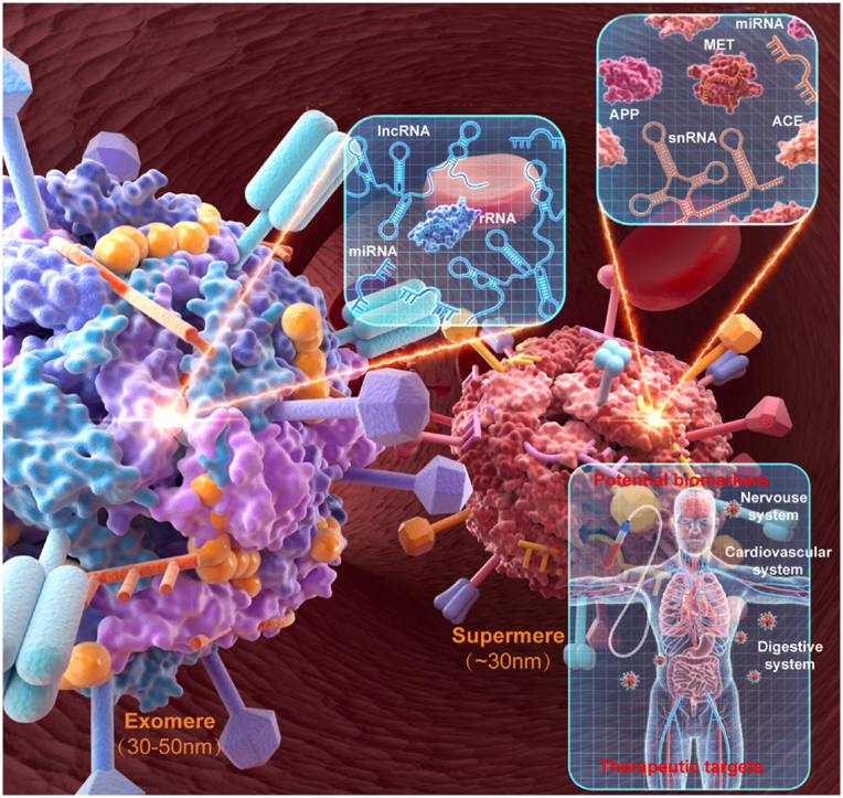
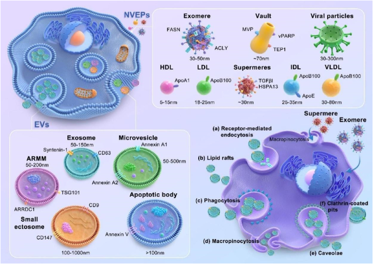
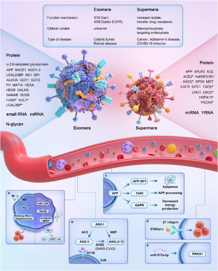
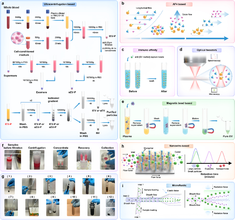
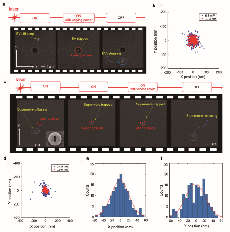
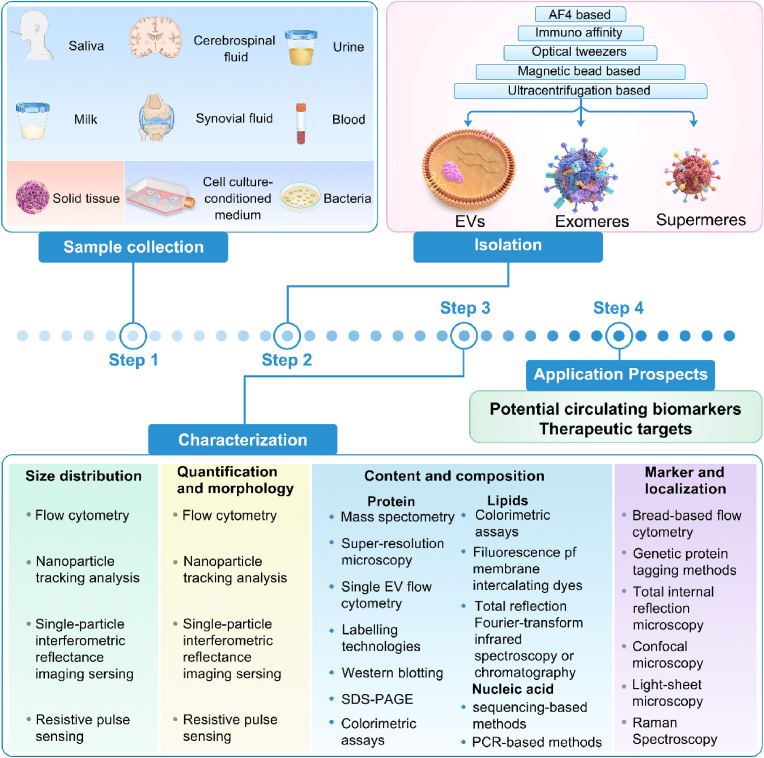
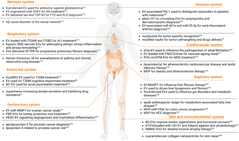
Recent studies have revealed a great diversity and complexity in extracellular vesicles and particles (EVPs). The developments in techniques and the growing awareness of the particle heterogeneity have spurred active research on new particle subsets. Latest discoveries highlighted unique features and roles of non-vesicular extracellular nanoparticles (NVEPs) as promising biomarkers and targets for diseases. These nanoparticles are distinct from extracellular vesicles (EVs) in terms of their smaller particle sizes and lack of a bilayer membrane structure and they are enriched with diverse bioactive molecules particularly proteins and RNAs, which are widely reported to be delivered and packaged in exosomes. This review is focused on the two recently identified membraneless NVEPs, exomeres and supermeres, to provide an overview of their biogenesis and contents, particularly those bioactive substances linked to their bio-properties. This review also explains the concepts and characteristics of these nanoparticles, to compare them with other EVPs, especially EVs, as well as to discuss their isolation and identification methods, research interests, potential clinical applications and open questions.

Keywords: Disease diagnosis and treatment; Exomeres; Nanoparticles; Separation and extraction; Supermeres.


Conflict of interest statement

The authors declare that they have no known competing financial interests or personal relationships that could have appeared to influence the work reported in this paper

Figures

Image 1
Graphical abstract
Fig. 1
Fig. 1
Extracellular vesicles and nanoparticles secreted by cells. The schematic provides a rough estimate of the size of various substances. While the entry mechanisms of EVs into cells have been extensively studied, those of exomeres and supermeres remain less understood. The caption discusses exosomes as a case study to elucidate the different modalities through which EVs are internalized by cells [18,20]. Recent studies suggest macropinocytosis as a potential pathway for the cellular uptake of supermeres, highlighting ongoing research in this area [21]. Abbreviations: HDL, high-density lipoprotein; LDL, low-density lipoprotein; IDL, intermediate-density lipoprotein; VLDL, very low-density lipoprotein; ARMM, arrestin domain-containing protein 1-mediated microvesicle; TGFBI, transforming growth factor beta induced; TSP1, thrombospondin 1; HSPA13, heat shock protein family A (Hsp70) member 13; FASN, fatty acid synthase; ACLY, ATP citrate lyase.
Fig. 2
Fig. 2
The composition, function and related pathways of exomeres and supermeres. Exomeres (left) and supermeres (right) precent distinct particle size and components, reflecting their unique particle populations. Key markers are highlighted with an asterisk (∗). a. MET and miR-1246 in supermere are related to cancer. HGF-MET pathway is closely related many solid tumors [27]. MiR-1246 is involved in lymphangiogenesis and CD8+ T cell apoptosis during cancer [28]. b. Supermeres are also reported to be featured by ACE2 enrichment, which make them possible targets in cardiovascular diseases and coronavirus related diseases [29]. c. Supermeres convey adequate level of APP, which promote the Alzheimer's disease development [30]. d. ST6Gal-I in exomere proved to promote the metastasis of colorectal tumors [31]. e. Exomere derived miR-517a-3p significantly reduced the expression of PRKG1 in miR-517a-3p-inhibitor (−) Jurkat cells [32].
Fig. 3
Fig. 3
Exomeres and supermeres are distinct extracellular nanoparticles. a. Negative stain transmission electron microscopy of DiFi-derived sEVs, NV, exomeres and supermeres. b. Venn diagram of unique and common proteins identified in DiFi-derived sEVs, NV fractions, exomeres and supermeres. c. Whole-organ imaging (top). Male C57BL/6 mice were intraperitoneally injected with labelled sEVs, exomeres or supermeres derived from DiFi cells. Their organs were harvested and analyzed after 24 h. Data are the mean ± s.e.m. of n = 3 animals (bottom). The sEVs and extracellular nanoparticles derived from DiFi cells were labelled with Alexa Fluor-647 (Invitrogen, A20173) according to the manufacturer's instructions. d. Percentage of small-RNA reads mapped small noncoding RNA for DiFi cells, the sEV-P, exomeres and supermeres following RNA-seq. Misc RNA, miscellaneous RNA; mt tRNA, mitochondrial tRNA; rRNA, ribosomal RNA; snoRNA, small nucleolar RNA; n = 3 independent samples. e-f. Inhibition of cellular supermere uptake. Cells were pre-incubated with uptake inhibitors for 30 min before the addition of labelled supermeres. After a 24 h incubation, images were acquired using an iSIM imaging system (bottom). scale bar, 20 μm. Data are the mean ± s.e.m. of n = 30 (MDA-MB-231) and 27 (HeLa) cells (top). Images are representative of three independent experiments. Adapted with permission from Ref. [21].Copyright © 2021, Qin zhang et al…
Fig. 4
Fig. 4
Isolation methods for EVs and NVEPs from whole blood and cell-conditioned medium. a. Ultracentrifugation is one of the most common methods. It is a method that can separate EVs, exomeres and supermeres. The separated samples can be washed by resuspension in PBS and centrifugation. There are some representative photographs of the most important steps during concentrator procedure(f) and the high-resolution gradient fractionation procedure(g). Adapted with permission from Ref. [143]. Copyright © 2023, Springer Nature Limited (1) Transfer the 36 %(wt/vol) iodixanol EV-P suspension to the centrifuge tube. (2) Tilt the centrifugation tube sideways and carefully dispense 2.4 ml of the 30 % and 24 %(wt/vol) iodixanol solution to the centrifuge tube. (3–4) Weigh the loaded centrifugation tube containing 12 ml of 12–36 %(wt/vol) iodixanol gradient on a balance. (5) Centrifuge at 120,000g for 15h at 4 °C. (6–9) Carefully collect fractions of 1 ml each from the top, not to disturb the underlying fractions. (10) Add 11 ml of PBS and mix until the solution appears to be homogenous. (11) Centrifuge at 120,000g for ≥4h at 4 °C. (12) Collect lEVs/sEVs. b. AF4-based methods have been used to separate EVs and exomeres. c and e. Immuno-affinity and magnetic bead are two commonly used methods for separating EVs. d. Optical tweezers has been able to successfully capture supermere, and may be a promising tool for single molecule analysis in the future. h and i. Nanowire-based and microfluidic are two relatively new methods for separating EVs.
Fig. 5
Fig. 5
Trapping mechanism of EVs and supermeres. a. The EV trapping experiment shows that the diffusing EV is trapped on the anapole nanoantenna, and then the EV is released when the 973 nm laser is turned off. b. The scatter plot shows the EV's trajectory when the EV is trapped on the anapole nanoantenna. The highest estimated stiffness along the x- and y-axis is 0.347 fN/nm and 0.329 fN/nm under 10.8 mW incident laser power, respectively. c. The frame sequence of supermere trapping experiment shows how the diffusing supermere is trapped near the anapole nanoantenna until the laser is off. d. The scatter plot shows the supermere's trajectory when the supermere is trapped at the anapole structure. The estimated stiffness for the trapped supermere under 19 mW incidence along the x- and y-axis are 0.215 fN/nm and 0.205 fN/nm, respectively. e,f. Histogram of the trapped supermere particle's position under 19 mW incident laser power along x and y directions, respectively. The red curve is the Gaussian fitting to estimate the trap stiffness. Reprinted with permission from Ref. [146]. Copyright 2023 American Chemical Society.
Fig. 6
Fig. 6
Workflow of exomeres and supermeres isolation and characterization. The primary sample types include body fluids, tissues, and cell cultures. These samples undergo processes to isolate sEVs, exomeres, and supermeres, which are then subjected to further characterization. This characterization primarily focuses on assessing size distribution, quantification, morphology, content and composition, as well as identifying specific markers and localizations of these particles. The EVs and NPs extracted through this workflow are utilized for subsequent research purposes. It is important to note that while the schematic provides a structured overview, it does not encompass all possible methodologies and variations in the field, as indicated in Ref. [4].
Fig. 7
Fig. 7
Extracellular particles and their associated diseases. There is an ever-increasing researches on EVs and NVEPs in various diseases, including nervous system, respiratory system, endocrine system, skin and musculoskeletal system, immune system, cardiovascular system, digestive system and genitourinary system. Summarization in the schematic is not exhaustive [21,79,90,104,[211], [212], [213], [214], [215], [216], [217], [218], [219], [220], [221], [222], [223], [224], [225], [226], [227], [228], [229], [230], [231], [232], [233], [234], [235], [236], [237], [238], [239], [240], [241], [242], [243]].

Similar articles

Cited by

References

    1. van Niel G., D'Angelo G., Raposo G. Shedding light on the cell biology of extracellular vesicles. Nat. Rev. Mol. Cell Biol. 2018;19:213–228. doi: 10.1038/nrm.2017.125. - DOI - PubMed
    1. Deatherage B.L., Cookson B.T., Andrews-Polymenis H.L. Membrane vesicle release in bacteria, eukaryotes, and archaea: a conserved yet underappreciated aspect of microbial life. Infect. Immun. 2012;80:1948–1957. doi: 10.1128/iai.06014-11. - DOI - PMC - PubMed
    1. Liu H., et al. The potential role of extracellular vesicles in bioactive compound-based therapy: a review of recent developments. Crit. Rev. Food Sci. Nutr. 2023;63:10959–10973. doi: 10.1080/10408398.2022.2081667. - DOI - PubMed
    1. Welsh J.A., et al. Minimal information for studies of extracellular vesicles (MISEV2023): from basic to advanced approaches. J. Extracell. Vesicles. 2024;13 doi: 10.1002/jev2.12404. - DOI - PMC - PubMed
    1. Jeppesen D.K., et al. Reassessment of exosome composition. Cell. 2019;177:428–445.e418. doi: 10.1016/j.cell.2019.02.029. - DOI - PMC - PubMed

LinkOut - more resources