Case Reports
. 2025 Jul;40(7):1388-1400.
doi: 10.1002/mds.30209. Epub 2025 Apr 25.

Expanding the Allelic and Clinical Heterogeneity of Movement Disorders Linked to Defects of Mitochondrial Adenosine Triphosphate Synthase

Affiliations
Case Reports

Expanding the Allelic and Clinical Heterogeneity of Movement Disorders Linked to Defects of Mitochondrial Adenosine Triphosphate Synthase

Philip Harrer et al. Mov Disord. 2025 Jul.

Abstract

Background: Defects of mitochondrial ATP synthase (ATPase) represent an emerging, yet incompletely understood group of neurodevelopmental diseases with abnormal movements.

Objective: The aim of this study was to redefine the phenotypic and mutational spectrum of movement disorders linked to the ATPase subunit-encoding genes ATP5F1A and ATP5F1B.

Methods: We recruited regionally distant patients who had been genome or exome sequenced. Fibroblast cultures from two patients were established to perform RNA sequencing, immunoblotting, mass spectrometry-based high-throughput quantitative proteomics, and ATPase activity assays. In silico three-dimensional missense variant modeling was performed.

Results: We identified a patient with developmental delay, myoclonic dystonia, and spasticity who carried a heterozygous frameshift c.1404del (p.Glu469Serfs*3) variant in ATP5F1A. The patient's cells exhibited significant reductions in ATP5F1A mRNA, underexpression of the α-subunit of ATPase in association with other aberrantly expressed ATPase components, and compromised ATPase activity. In addition, a novel deleterious heterozygous ATP5F1A missense c.1252G>A (p.Gly418Arg) variant was discovered, shared by three patients from two families with hereditary spastic paraplegia (HSP). This variant mapped to a functionally important intersubunit communication site. A third heterozygous variant, c.1074+1G>T, affected a canonical donor splice site of ATP5F1B and resulted in exon skipping with significantly diminished ATP5F1B mRNA levels, as well as impaired ATPase activity. The associated phenotype consisted of cerebral palsy (CP) with prominent generalized dystonia.

Conclusions: Our data confirm and expand the role of dominant ATP5F1A and ATP5F1B variants in neurodevelopmental movement disorders. ATP5F1A/ATP5F1B-related ATPase diseases should be considered as a cause of dystonia, HSP, and CP. © 2025 The Author(s). Movement Disorders published by Wiley Periodicals LLC on behalf of International Parkinson and Movement Disorder Society.

Keywords: ATP synthase; ATP5F1A; ATP5F1B; cerebral palsy; dominant variant; dystonia; mitochondrial disease; spasticity.


Figures

FIG. 1
FIG. 1
Pedigree drawings for families with heterozygous variants in ATP5F1A or ATP5F1B. (A) Family A with patient 1 (P1). (B) Family B with P2. (C) Family C with P3 (daughter) and P4 (mother). (D) Family D with P5. Closed symbols indicate the individuals affected by movement disorders; mut/wt denotes the reported ATP5F1A/ATP5F1B variants in the heterozygous state; and wt/wt denotes a homozygous wild‐type allele. Index patients are marked with arrows. The variant in P2 was confirmed to be de novo by targeted Sanger testing of the parents.
FIG. 2
FIG. 2
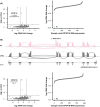
RNA sequencing (RNAseq) studies showing reduced expression of ATP5F1A and ATP5F1B. (A) Significantly reduced ATP5F1A mRNA levels in patient 1 (P1; family A) affected by the heterozygous frameshift variant c.1404del (p.Glu469Serfs*3). Transcriptomics volcano and sample rank plots are shown. In the volcano plot, the underexpression outlier ATP5F1A is indicated in blue; the horizontal line represents a P value of 2.5 × 10−6 (corrected P value for 20,000 hypotheses corresponding to the number of theoretically identifiable gene‐derived RNAs), and the vertical line represents log2fold changes of ±1. In comparison with all in‐house RNAseq control samples (n > 750), P1 had the lowest amounts of ATP5F1A mRNA (blue data point in the sample rank plot). (B) Missplicing and significantly reduced ATP5F1B mRNA levels in P5 (family D) affected by the heterozygous splice‐site variant c.1074+1G>T. Transcriptomics sashimi, volcano, and sample rank plots are shown. Sashimi plots for P5 and a representative control subject demonstrate the occurrence of exon skipping (exon 7, exons 6 plus 7) as a result of the patient‐specific G>T substitution at the donor splice site of exon 7. The number of reads spanning each junction is highlighted by the size of the sashimi‐plot curve. In the volcano plot, the underexpression outlier ATP5F1B is indicated in pink; the horizontal line represents a P value of 2.5 × 10−6 (corrected P value for 20,000 hypotheses corresponding to the number of theoretically identifiable gene‐derived RNAs), and the vertical line represents log2fold changes of ±1. In comparison with all in‐house RNAseq control samples (n > 750), P5 had the lowest amounts of ATP5F1B mRNA (pink data point in the sample rank plot). [Color figure can be viewed at wileyonlinelibrary.com]
FIG. 3
FIG. 3
Effect of variants in ATP5F1A and ATP5F1B on ATP synthase subunit expression. Immunoblot analysis of ATP5F1A, ATP5F1B, ATP5PD, ATP5PO, ATP5F1C, and ATP5F1D was performed in fibroblast lysates from three unrelated control subjects (CTRL), patient 1 (P1; family A) affected by the heterozygous ATP5F1A frameshift variant c.1404del (p.Glu469Serfs*3), and P5 (family D) affected by the heterozygous ATP5F1B splice‐site variant c.1074+1G>T. Three biological replicates were analyzed, and representative images are shown in (A). In the quantifications (B), the values shown represent means ± standard deviations. Significances were assessed by Student's t test; *P ≤ 0.05, **P ≤ 0.01. (C) Proteomics volcano plot for P1 (family A) is shown. Darker gray dots represent proteins whose expression was significantly altered (adjusted P value threshold of 0.05), and lighter gray dots represent proteins whose expression was not significantly altered (adjusted P > 0.05). The significant underexpression outliers ATP5F1A, ATP5F1B, ATP5PD, ATP5PO, ATP5PB, and ATP5ME are highlighted in different colors, as indicated. [Color figure can be viewed at wileyonlinelibrary.com]
FIG. 4
FIG. 4
ATP synthase activity studies using fibroblasts from patients with variants in ATP5F1A and ATP5F1B. (A) Representation of ATPase activity, evaluated by the ATP synthase Activity Microplate Assay Kit (Abcam), in the fibroblasts from two age‐ and sex‐matched control subjects (CTRL 1/2), patient 1 (P1; family A) affected by the heterozygous ATP5F1A frameshift variant c.1404del (p.Glu469Serfs*3), and P5 (family D) affected by the heterozygous ATP5F1B splice‐site variant c.1074+1G>T. Data represent three biological replicates with three technical replicates per biological replicate. Boxplots (B) represent the median, first quartile, and third quartile, and whiskers extend to a maximum of 1.5× interquartile range. Significances were assessed by Student's t test. [Color figure can be viewed at wileyonlinelibrary.com]
FIG. 5
FIG. 5
Location and structural modeling of the ATP5F1A missense variant p.Gly418Arg. (A) A three‐dimensional representation of the human ATPase F1 domain (PDB: 8H9I) is shown, with major subunits highlighted in the indicated colors. The residue Gly418 mutated in families B and C is situated at the α‐β intersubunit interaction space (ATP5F1A‐ATP5F1B). Moreover, Gly418 is in close proximity to the ADP binding site, as highlighted in the magnified view of the sequence containing this amino acid; ADP is shown in stick‐and‐ball representation (gray). (B) Model of ATPase and magnified view of the predicted p.Gly418Arg substitution in the α‐β communication space (PDB: 6ZPO). Substitution of the nonpolar Gly418 by a positively charged arginine may perturb functionally important subunit interactions and/or structural features that sustain binding (or nonbinding) behaviors. As illustrated by blue dashed lines, Arg418 may introduce new intermolecular bonding interactions (H‐bonds) involving the α‐ and β‐subunits. [Color figure can be viewed at wileyonlinelibrary.com]

References

    1. Shigetomi E, Sakai K, Koizumi S. Extracellular ATP/adenosine dynamics in the brain and its role in health and disease. Front Cell Dev Biol 2023;11:1343653. - PMC - PubMed
    1. Jonckheere AI, Smeitink JA, Rodenburg RJ. Mitochondrial ATP synthase: architecture, function and pathology. J Inherit Metab Dis 2012;35(2):211–225. - PMC - PubMed
    1. Dautant A, Meier T, Hahn A, Tribouillard‐Tanvier D, di Rago JP, Kucharczyk R. ATP synthase diseases of mitochondrial genetic origin. Front Physiol 2018;9:329. - PMC - PubMed
    1. Hamosh A, Scott AF, Amberger JS, Bocchini CA, McKusick VA. Online Mendelian inheritance in man (OMIM), a knowledgebase of human genes and genetic disorders. Nucleic Acids Res 2005;33(Database issue):D514–D517. - PMC - PubMed
    1. Indelicato E, Schlieben LD, Stenton SL, et al. Dystonia and mitochondrial disease: the movement disorder connection revisited in 900 genetically diagnosed patients. J Neurol 2024;271(7):4685–4692. - PMC - PubMed

Publication types

Substances