Review
. 2025 Mar 31;15(4):222.
doi: 10.3390/bios15040222.

A Review on Optical Biosensors for Monitoring of Uric Acid and Blood Glucose Using Portable POCT Devices: Status, Challenges, and Future Horizons

Affiliations
Review

A Review on Optical Biosensors for Monitoring of Uric Acid and Blood Glucose Using Portable POCT Devices: Status, Challenges, and Future Horizons

Kermue Vasco Jarnda et al. Biosensors (Basel). .

Abstract

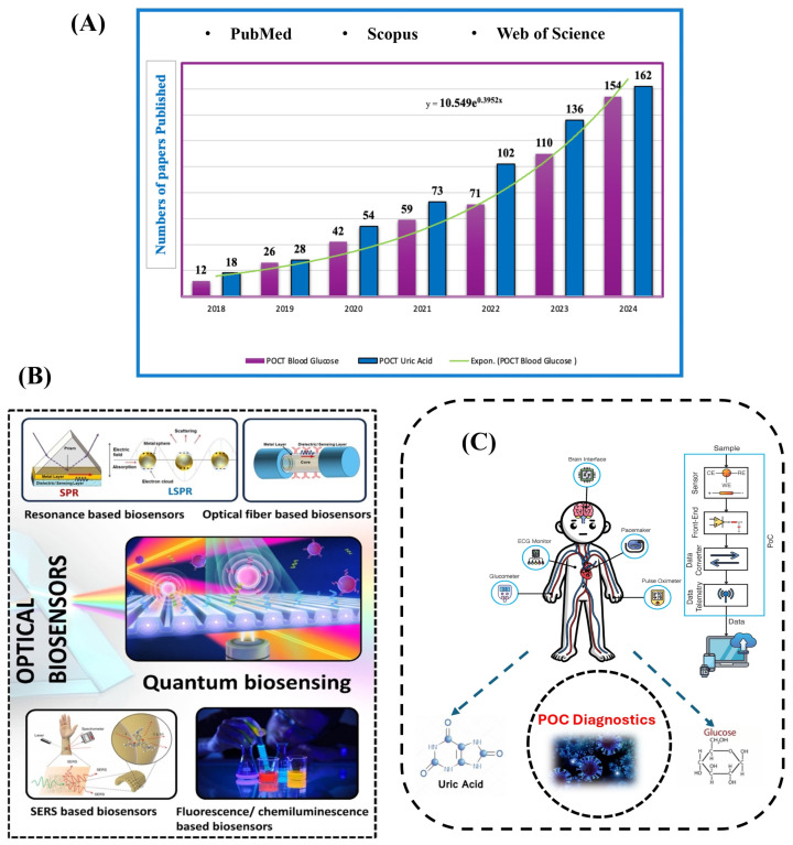
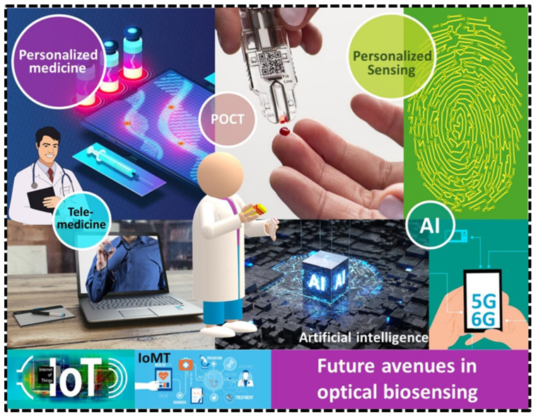
The growing demand for real-time, non-invasive, and cost-effective health monitoring has driven significant advancements in portable point-of-care testing (POCT) devices. Among these, optical biosensors have emerged as promising tools for the detection of critical biomarkers such as uric acid (UA) and blood glucose. Different optical transduction methods, like fluorescence, surface plasmon resonance (SPR), and colorimetric approaches, are talked about, with a focus on how sensitive, specific, and portable they are. Despite considerable advancements, several challenges persist, including sensor stability, miniaturization, interference effects, and the need for calibration-free operation. This review also explores issues related to cost-effectiveness, data integration, and wireless connectivity for remote monitoring. The review further examines regulatory considerations and commercialization aspects of optical biosensors, addressing the gap between research developments and clinical implementation. Future perspectives emphasize the integration of artificial intelligence (AI) and healthcare for improved diagnostics, alongside the development of wearable and implantable biosensors for continuous monitoring. Innovative optical biosensors have the potential to change the way people manage their health by quickly and accurately measuring uric acid and glucose levels. This is especially true as the need for decentralized healthcare solutions grows. By critically evaluating existing work and exploring the limitations and opportunities in the field, this review will help guide the development of more efficient, accessible, and reliable POCT devices that can improve patient outcomes and quality of life.

Keywords: blood glucose; optical biosensors; uric acid; μPADs.


Conflict of interest statement

None declared. All authors had full access to all the study data, and take responsibility for the integrity of the data and accuracy of the data analysis.

Figures

Figure 1
Figure 1
(A) Annual publication counts of publications utilizing the specified keywords as of 31 October 2024. (B) Cutting-edge optical biosensors: The figure illustrates many commonly utilized optical biosensing platforms, including resonance-based biosensors, optical fiber-based biosensors, quantum-based biosensors, and surface-enhanced Raman scattering (SERS) biosensors [55]. (C) Various point-of-care devices, illustrating the integration of sensors for different health-monitoring applications.
Figure 5
Figure 5
Comparative diagram of different biosensor labels and sub-classes based on the type of transducer [68].
Figure 6
Figure 6
(A) A typical optical fiber biosensor [23]. (B) The generation of localized surface plasmon resonance in metallic nanostructures along with the simulation outcomes [88]. (C) The excitation of surface plasmon resonance by different light coupling methods for SPR biosensing [88]. (D) Localized surface plasmon resonance [89]. (E) LSPR biosensing methods, including extinction, dark-field, and prism coupler on the plasmonic nanostructured surface [89].
Figure 18
Figure 18
Portable devices for UA on-site detection. (A) Multiplex colorimetric bioassay platform based on single-droplet samples [191]. (B) The structural design of the centrifugal microfluidic device and its microfluidic chips [192]. (C) The structural design and schematic of the multi-channel handheld automatic photometer [193]. (D) Schematic illustration of smartphone-based fiber optic-LRSPR sensor [194]. (E) The schematic and the physical picture of the colorimetric analysis platform are based on paper-based microfluidic [195]. (F) Schematic representation of the mobile healthcare system for the detection of UA in whole blood [190]. (G) A schematic and physical representation of a colorimetric dermal tattoo biosensor utilizing a microneedle patch for uric acid detection [196].
Figure 21
Figure 21
Simple classification of blood glucose detection technology [200].
Figure 22
Figure 22
(A) Paper membrane-based SERS platform for the determination of glucose in blood samples [208]. (B) Quantum dot-modified paper-based assay for glucose screening [209]. (C) Turn-on paper-based phosphorescence biosensor for detection of glucose in serum [217].
Figure 23
Figure 23
(A) Photo of the assembled sampling apparatus and paper chip. (B) Schematic illustration for the G&L@ZIF@Paper. (C) The operation process of the “Glucose Sensor” app used to detect glucose level [211].
Figure 2
Figure 2
Diagrammatic representation of clinical biosensor application.
Figure 3
Figure 3
Types of nanomaterial-based biosensors [62].
Figure 4
Figure 4
Schematic diagram of a typical biosensor consisting of the bioreceptor, transducer, electronic system (amplifier and processor), and display (PC or printer), and various types of bioreceptors and transducers used in the biosensors are also shown [62].
Figure 7
Figure 7
(A) Glucose binding with bisboronic acid functionalized the Au surface and helped in distinguishing from hypoglycemia. (B) Log-scale glucose concentration versus integrated SERS intensity from the concentration-dependent SERS difference spectra [92].
Figure 8
Figure 8
Different types of materials utilized in optical sensors on the basis of the underlying phenomenon.
Figure 9
Figure 9
(A) Graphic representation of the microfluidic chips. (B) Types of microfluidic chips and (C) applications of microfluidic chips [105].
Figure 10
Figure 10
(A) Role of microfluidics in the general workflow of biomarker quantitation [114]. (B) Advantages of microfluidic chips in the field of biomarker quantitation [114]. (C) Key features of microfluidics-based paper analytical devices [115].
Figure 11
Figure 11
An overview of the µPAD process and its applications. Usually, µPAD-based biosensors are influenced by several development factors from (AD) when used in numerous applications in healthcare, food, agriculture, and energy harvesting [115].
Figure 12
Figure 12
(A). A roll-to-roll machine is used to perform the slot-die technique through which the ink leaks through a shim and is pumped to the slot-die head [137]. (B) A schematic view of a fully printed electrochemical sensor with slot die coating by roll-to-roll processing for screen printing [137]. (C) A flexo printing unit [143].
Figure 13
Figure 13
(A) Photolithographic method: the diagram shows the patterning method using the photolithography process, which embeds the SU-8 photoresist into the paper [115]. (B) Wax dipping fabrication process: The procedure for patterning paper by wax-dipping, with top and lateral view. (C) One-step laser cutting for creating the pattern.
Figure 14
Figure 14
(A) RGB color space [144]. (B) CMY/CMYK color space [145]. (C) HSV (left) and HSL (right) color spaces [146]. (D) CIE XYZ color space [145]. (E) L*a*b* color space [145]. (F) YUV color space [145].
Figure 15
Figure 15
The summary of UA-related diseases and the classification of optical sensors for UA detection. (A) Diseases caused by abnormal UA concentration. (B) The classification of optical sensors for UA detection [95].
Figure 16
Figure 16
The correlation of serum UA with multiple diseases (A); dual-FRET aptasensor for UA detection (B); and facilitating UA detection with a portable 3D-printed device and RGB-based cell phone App (C) [187].
Figure 17
Figure 17
(A) Sketch map for UA detection. (B) Structural demonstration of the main components and their locations in a 3D-printed device. (C) Demonstration of the portable box for UA detection. (D) Analysis of UA from 0 to 300 μM with a 3D-printed device and RGB-based app in the buffer. (E) Calibration plots of human serum samples with different concentration of UA. (F) Validation of the accuracy of the portable aptasensor with human serum samples [187].
Figure 19
Figure 19
Current challenges in UA detection using optical methods [95].
Figure 20
Figure 20
Schematic diagram of glucose biosensors used in detecting biological fluids, along with the mechanism and platform.
Figure 24
Figure 24
Challenges faced by microfluidic-based paper analytical devices.
Figure 25
Figure 25
The future scope of optical biosensing—optical biosensors in future will be integrating with advanced technologies such as artificial intelligence (AI), Internet of Things (IoTs, IoMTs) and 5th/6th generation technology, which will contribute to an advanced ecosystem of personalized medicines and personalized POC sensing [55].

Similar articles

Cited by

References

    1. Huang X., Xu D., Chen J., Liu J., Li Y., Song J., Ma X., Guo J. Smartphone-based analytical biosensors. Analyst. 2018;143:5339–5351. - PubMed
    1. Du L., Zong Y., Li H., Wang Q., Xie L., Yang B., Pang Y., Zhang C., Zhong Z., Gao J. Hyperuricemia and its related diseases: Mechanisms and advances in therapy. Signal Transduct. Target. Ther. 2024;9:212. - PMC - PubMed
    1. Xiong Q., Liu J., Xu Y. Effects of uric acid on diabetes mellitus and its chronic complications. Int. J. Endocrinol. 2019;2019:9691345. - PMC - PubMed
    1. Bodaghi A., Fattahi N., Ramazani A. Biomarkers: Promising and valuable tools towards diagnosis, prognosis and treatment of Covid-19 and other diseases. Heliyon. 2023;9:e13323. - PMC - PubMed
    1. Lino C., Barrias S., Chaves R., Adega F., Martins-Lopes P., Fernandes J.R. Biosensors as diagnostic tools in clinical applications. Biochim. Biophys. Acta Rev. Cancer. 2022;1877:188726. - PubMed

LinkOut - more resources