. 2025 Apr 13;15(4):247.
doi: 10.3390/bios15040247.

Design and In Vivo Evaluation of an Intraocular Electrode for Ciliary Muscle Biopotential Measurement in a Non-Human Primate Model of Human Accommodation

Affiliations

Design and In Vivo Evaluation of an Intraocular Electrode for Ciliary Muscle Biopotential Measurement in a Non-Human Primate Model of Human Accommodation

Sven Schumayer et al. Biosensors (Basel). .

Abstract

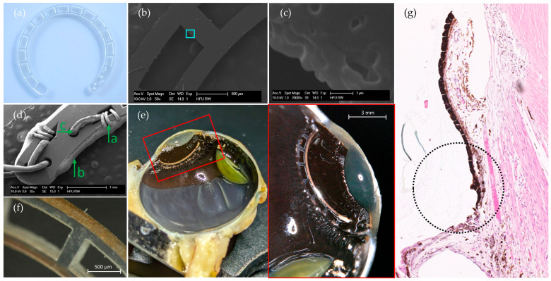
The measurement of electrical potentials in the human body is becoming increasingly important in healthcare as a valuable diagnostic parameter. In ophthalmology, while these signals are primarily used to assess retinal function, other applications, such as recording accommodation-related biopotentials from the ciliary muscle, remain poorly understood. Here, we present the development and evaluation of a novel implantable ring electrode for recording biopotentials from the ciliary muscle. Inspired by capsular tension rings, the electrode was fabricated using laser cutting, wiring, and physical vapor deposition coating. The constant impedance and weight over a simulated aging period of 391 days, demonstrated the electrode's stability. In vivo testing in non-human primates further validated the electrode's surgical handling and long-term stability, with no delamination or tissue ingrowth after 100 days of implantation. Recorded biopotentials from the ciliary muscle (up to 700 µV) exceeded amplitudes reported in the literature. While the results are promising, further research is needed to investigate the signal quality and origin as well as the correlation between these signals and ciliary muscle activity. Ultimately, this electrode will be used in an implanted device to record ciliary muscle biopotentials to control an artificial lens designed to restore accommodation in individuals with presbyopia.

Keywords: accelerated aging; biopotential; electrode conception; intraocular electrode; laser cutting.


Conflict of interest statement

The authors declare no conflict of interest.

Figures

Figure 1
Figure 1
Ring-shaped bipolar electrode (a) Schematic drawing of a cross-section of the eye, where the ring electrode is inserted by an incision at the limbus and placed, by rotating (white arrow) it into the ciliary sulcus. (b) Rendering of the electrode. The inner and outer faces of the electrode are mainly connected by spokes. The magnification (red) highlights the geometry of the conducting area, where the gold wires are manually attached, before coating.
Figure 2
Figure 2
Manufacturing process of the ring electrode. (a) The 5-axis laser cutting machine with the magnified experimental setup within the machine, and the utilized laser parameters (b). (c) The “sandwich-like” masking, with the ring blank between. The cross-section shows the two channels (red arrows) where the gelling sugar is applied. (d) Setup in the physical vapor deposition (PVD) machine with the skewed (20°) sample holder and rotation unit above the shutter, which covers the gold boat. The silver round deepening at the lower left-hand side is the Ti-target. (e) The coating parameters of the ring electrodes for Ti as a bonding agent and Au as the electrode surface. (f) The schematic functionalization process, where first (1) the electrically conductive glue (green) is applied on the winded gold wires and cured at 80 °C for at least 10 h. Secondly (2), gelling sugar shown in orange, as a temporary masking agent, is applied on both tips of the ring electrode. Before evaporating 120 nm electrode material (Au), 10 nm Ti is sputtered as an adhesion promotor. The masking agent is dissolved within an ultrasonic cleaner for 3 min at 35 °C.
Figure 3
Figure 3
Microscope images. (a) The laser cut ring blank before connecting. (b,c) An edge at a spoke at different magnifications shows that there is a laser-induced melting zone of less than 3 µm. (Images (bd) were taken with a SEM.) The light blue square shows the magnified area. (d) The wedge-shaped cut-out serves to temporarily fix the stripped gold wire (d-a) before it is bonded with an electrically conductive adhesive (d-b). The drill holes (d-c) serve as strain reliefs. (e) On the left-hand side, an overview of the left half of the enucleated eye (monkey 1) can be seen. The image on the right-hand side displays the magnification of the red box on the left-hand side and illustrates half of the electrode placed at the ciliary sulcus, anterior to the corona ciliaris, and posterior to the pupil. The yellow-greenish semi-circle is the lens, that slipped during cutting the eye. (f) An example of the outer, electrically conductive surface of the electrode #2 after the accelerated aging test. (g) Histological analysis of the ciliary sulcus (black dotted circle) in which the electrode was placed (cf. e). Above the circle is the posterior part of the iris and below, a base of the zonular fibers (ciliary process). The analysis, in the black dotted circle, shows a localized loss of ciliary body epithelium that may have occurred during surgery, although the ciliary body appears to be intact and unaffected.
Figure 4
Figure 4
The electrochemical impedance spectroscopy. Bode plot of the inner (left) and the outer (right) electrode surface is shown up to day 55 equal to 391 days in vivo. Solid lines represent the mean, whereas the 95% confidence interval (cyan-colored area) is shaded. The dashed lines logarithmically represent the magnitude of impedance over the logarithmical scaled frequency range, whereas the long-dashed lines illustrate the phase shift over the frequency range.
Figure 5
Figure 5
In vivo measurement. The raw measured biopotentials of the intraocular implant over a period of 25 s, with the approximate distance of the focus color-coded. The focus on the treat, which gradually moved towards the monkey, resulted in an increasing biopotential. A constant distance of about 15 cm (time: 11.5–14 s) caused a plateau-like shape while looking into the distance led to a voltage drop.

Similar articles

References

    1. AlGhatrif M., Lindsay J. A Brief Review: History to Understand Fundamentals of Electrocardiography. J. Community Hosp. Intern. Med. Perspect. 2012;2:14383. doi: 10.3402/jchimp.v2i1.14383. - DOI - PMC - PubMed
    1. Creel D.J. Visually Evoked Potentials. [(accessed on 1 September 2015)]. Available online: http://webvision.med.utah.edu/book/electrophysiology/visually-evoked-pot...
    1. Robson A.G., Nilsson J., Li S., Jalali S., Fulton A.B., Tormene A.P., Holder G.E., Brodie S.E. ISCEV Guide to Visual Electrodiagnostic Procedures. Doc. Ophthalmol. 2018;136:1–26. doi: 10.1007/s10633-017-9621-y. - DOI - PMC - PubMed
    1. Swegmark G., Olsson T. Impedance Cyclography a New Method for Accommodation Recording. Acta Ophthalmol. 1968;46:946–968. doi: 10.1111/j.1755-3768.1968.tb00933.x. - DOI - PubMed
    1. Bornschein H., Schubert G. Bestandpotential Und Akkommodationszustand Des Menschlichen Auges. Albrecht Von Graefes Arch. Ophthalmol. 1957;159:45–51. doi: 10.1007/BF00683597. - DOI - PubMed

LinkOut - more resources