Review
. 2025 Mar 24;10(4):196.
doi: 10.3390/biomimetics10040196.

The Right Tool for the Job: A Review of Insect Mouthparts as a Tool Kit for Biomimetic Studies

Affiliations
Review

The Right Tool for the Job: A Review of Insect Mouthparts as a Tool Kit for Biomimetic Studies

Matthew S Lehnert et al. Biomimetics (Basel). .

Abstract

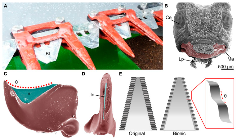
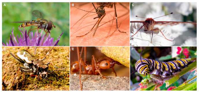
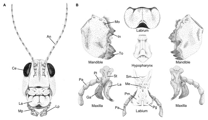
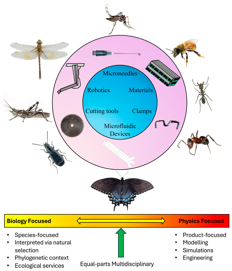
Few traits exhibit a more diverse collection of exemplary structure-function relationships than the mouthparts of insects. The global dominance of insects is attributed to their diverse food sources, which are matched by an array of morphological and chemical adaptations: a 'tool kit' for biomimicry. This review provides an overview of insect mouthparts that have contributed to biomimetics, including information about morphology and functionality in relation to particular feeding mechanisms. Themes in the groups of insects employed for particular biomimetic studies, including their lineages and feeding strategies, are identified along with suggestions for future studies, which together underscore the importance and promise of the development of novel engineered devices inspired by the unique 'tools' of insect mouthparts.

Keywords: feeding mechanisms; haustellate mouthparts; insect morphology; mandibulate mouthparts; structure–function relationships.


Conflict of interest statement

The authors declare no conflicts of interest.

Figures

Figure 4
Figure 4
Representative biomimetic hemp harvest blades inspired by the mandibles of longhorn beetles. (A) An image of bionic hemp cutting blades (Bl) on an agricultural harvester that was improved through biomimicry. (B) A scanning electron microscopy (SEM) image of the head of the flat faced longhorn beetle, Acanthocinus obsoletus, a closely related species to Leiopus nebulosus, which had its mandibles used as inspiration for the new blade. The image shows the compound eye (Ce), labial palpi (Lp), and mandibles (Ma, false colored) similar to those used for biomimetic studies. (C) A false-colored SEM image of the ventral side of the mandibles of A. obsoletus. The incisor (In) region is false-colored blue, and the red dotted line represents the contour angle (θ) used to develop the serration pattern of the bionic blades. (D) False-colored SEM image showing the sharp cutting edge of the incisor on the medial side of the mandibles of A. obsoletus. (E) Schematic showing a representative original (left) and bionic (right) hemp harvesting blade with the different serration patterns. The bionic blades use the same contour angle (θ, shown in the red box) as the mandibles of L. nebulosus (based on [78]). The image in (A) was used with permission by Forever Green Worldwide Corporation at Hempcutter.com.
Figure 1
Figure 1
General growth of biomimicry of insects and insect mouthparts as determined by publication quantity per decade. The orange markers represent the number of publications using the keywords “Insect biomimicry” in Google Scholar. The blue markers represent a search using the keywords “Insect mouthparts biomimicry”. Both topics are experiencing growth, as indicated by the increasing number of publications.
Figure 2
Figure 2
Diversity of insect feeding habits. (A) Image of a hoverfly, Allograpta obliqua (Diptera: Syrphidae) feeding from a flower with mouthparts adapted for fluid uptake. (B) The mosquito, Ochlerotatus notoscriptus (Diptera: Culicidae), piercing human skin for a blood meal. (C) A great spangled fritillary, Speyeria cybele (Lepidoptera: Nymphalidae) using its proboscis to feed from fluids inside a flower. (D) Image of a tiger beetle, Neocicindela tuberculata (Coleoptera: Cicindelidae) showing its mandibles adapted for biting and handling prey. (E) A leaf cutter ant, Atta sp. (Hymenoptera: Formicidae) using its mandibles to transport a leaf. (F) A larva of a monarch butterfly, Danaus plexippus (Lepidoptera: Nymphalidae) using its mandibles to feed on milkweed. (A) Image was acquired by John Chapman and used under the CC Attribution-Share Alike 4.0 International license. (B) Image is used under the CC Attribution-Share Alike 3.0 Unported license and was acquired by J.J. Harrison. (C) Image acquired by Sharon Mollerus and used under the CC Attribution 2.0 Generic. (D) Image used under CC Attribution-Share Alike 4.0 International license and acquired by John Marris. (E) Image was acquired by Malin Björnsdotter Åber and used under the CC Attribution-Share Alike 3.0 Unported license. (F) Image was used under the CC Attribution-Share Alike 3.0 Unported license and acquired by Ser Marr.
Figure 3
Figure 3
Illustrations of the head and mouthparts of the lubber grasshopper, Romalea microptera (Orthoptera), showing the plesiomorphic mouthpart condition. (A) Anterior view of the head showing the labrum (La), maxillary palpi (Mp), and labial palpi (Lp) mouthparts along with the compound eyes (Ce) and antennae (An) for reference. (B) Detailed illustrations of the different mouthparts, including the single labrum, pair of mandibles, hypopharynx, pair of maxillae, and the labium. The mandible shows three different regions, including the molar (Mo), incisor (In), and tooth (To) regions. The maxilla is composed of several parts, including the stipes (St), lacinia (La), galea (Ga), and of the maxillary palpi, the palpifer (Pl), and palpus (Pa). The labium also consists of several parts, including the submentum (Sm), mentum (Me), prementum (Pm), paraglossa (Pg), and a palpus (Pa). All illustrations were drawn by Erin Kelly.
Figure 5
Figure 5
Metal presence in the cuticle of the mandibles of the leafcutter ant, Atta cephalotes (Formicidae). (A) SEM image of leafcutter ant head showing the mandible (Ma) and the compound eye (Ce) for reference. (B) Energy dispersive X-ray spectroscopy image of the mandible, showing regions of high zinc concentrations (yellow). The inset shows the same image without the SEM overlay and the white box shows where the elemental analysis was performed for image (C). (C) Output of X-ray spectroscopy showing 5% zinc in studied region.
Figure 6
Figure 6
Morphology and anti-adhesive properties of the mandibles of honey bees, Apis mellifera (Apidae). (A) A confocal laser scanning microscopy (CLSM) image of the distal medial tip of the mandible of an adult honey bee, Apis mellifera, used to forage pollen (shown in (B)). (C) A false-colored SEM image of the microtopography and cuticle pattern on the distal medial tip of the mandibles of a honeybee. (D) Representative illustration of the anti-adhesive properties of the mandibles of honeybees. The uneven surface topography and thin liquid layer (Li) on the surface provide anti-adhesive properties, preventing propolis (Ps) from sticking and allowing it to slide off the mandible surface. (B) Image was acquired by Flocci Nivis and used under the CC Attribution-Share Alike 4.0 International license.
Figure 7
Figure 7
Mosquito mouthparts as a model for biomimetic needles. (A) A mosquito (Ochlerotatus sp.) using its mouthparts, which consist of six stylets and a sheath (Sh), to acquire a blood meal from a human. (B,C) SEM images of the sheath and proboscis of a mosquito (Aedes sp.), respectively. The proboscis is composed of the elongated maxillae (Mx), mandibles (Ma), hypopharynx (Hy), and labrum (La). The inset represents a schematic of a biomimetic needle inspired by mosquito stylets (based on [137]). (A) Image was acquired by Dunpharlain and used under the Creative Commons Attribution-Share Alike 4.0 International license.
Figure 8
Figure 8
The lepidopteran proboscis as a model for microfluidic devices. (A) False-colored SEM image of the coiled proboscis of the red-spotted purple butterfly, Limenitis arthemis astyanax (Nymphalidae), which has enlarged sensilla styloconica (Ss) (false-colored blue). (BD) SEM images of the proboscis of the monarch butterfly, Danaus plexippus (Nymphalidae). (B) The dorsal legulae (false-colored yellow, Dl) link the two c-shaped galeae (purple) together and their porosity allows liquids to travel across them via capillary action, which served as inspiration for a biomimetic fiber. (C,D) SEM images showing the porous arrangement of dorsal legulae in the nondrinking and drinking region, respectively. (E) Schematic of the nanopores in the fibers of a proboscis-inspired probe (based on [151]).
Figure 9
Figure 9
Images of the mouthparts of fluid-feeding insects with potential biomimetic applications. (AC) Confocal laser scanning microscope (CLSM) images of the distal region of a single galea of lepidopteran proboscises. (A) The spicebush swallowtail, Papilio troilus (Papilionidae), has a smooth proboscis surface with relatively small chemosensilla (sensilla basiconica, Sb) and smooth dorsal legulae (Dl). (B) The proboscis of the red-spotted purple butterfly, Limenitis arthemis astyanax (Nymphalidae), uses its large sensilla styloconica (Ss) similar to a mop. (C) The proboscis of the vampire moth, Calyptra lata (Noctuidae), is armed with sensory cones (Sc), erectile barbs (Eb), and tearing hooks (Th) that aid in piercing tissues, such as the skin of fruit or vertebrates for a blood meal. (D) A CLSM image of the labellum of the house fly, Musca domestica (Diptera: Muscidae) that has an array of conduits, the pseudotrachea (Ps), that act similar to a sponge. (E) False-colored scanning electron microscope image showing the pseudotracheae (green) that are interspersed by cuticle with elastic properties (pink).
Figure 10
Figure 10
Representative diagram of patterns in biomimicry studies that feature insect mouthparts. The top image shows the main categories of tools that have been impacted by biomimetics of insect mouthparts. Outside of the circle shows representative insect orders that correspond to each tool. The bottom image is a schematic showing two ends of a spectrum in themes in biomimetics. We found that some papers were biology focused and lacked the development of a tool, whereas other papers were engineering-heavy, but lacked important biology details. Image of the mosquito (Aedes aegypti) (Diptera: Culicidae) was acquired by Muhammad Mahdi Karim and used under the GNU free documentation 1.2 license. The photograph of Apis mellifera was used under the CC attribution 3.0 unported license and acquired by CSIRO. Image of Dinoponera quadriceps (Hymenoptera: Formicidae) was acquired by Dider Descouens and used under the CC Attribution-Share Alike 4.0 International license. The image of an ant was used under CC attribution 2.0 generic license and acquired by Micheal Caven. The photograph of the dark morph tiger swallowtail, Papilio glaucus (Lepidoptera: Papilionidae) was taken by khteWisconsin and used under CC public domain. Image of the ground beetle Microlestes minutulus (Coleoptera: Carabidae) was used under the CC Attribution-Share Alike 4.0 International license and acquired by URSchmidt. The photograph of the bark mimicking grasshopper, Coryphistes ruricola (Orthoptera: Acrididae) was taken by Flagstaffotos and used under the GNU free documentation license. The image of the vagrant emperor dragonfly (Anax ephippiger) (Odonata: Aeshnidae) was used under the CC Attribution-Share Alike 4.0 International license and acquired by Alvesgaspar. The photograph of the needle was acquired by Nicola Sap De Mitri and used under CC Attribution Share Alike 2.0 generic license. The surgical staple clamp image was used under CC Attribution Share Alike 3.0 unported license and was acquired by Jakubtr. The yarn schematic was created by Paulo H. T. F. Alves and used under CC Attribution-Share Alike 4.0 International license. Image of the saw blade was taken by the Missouri Historical Society and used under public domain.

Similar articles

References

    1. Phan H.V., Park H.C. Mimicking nature’s flyers: A review of insect-inspired flying robots. Curr. Opin. Insect Sci. 2020;42:70–75. doi: 10.1016/j.cois.2020.09.008. - DOI - PubMed
    1. Manoonpong P., Patanè L., Xiong X., Brodoline I., Dupeyroux J., Viollet S., Arena P., Serres J.R. Insect-inspired robots: Bridging biological and artificial systems. Sensors. 2021;21:7609. doi: 10.3390/s21227609. - DOI - PMC - PubMed
    1. Li Q., Li H., Shen H., Yu Y., He H., Feng X., Sun Y., Mao Z., Chen G., Tian Z., et al. An aerial-wall robotic insect that can land, climb, and take off from vertical surfaces. Research. 2023;6:0144. doi: 10.34133/research.0144. - DOI - PMC - PubMed
    1. French J.R.J., Ahmed B.M. The challenge of biomimetic design for carbon-neutral buildings using termite engineering. Insect Sci. 2010;17:154–162. doi: 10.1111/j.1744-7917.2009.01306.x. - DOI
    1. Claggett N., Surovek A., Streeter B., Nam S., Bardunias P., Cetin B. Insights and Innovations in Structural Engineering, Mechanics and Computation. CRC Press; Boca Raton, FL, USA: 2016. Biomimicry and locally responsive construction: Lessons from termite mounds for structural sustainability.

LinkOut - more resources