. 2025 Apr 12;14(8):582.
doi: 10.3390/cells14080582.

Neuroprotection by Mitochondrial NAD Against Glutamate-Induced Excitotoxicity

Affiliations

Neuroprotection by Mitochondrial NAD Against Glutamate-Induced Excitotoxicity

Bruna S Paiva et al. Cells. .

Abstract

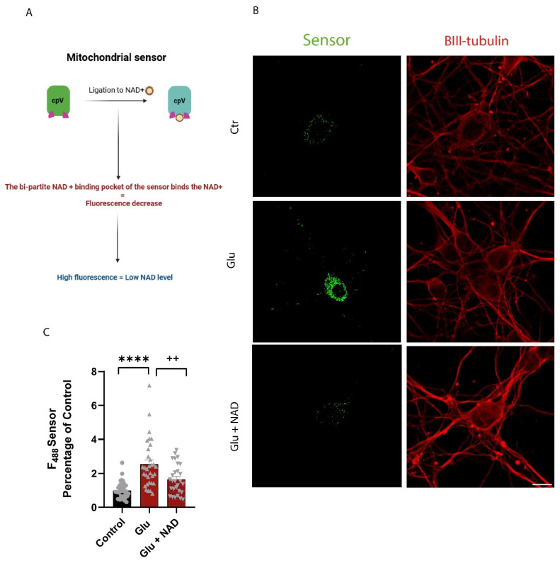
Excitotoxicity is a pathological process that occurs in many neurological diseases, such as stroke or epilepsy, and is characterized by the extracellular accumulation of high concentrations of glutamate or other excitatory amino acids (EAAs). Nicotinamide adenine dinucleotide (NAD) depletion is an early event following excitotoxicity in many in vitro and in vivo excitotoxic-related models and contributes to the deregulation of energy homeostasis. However, the interplay between glutamate excitotoxicity and the NAD biosynthetic pathway is not fully understood. To address this question, we used a primary culture of rat cortical neurons and found that an excitotoxic glutamate insult alters the expression of the NAD biosynthetic enzymes. Additionally, using a fluorescent NAD mitochondrial sensor, we observed that glutamate induces a significant decrease in the mitochondrial NAD pool, which was reversed when exogenous NAD was added. We also show that exogenous NAD protects against the glutamate-induced decrease in mitochondrial membrane potential (MMP). Glutamate excitotoxicity changed mitochondrial retrograde transport in neurites, which seems to be reversed by NAD addition. Finally, we show that NAD and NAD precursors protect against glutamate-induced cell death. Together, our results demonstrate that glutamate-induced excitotoxicity acts by compromising the NAD biosynthetic pathway, particularly in the mitochondria. These results also uncover a potential role for mitochondrial NAD as a tool for central nervous system (CNS) regenerative therapies.

Keywords: NAD metabolism; excitotoxicity; glutamate; mitochondria.


Conflict of interest statement

The authors declare that they have no conflicts of interest.

Figures

Figure 1
Figure 1
Alterations in the expression of NAD biosynthetic enzymes following glutamate-induced excitotoxicity. (AC) Representative images and quantitative data of NAMPT and NAPRT enzymes. Analysis of NAMPT and NAPRT protein expression. Rat cortical neurons were exposed to 50 µM glutamate for 15 min and protein levels were determined at the indicated times by Western blot. β-Actin was used as a loading control (A). We observed an increase in NAMPT and no alterations in NAPRT protein expression after glutamate stimulation (B,C). (D) Quantitative data of NAMPT and NNMAT enzymes mRNA expression. Evaluation of NAMPT, NMNAT1, NMNAT2, and NMNAT3 mRNA expression. Rat cortical neurons were exposed to 50 µM glutamate for 15 min and mRNA expression was analyzed after 4 h and 24 h. RT-PCR results indicated an increase in gene expression levels for NAMPT and NMNAT1 after glutamate stimulation and a decrease in NMNAT3 levels following 24 h of glutamate stimulation. 18s rDNA was used as an internal control gene. The dashed line represents normalization to the control condition for each gene. Bars represent the mean ± SEM of 3 independent experiments; * represents p < 0.05; ** represents p < 0.01 and *** represents p < 0.001 by ANOVA followed by Dunnett’s multiple comparisons test when compared to control.
Figure 2
Figure 2
Exogenous NAD+ rescues the decrease in mitochondrial NAD+ following glutamate-induced excitotoxicity. (A) Schematic depiction illustrating the operational mechanism of the mitochondrial sensor. (B) Representative images of mitochondrial NAD sensor fluorescence. Sum intensity projections of Z-stacks from cortical neurons transfected with 3 µM mitochondrial NAD sensor (green) and immunolabeled for the neuronal marker βIII-tubulin (red). Sum intensity projections of Z-stacks were assembled to generate a single image. An increase in cell fluorescence represents a decrease in free NAD in the mitochondria. Scale bar: 10 µm. (C) Quantitative data of mitochondrial NAD sensor fluorescence. Relative corrected total cell fluorescence was quantified in cortical neurons exposed to glutamate in the presence or absence of NAD. Glutamate (Glu) induces an increase in mitochondrial NAD sensor fluorescence intensity, which is reversed by 5 mM NAD+ addition prior to Glu, indicating an increase in free NAD+ in the mitochondria. Control (circles); Glutamate (triangles); Glutamate + NAD (upside down triangle) Bars represent the mean ± SEM of at least 30 neurons randomly selected from 3 independent experiments. Statistical significance was assessed by ANOVA followed by Sidak’s multiple comparison test. **** represents p < 0.0001 when compared to control. ++ represents p < 0.01 when compared to glutamate.
Figure 3
Figure 3
NAD reverts the decrease in MMP induced by glutamate excitotoxicity. Mitochondrial membrane potential (MMP) was measured in cortical neurons with the JC-10 probe. Exposure to 50 µM glutamate (Glu) results in a loss of MMP that is prevented by 5 mM NAD+ addition. Control (circles); Glutamate (triangles); Glutamate + NA (upside down triangle); FCCP (empty diamondsBars represent the mean ± SEM from 6 independent experiments. Statistical significance was assessed by ANOVA followed by Sidak’s multiple comparison test. * represents p < 0.05; **** represents p < 0.0001 when compared to control. ++ represents p < 0.01 when compared to glutamate.
Figure 4
Figure 4
Glutamate excitotoxicity promotes mitochondrial anterograde movement in neurites. (AC) Analysis of mitochondrial velocity and displacement in the neurites of cortical neurons. Neurons were exposed to 50 µM glutamate for 15 min in the presence or absence of 5 mM NAD, stained with TMRM, and live imaged by confocal microscopy after 24 h. The mitometer MATLAB algorithm was used to quantify mitochondrial movement in the neurites (i.e., area outside the red circle in (A)). Scale bar: 10 µm. Glutamate increased mitochondrial velocity and displacement (B,C), which were reversed by the presence of NAD. Bars represent the mean ± SEM of 4 independent experiments. * represents p < 0.05 when compared to control. + represents p < 0.05 when compared to control. Statistical analysis was conducted with ANOVA test (Sidak’s multiple comparison test). (DG) Quantitative data of mitochondrial movement in neurites. FIJI software was used to generate kymographs of mitochondria in neurites. Neurite selection was made from the cellular body to the end of the neurite (D). Scale bar: 10 µm. While glutamate appears to induce a directional anterograde movement, exogenous NAD preferentially promotes mitochondrial retrograde transport (quantified in (E,F)). Bars represent the mean ± SEM of five independent experiments. * represents p < 0.05 when compared to control. Statistical analysis was conducted with ANOVA test (Sidak’s multiple comparison test) and two-way ANOVA.
Figure 5
Figure 5
Stimulation with NAD or NAD biosynthetic pathway precursors protects against glutamate-induced excitotoxicity. (A) Representative images of Hoechst staining of cortical neurons. Cortical neurons were exposed to 50 µM glutamate in the presence or absence of NAD or its precursors. Cell death was evaluated by counting the number of condensed nuclei stained with the fluorescent dye Hoechst 33342. Scale bar: 30 µm. (B) Quantitative data of cell death. Glutamate exposure increased the percentage of condensed nuclei in cortical neurons, which was reverted by NA, NAM, and NAD. Results are expressed as a percentage of control. Control (circles); Glutamate (triangles); Glutamate + NA (upside down triangle); Glutamate + NAM (filled diamonds); Glutamate + NAD (Unfilled diamonds); NA(triangle filled only on the left side); NAM (triangle filled only on the right side); NAD(empty circle) Bars represent the mean ± SEM of three independent experiments. Statistical significance was assessed by ANOVA followed by the Bonferroni multiple comparison test. **** represents p < 0.0001 when compared to control. + represents p < 0.05; ++ represents, and ++++ represents p < 0.0001 when compared to glutamate.
Figure 6
Figure 6
Stimulation with NAD or NAD biosynthetic pathway precursors protects against glutamate-induced metabolic dysfunction. Cortical neurons were exposed to 50 µM glutamate in the presence or absence of NAD or its precursors, and cell metabolic activity was measured by the Alamar Blue assay. Glutamate decreased cell metabolic activity, which was reverted by NAD or its precursors in both types of neurons. Control (circles); Glutamate (triangles); Glutamate + NA (upside down triangle); Glutamate + NAM (filled diamonds); Glutamate + NAD (Unfilled diamonds); NA(triangle filled only on the left side); NAM (triangle filled only on the right side); NAD(empty circle) Statistical significance was assessed by ANOVA followed by the Bonferroni multiple comparison test. **** represents p < 0.0001 when compared to control. ++++ represents p < 0.0001 when compared to glutamate.

Similar articles

References

    1. Neves D., Salazar I.L., Almeida R.D., Silva R.M. Molecular Mechanisms of Ischemia and Glutamate Excitotoxicity. Life Sci. 2023;328:121814. doi: 10.1016/j.lfs.2023.121814. - DOI - PubMed
    1. Mao R., Zong N., Hu Y., Chen Y., Xu Y. Neuronal Death Mechanisms and Therapeutic Strategy in Ischemic Stroke. Neurosci. Bull. 2022;38:1229–1247. doi: 10.1007/s12264-022-00859-0. - DOI - PMC - PubMed
    1. Shen Z., Xiang M., Chen C., Ding F., Wang Y., Shang C., Xin L., Zhang Y., Cui X. Glutamate Excitotoxicity: Potential Therapeutic Target for Ischemic Stroke. Biomed. Pharmacother. 2022;151:113125. doi: 10.1016/j.biopha.2022.113125. - DOI - PubMed
    1. Carvajal F.J., Mattison H.A., Cerpa W. Role of NMDA Receptor-Mediated Glutamatergic Signaling in Chronic and Acute Neuropathologies. Neural Plast. 2016;2016:1–20. doi: 10.1155/2016/2701526. - DOI - PMC - PubMed
    1. Deep S.N., Mitra S., Rajagopal S., Paul S., Poddar R. GluN2A-NMDA Receptor–Mediated Sustained Ca2+ Influx Leads to Homocysteine-Induced Neuronal Cell Death. J. Biol. Chem. 2019;294:11154–11165. doi: 10.1074/jbc.RA119.008820. - DOI - PMC - PubMed

Publication types

LinkOut - more resources