. 2025 Apr 14;14(8):593.
doi: 10.3390/cells14080593.

LRG1 Alters Pericyte Phenotype and Compromises Vascular Maturation

Affiliations

LRG1 Alters Pericyte Phenotype and Compromises Vascular Maturation

Alexandra E Hoeh et al. Cells. .

Abstract

Upregulation of leucine-rich alpha-2-glycoprotein-1 (LRG1) contributes to aberrant neovascularization in many different diseases. In contrast, LRG1 is not involved in developmental angiogenesis. Here, we investigated the vasculopathic properties of LRG1 by examining its effect on developing retinal blood vessels. By injecting recombinant protein or an expression vector into the mouse retina during vascular development, we showed that exogenous LRG1 reduces pericyte coverage and NG2 expression. It leads to diminished collagen IV sheathing, fewer adhesion and gap junctions, and reduced vessel calibre and vascular density. Moreover, in mouse retinae containing exogenous LRG1, the developing blood-retinal barrier remains more permeable with significantly higher numbers of transcytotic vesicles present in microvascular endothelial cells. These results reveal that exogeneous LRG1 is sufficient to interfere with the maturation of developing retinal vessels and drive vessel development towards a dysfunctional phenotype. These observations deliver further evidence that LRG1 is an angiopathic factor and highlight the therapeutic potential of blocking LRG1 in diseases characterized by pathogenic angiogenesis or vascular remodelling.

Keywords: angiogenesis; blood vessel maturation; blood–retinal barrier; leucine-rich alpha-2-glycoprotein-1 (LRG1); pericyte.


Conflict of interest statement

J.G. and S.E.M. are founders and members of the scientific advisory board of a company spun out by UCL Business to commercialize a LRG1 function-blocking therapeutic antibody developed through the UK Medical Research Council DPFS funding scheme. J.G. and S.E.M. are shareholders of this company and named inventors on three patents related to LRG1 as a therapeutic target. All other authors have declared that no conflicts of interest exist.

Figures

Figure 1
Figure 1
Lrg1 is expressed at very low levels postnatally in the retina. Single-cell RNA sequencing data from mouse retina on postnatal day (P)6. A colour-coded UMAP of cell type identity is shown alongside UMAP illustrating the expression levels of the indicated genes per cell in each cluster. Each UMAP plot names the cluster(s) expressing the indicated gene. The corresponding violin plots illustrate the expression levels of each cell cluster. Each data point represents the value for one cell.
Figure 2
Figure 2
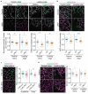
LRG1 alters the expression of pericyte markers and reduces pericyte coverage. (a) Representative micrographs of transgenic NG2 reporter mice expressing DsRed under the control of the NG2 promoter at P5. Pericytes were counted and normalized to the vessel area of each image, with lower pericyte density observed in LRG1-treated vessels; n = 19–26 eyes per group. Reduced expression of DsRed in pericytes at P5 after intravitreal injection of LRG1 protein; n = 18–28 eyes per group. (b) Representative micrographs demonstrating reduced expression of NG2 in microvascular pericytes of the central retina and at the leading edge of the growing vasculature in LRG1 protein-treated and control retinas. Quantitation shows significant differences between control and LRG1-treated retinas at P5; n = 20–23 eyes per group. (c) Representative images of the deep vascular plexus at P16 demonstrate weaker staining for NG2 in LRG1 overexpressing retinas (7m8-LRG1) compared to null vector controls (7m8-null). Quantitation shows significantly reduced NG2 expression in the deep vessel plexus after injection of the LRG1 overexpression vector and non-significant reduction in NG2 expression in the superficial plexus; n = 35–44 eyes per group (superficial plexus) and n = 17–23 eyes per group (deep plexus). (d) Images and dot plots showing reduced expression of desmin in capillaries in the retinal centre, but not the leading edge, at P5 following LRG1 protein treatment; n = 20–21 eyes per group. Unpaired t-test. Mean ± SEM of n ≥ 3 independent experiments. * p ≤ 0.05, ** p ≤ 0.01, *** p ≤ 0.001. Scale bars 25 μm.
Figure 3
Figure 3
LRG1 impairs collagen IV ensheathing of retinal microvessels. (a) Illustrative micrographs of retinal vascular collagen IV expression at P5 following intravitreal injection with LRG1 protein or denatured LRG1 protein (control). Quantitation demonstrates reduced collagen IV expression in LRG1-injected eyes; n = 26–31 eyes per group. (b) Images of retinal vascular collagen IV expression at P16 following delivery of LRG1 overexpression (7m8-LRG1) or null (7m8-null) vectors. Collagen IV expression was reduced in the deep vessel plexus in the LRG1 overexpression vector group, but no difference was found in the superficial plexus; n = 34–45 eyes per group. Unpaired t-test or Mann–Whitney test was used depending on normality. Mean ± SEM of n ≥ 3 independent experiments. * p ≤ 0.05, ** p ≤ 0.01, **** p ≤ 0.0001. Scale bars 25 μm.
Figure 4
Figure 4
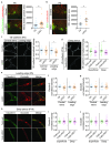
LRG1 increases vascular permeability without changing tight junction expression. (a) Images of Dextran-488 (10 kDa) and (b) bovine serum albumin (BSA-488) vascular leakage and associated quantification showing a significant increase in permeability at the leading edge of P5 eyes treated with LRG1; n = 21 eyes per group (Dextran-488) and n = 10 eyes per group (BSA-488). Scale bar 100 μm. Wilcoxon test. Mean ± SEM, * p ≤ 0.05. (c) Representative micrographs and quantified pixel intensity of junctional VE-cadherin showing reduced expression at the leading edge, but not in the central retina, of LRG1-treated eyes at P5; n = 24 eyes per group. Unpaired t-test. Mean ± SEM of n ≥ 3 independent experiments. * p ≤ 0.05. Scale bars 5 μm. (d) Significant reduction in VE-cadherin expression in the deep vessel plexus in LRG1 overexpressing retinas (7m8-LRG1) at P16. No significant change was observed in the superficial plexus. n = 20–23 eyes per group. Unpaired t-test. Mean ± SEM of n ≥ 3 independent experiments. * p ≤ 0.05. Scale bars 5 μm. (e) Representative images of vascular claudin-5 and occludin expression at P5 following administration of LRG1 protein (scale bars 5 μm). Quantification revealed no change in expression of claudin-5 ((f); n = 18–19 eyes per group) or occludin ((g); n = 18–20 eyes per group). Bright luminal autofluorescent signal from erythrocytes was excluded from the analysis. Unpaired t-test. Mean ± SEM of n ≥ 3 independent experiments. (h) Representative micrographs of claudin-5 and occludin staining in the deep vessel plexus at P16 (scale bars 5 μm) following LRG1 gene or empty vector delivery. Quantification revealed no change in expression of claudin-5 ((i); n = 19–22 eyes per group) or occludin ((j); 16–21 eyes per group) expression in the superficial or deep retinal vascular plexus. Unpaired t-test. Mean ± SEM of n ≥ 3 independent experiments.
Figure 5
Figure 5
LRG1 increases transcellular transport at the developing BRB. Representative images and quantification of PLVAP (a) and MFSD2A (b) expression in retinal vessels at P5 following intravitreal injection of LRG1 protein. Quantification shows that LRG1 administration results in higher levels of PLVAP centrally and at the leading edge, and lower levels of MFSD2A expression at the leading edge compared to controls (scale bar, 25 μm); n = 17–19 eyes per group (PLVAP) and n = 9–14 eyes per group (MFSD2A). Unpaired t-test, mean ± SEM, * p ≤ 0.05, ** p ≤ 0.01. (c) Transmission electron microscopy was performed on LRG1 overexpressing (7m8-LRG1) and control (7m8-null) retinas at P10 following intraperitoneal delivery of the tracer HRP. Areas of the leading edge microvessels were analyzed. Images show transverse sections through representative microvessels in lower magnification (left) and higher magnification (right). Lumen (L), endothelial cell (E), HRP-filled intraendothelial vesicles <100 nm (arrows), empty intraendothelial vesicles <100 nm (arrowheads), HRP-filled intraendothelial vesicles >100 nm (star). Quantification of HRP-filled vesicles <100 nm in diameter (arrows) which were associated with the plasma membrane revealed significantly higher number of vesicles in LRG1 overexpressing eyes compared to controls. Data from a total of 48 retinal vessels and 6 eyes. Unpaired t-test. Mean ± SEM. * p ≤ 0.05. Scale bars in overview images 2 μm and 0.1 μm in magnified images.
Figure 6
Figure 6
LRG1 gene overexpression reduces expression of Cx43 on arteries and superficial capillaries. Representative micrographs of retinal vasculature from P16 eyes treated with LRG1 overexpression (7m8-LRG1) or null vector (7m8-null). The proportion of the vessels overlapping with Cx43 staining was quantified and shows a significant reduction in Cx43 on arteries and the capillaries of the superficial plexus in LRG1 overexpressing retinas; n = 15–19 eyes per group. Unpaired t-test. Mean ± SEM of n ≥ 3 independent experiments. * p ≤ 0.05. Scale bar 25 μm.
Figure 7
Figure 7
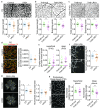
LRG1 alters the vascular growth pattern. Reduced retinal vessel density in LRG1 protein-treated retinas at P5 (a) and in LRG1 overexpressing retinas at P16 (b,c); n = 23–25 eyes per group (a), n = 16–19 eyes per group (b), n = 15–21 eyes per group (c). Scale bars 100 μm. (d) Representative image of the superficial plexus at P5 showing empty (non-endothelialised, collagen IV positive) vessel sleeves (arrow heads). Scale bar 25 μm. Quantification reveals no difference in the amount of vessel regression at P5 between control and LRG1 protein treatment (n = 17–19 eyes per group), and at P16 (e) in the superficial plexus and deep plexus between control (7m8-null) and LRG1 overexpressing (7m8-LRG1) retinas; n = 23–30 eyes per group. (f) Images and dot plot of quantified data showing reduced number of vessel sprouts per field at the leading edge in LRG1-treated retinas at P5; n = 29–30 eyes per group. Scale bar 25 μm. (g) Representative micrographs and dot plots showing significant reduction in venous, but not arterial, vessel calibre at P5 in LRG1-treated retinas. Arteries (a), veins (v); n = 17–19 eyes per group. Scale bar 500 μm. (h) Representative images of the capillaries of the deep vessel plexus (scale bar 25 μm). Dot plot of significant reduction in capillary calibre in the deep vessel plexus at P16 in LRG1 overexpressing retinas, but no change in capillary thickness of the superficial plexus; n = 18–25 eyes per group. Unpaired t-test or Mann–Whitney test depending on normality. Mean ± SEM, * p ≤ 0.05, ** p ≤ 0.01.

References

    1. Chung A.S., Ferrara N. Developmental and pathological angiogenesis. Annu. Rev. Cell Dev. Biol. 2011;27:563–584. doi: 10.1146/annurev-cellbio-092910-154002. - DOI - PubMed
    1. Daneman R., Zhou L., Kebede A.A., Barres B.A. Pericytes are required for blood-brain barrier integrity during embryogenesis. Nature. 2010;468:562–566. doi: 10.1038/nature09513. - DOI - PMC - PubMed
    1. Ben-Zvi A., Lacoste B., Kur E., Andreone B.J., Mayshar Y., Yan H., Gu C. Mfsd2a is critical for the formation and function of the blood-brain barrier. Nature. 2014;509:507–511. doi: 10.1038/nature13324. - DOI - PMC - PubMed
    1. Chow B.W., Gu C. Gradual Suppression of Transcytosis Governs Functional Blood-Retinal Barrier Formation. Neuron. 2017;93:1325–1333.e1323. doi: 10.1016/j.neuron.2017.02.043. - DOI - PMC - PubMed
    1. Stratman A.N., Davis G.E. Endothelial cell-pericyte interactions stimulate basement membrane matrix assembly: Influence on vascular tube remodeling, maturation, and stabilization. Microsc. Microanal. 2012;18:68–80. doi: 10.1017/S1431927611012402. - DOI - PMC - PubMed

Publication types