. 2025 Apr 21;14(8):621.
doi: 10.3390/cells14080621.

Mimicking the Liver Sinusoidal Endothelial Cell Niche In Vitro to Enhance Fenestration in a Genetic Model of Systemic Inflammation

Affiliations

Mimicking the Liver Sinusoidal Endothelial Cell Niche In Vitro to Enhance Fenestration in a Genetic Model of Systemic Inflammation

Dibakar Borah et al. Cells. .

Abstract

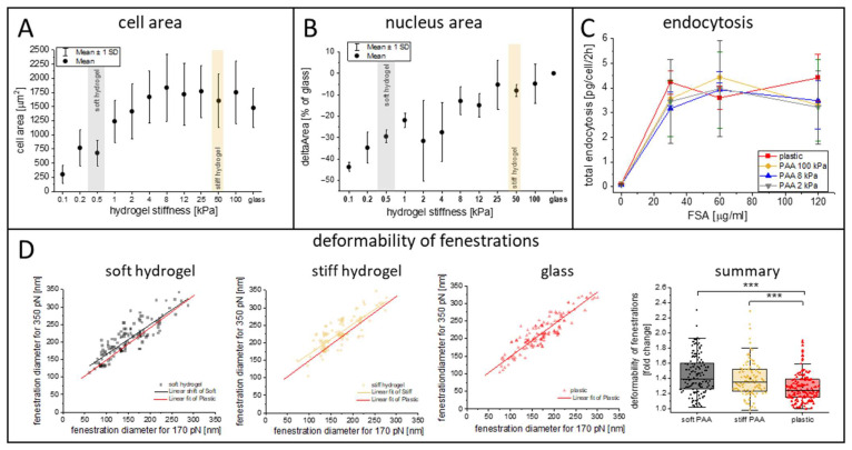
Liver sinusoidal endothelial cells (LSECs) play a crucial role in hepatic homeostasis, clearance, and microcirculatory regulation. Their fenestrations-patent transcellular pores-are essential for proper liver function, yet disappear in pathological conditions such as liver fibrosis and inflammation through a process known as defenestration. Defenestrated sinusoids are often linked to the liver stiffening that occurs through mechanotransduction-regulated processes. We performed a detailed characterization of polyacrylamide (PAA) hydrogels using atomic force microscopy (AFM), rheometry, scanning electron microscopy, and fluorescence microscopy to assess their potential as biomimetic substrates for LSECs. We additionally implemented AFM; quantitative fluorescence microscopy, including high-resolution structured illumination microscopy (HR-SIM); and an endocytosis assay to characterize the morphology and function of LSECs. Our results revealed significant local variations in hydrogel stiffness and differences in pore sizes. The primary LSECs cultured on these substrates had a range of stiffnesses and were analyzed with regard to their number of fenestrations, cytoskeletal organization, and endocytic function. To explore mechanotransduction in inflammatory liver disease, we investigated LSECs from a genetic model of systemic inflammation triggered by the deletion of Mcpip1 in myeloid leukocytes and examined their ability to restore their fenestrations on soft substrates. Our study demonstrates the beneficial effect of soft hydrogels on LSECs. Control cells exhibited a similar fenestrated morphology and function compared to cells cultured on plastic substrates. However, the pathological LSECs from the genetic model of systemic inflammation regained their fenestrations when cultured on soft hydrogels. This observation supports previous findings on the beneficial effects of soft substrates on LSEC fenestration status.

Keywords: actin cytoskeleton; atomic force microscopy; elastic properties; fenestrations; liver sinusoidal endothelial cells; mechanotransduction; polyacrylamide.


Conflict of interest statement

The authors declare no conflicts of interest.

Figures

Figure 1
Figure 1
Characteristics of synthesized polyacrylamide (PAA) hydrogels. (A) Representative SEM images of dehydrated hydrogels. (B) Representative AFM images of the topography of a hydrated stiff hydrogel and stiff hydrogel with collagen III deposited on its surface. Similar images for soft hydrogels are presented in Figure S1. (C) AFM spectroscopy showing the distribution of the apparent Young’s modulus for stiff and soft hydrogels, both bare and with 0.1 mg/mL collagen III deposited on their surface. The box range represents the standard deviation, the bar represents the median, and the open square represents the mean value; *** p < 0.001. (D) Rheological properties of soft and stiff hydrogels. Storage (G′, filled points) and loss (G″, hollow points) moduli were obtained. LVER lines were fit to the plateau of G′ and G″ and used to calculate the loss factor. (E) Representative fluorescence images of the collagen III deposited on the surface of glass and stiff and soft hydrogels. Negative images are presented.
Figure 2
Figure 2
Morphology and ultrastructure of LSECs cultured on plastic and stiff and soft PAA hydrogels. Phase contrast images (top panel) and AFM topography (bottom panel) present a spread of cells with LSEC characteristics, such as the formation of tight groups, a flat cell periphery and bulging nuclei, and sieve plates filled with fenestrations. AFM images are 35 × 40 µm2 and 405 × 463 pixels. Insets (corresponding black boxes in each column) represent a digital enlargement of the area with fenestrations. All groups have the same magnification and scale in their corresponding images.
Figure 3
Figure 3
Comparison of changes in cell area (A), cell nuclei area (B), and endocytosis (C) of LSECs cultured on commercial PAA hydrogels with varying elastic properties. The stiffness ranges of the soft and stiff hydrogels are marked in gray and bright orange, respectively. (D) The deformability of fenestrations calculated from living LSECs cultured on substrates with varying elastic properties, based on AFM imaging performed according to the methodology described in the Materials and Methods. That summary presents the final deformability parameter, which is calculated as a ratio of the diameter of the fenestrations measured at 300 pN to the diameter measured at 170 pN. More than 100 fenestrations were measured per group in a one-to-one manner; *** p < 0.001.
Figure 4
Figure 4
Representative LSECs cultured on stiff and soft PAA hydrogels, visualized using super-resolution structured illumination microscopy (SR-SIM). Actin shows distinct levels of organization, which are translated to an F-actin polymerization degree, and a similar organization of tubulin was seen when LSECs were cultured on stiff and soft substrates. Actin (phalloidin-Alexa Fluor 488, green) and tubulin (immunofluorescence, Alexa Fluor 555, purple) are both visible. The lower panel is a 3D projection (cross-section) of an LSEC cultured on a soft hydrogel, with the elevated edges of the cell, which is embedded within the hydrogel structure (white arrows) (Supplementary Video S1). Image size: 40.96 µm × 40.96 µm.
Figure 5
Figure 5
Representative AFM images of LSECs isolated from Mcpip1 control and Mcpip1 KO mice cultured on glass and stiff and soft hydrogels. Selected areas in the cells’ peripheries were digitally magnified to depict fenestrations in corresponding groups (gray square for stiff hydrogel and black for soft hydrogel). Images are presented at the same scale for comparison. Image size: 25 × 30 µm2 and 300 × 360 pixels. The chart presents LSEC porosity, expressed as the number of fenestrations per cell area (fen./µm2). The box range represents the standard deviation, the bar represents the median, and the open square represents the mean value; *** p < 0.001.
Figure 6
Figure 6
(A) Representative fluorescence images of actin and cell nuclei of LSECs isolated from Mcpip1fl/fl and Mcpip1fl/fl LysMCre mice and cultured on glass and stiff and soft PAA hydrogels. Image size: 133.1 × 133.1 µm2. (B) Actin filaments detected using free FilamentSensor 2.0 software. Representative images containing detected filaments are presented in Figure S5. (C) Cell stiffness of LSECs expressed as apparent Young’s modulus and calculated for each group. Box range represents standard deviation, the bar represents the median, and the open square represents the mean value; * p < 0.05, *** p < 0.001.

Similar articles

Cited by

References

    1. Li N., Zhang X., Zhou J., Li W., Shu X., Wu Y., Long M. Multiscale Biomechanics and Mechanotransduction from Liver Fibrosis to Cancer. Adv. Drug Deliv. Rev. 2022;188:114448. doi: 10.1016/j.addr.2022.114448. - DOI - PubMed
    1. Kang N. Mechanotransduction in Liver Diseases. Semin. Liver Dis. 2020;40:84–90. doi: 10.1055/s-0039-3399502. - DOI - PMC - PubMed
    1. Le Couteur D.G., Warren A., Cogger V.C., Smedsrød B., Sørensen K.K., De Cabo R., Fraser R., McCuskey R.S. Old Age and the Hepatic Sinusoid. Anat. Rec. 2008;291:672–683. doi: 10.1002/ar.20661. - DOI - PubMed
    1. Fraser R., Cogger V.C., Dobbs B., Jamieson H., Warren A., Hilmer S.N., Le Couteur D.G. The Liver Sieve and Atherosclerosis. Pathology. 2012;44:181–186. doi: 10.1097/PAT.0b013e328351bcc8. - DOI - PubMed
    1. Wisse E., de Zanger R.B., Charels K., Van Der Smissen P., McCuskey R.S. The Liver Sieve: Considerations Concerning the Structure and Function of Endothelial Fenestrae, the Sinusoidal Wall and the Space of Disse. Hepatology. 1985;5:683–692. doi: 10.1002/hep.1840050427. - DOI - PubMed

Publication types

LinkOut - more resources