Comparative Study
. 2025 Apr 3;17(4):177.
doi: 10.3390/toxins17040177.

Establishment and Comparison of Detection Methods for Ricin and Abrin Based on Their Depurination Activities

Affiliations
Comparative Study

Establishment and Comparison of Detection Methods for Ricin and Abrin Based on Their Depurination Activities

Lina Dong et al. Toxins (Basel). .

Abstract

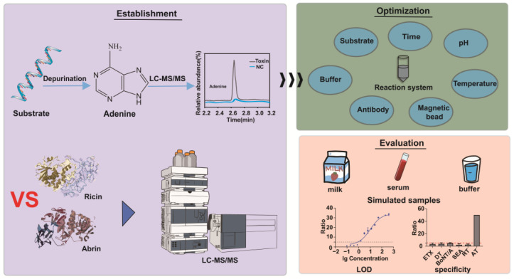
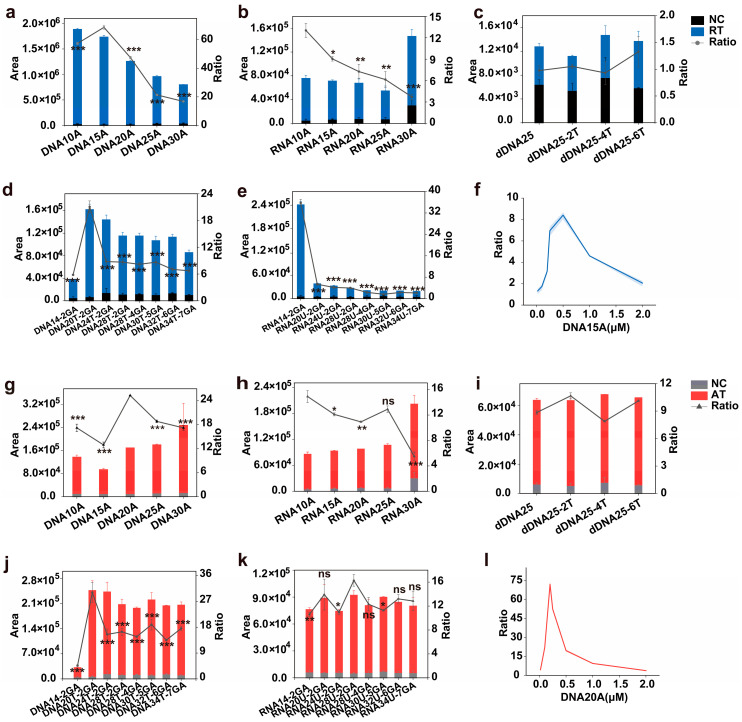
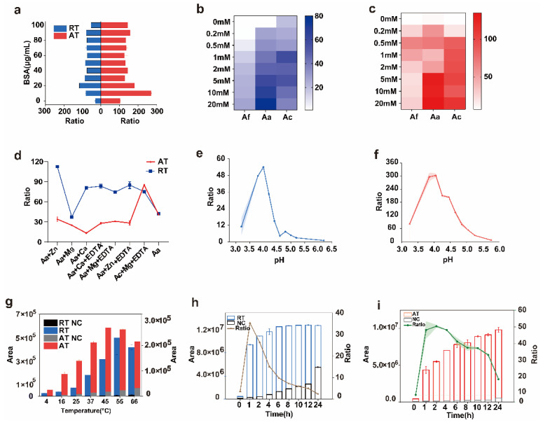
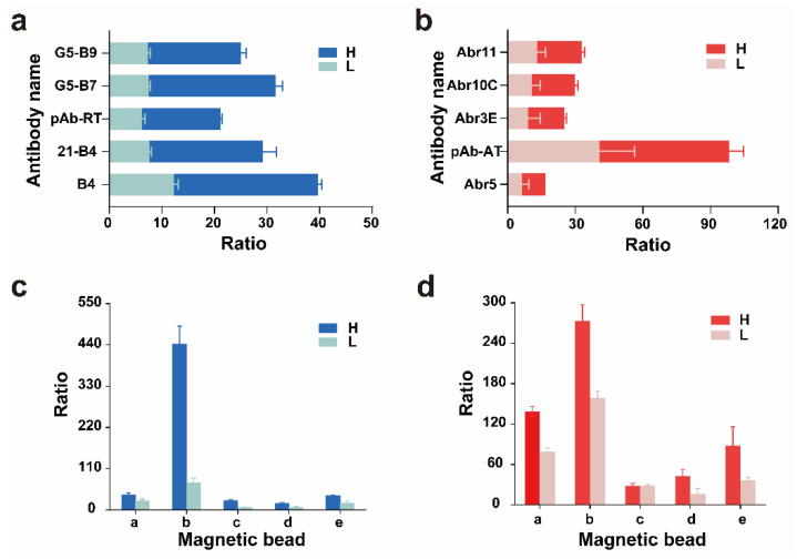
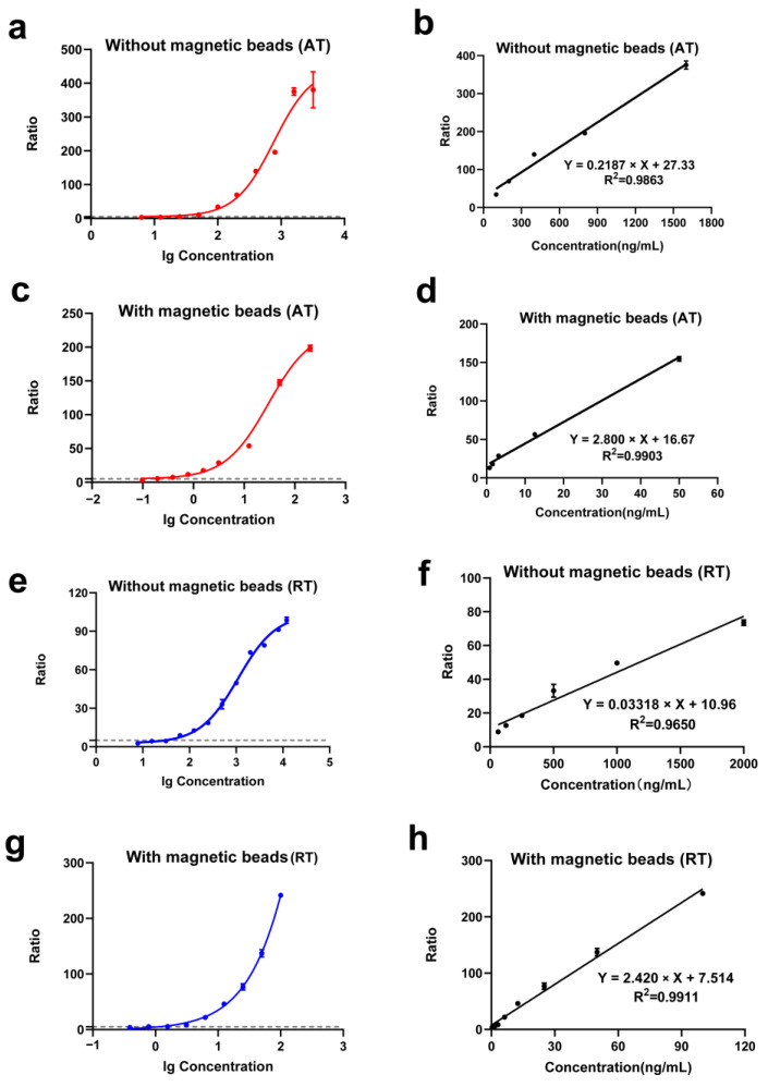
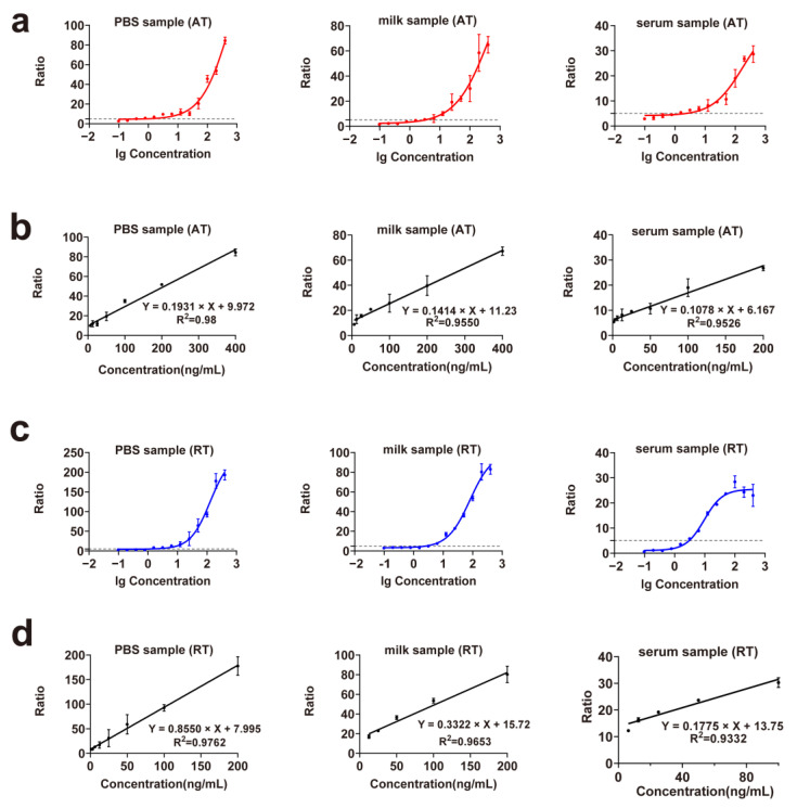
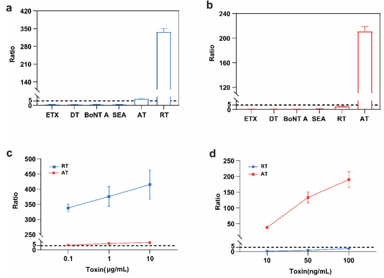
Ricin (RT) and abrin (AT) are plant toxins extracted from Ricinus communis and Abrus precatorius, respectively, and both have N-glycosidase activity. The detection of these toxins is vital because of their accessibility and bioterrorism potential. While ricin can be effectively detected based on its depurination activity, only a few tests are available for detecting the depurination activity of abrin. Therefore, it is unclear whether they share the same optimal reaction substrate and conditions. Here, we established optimum depurination conditions for ricin and abrin, facilitating the in vitro detection of their depurination activity using high-performance liquid chromatography-tandem mass spectrometry. The parameters optimized were the reaction substrate, bovine serum albumin (BSA), buffer, pH, temperature, time, antibodies, and magnetic beads. Both toxins showed better depurination with single-stranded DNA. However, substrate length, adenine content, BSA concentration, buffer concentration, reaction temperature, and reaction time differed between the two toxins. The optimal conditions for ricin depurination involved a reaction in 1 mM ammonium acetate solution (0.5 μM DNA15A, 20 μg/mL BSA, and 1 mM Zn2+, with pH 4.0) at 55 °C for 1 h. The optimal conditions for abrin depurination involved a reaction in 1 mM ammonium citrate solution (0.2 μM DNA20A, 10 μg/mL BSA, 1 mM Mg2+, and 0.5 mM EDTA, with pH 4.0) at 45 °C for 2 h. After optimization, the limits of detection (LOD) for ricin and abrin were 0.506 ng/mL and 0.168 ng/mL, respectively. The detection time was also significantly reduced.

Keywords: HPLC–MS/MS; N-glucosidase activity; abrin; detection; ricin.


Conflict of interest statement

The authors declare no conflicts of interest.

Figures

Figure 1
Figure 1
An illustration of the overall research strategy in this study.
Figure 2
Figure 2
Ricin and abrin reaction substrates and their optimal concentrations. (ae) Ricin (5 μg/mL) reacts with single-stranded DNA, single-stranded RNA, double-stranded DNA, neck-ring DNA, and neck-ring RNA structures. (f) Ricin (0.2 μg/mL) reacts with varying DNA15A concentrations. (gk) Abrin (5 μg/mL) reacts with single-stranded DNA, single-stranded RNA, double-stranded DNA, neck-ring DNA, and neck-ring RNA structures. (l) Abrin (0.02 μg/mL) reacts with varying DNA20A concentrations. Peak adenine detection levels are shown. They y-axis represents the ratio of substrate depurination due to toxin activity versus substrate self-depurination. Sterile water replaced the toxin as a negative control, with all other reaction components unchanged. The amount of adenine shed by the substrate itself was assessed. ns: p > 0.05; *: p < 0.05; **: p < 0.01; ***: p < 0.001.
Figure 3
Figure 3
The optimization of the reaction conditions for ricin and abrin. (a) The depurination of ricin and abrin in different concentrations of BSA. The concentrations of ricin and abrin were 2 μg/mL and 0.4 μg/mL; (b) the depurination of ricin with different buffer solution concentrations. The concentration of ricin was 5 μg/mL; (c) the depurination of abrin with different concentrations of the buffer solution. The concentration of abrin was 5 μg/mL; (d) the depurination of ricin and abrin in buffered solutions containing Zn2+, Mg2+, Ca2+, or EDTA. The concentration of ricin and abrin was 5 μg/mL; (e) ricin depurination activity at different pH levels. The concentration of ricin was 2 μg/mL; (f) abrin depurination activity at different pH levels. The concentration of ricin was 0.4 μg/mL; (g) the depurination of ricin and abrin at different temperatures. The concentrations of ricin and abrin were 10 μg/mL and 5 μg/mL, respectively; (h) ricin depurination with different incubation times. The concentration of ricin was 0.2 μg/mL; (i) abrin depurination with different incubation times. The concentration of abrin was 0.04 μg/mL. Aa, ammonium acetate; Aa + EDTA, ammonium acetate + 5 mM EDTA; Ac, ammonium citrate; Ac + EDTA, ammonium citrate + 5 mM EDTA; Af, ammonium formate; BSA, bovine serum albumin; EDTA, ethylene diamine tetraacetic acid.
Figure 4
Figure 4
Effects of different magnetic beads and trapping antibodies on ricin and abrin depurination. (a) Five ricin antibodies were coated with magnetic beads and depurination was performed; (b) five abrin antibodies were coated with magnetic beads and depurination was performed; (c) five types of magnetic beads were coated with the best antibodies of ricin and depurination was performed; (d) five types of magnetic beads were coated with the best antibodies of abrin and depurination was performed. The magnetic-bead amount was 30 μL (1 mg/mL), and the amount of antibody used was 30 μg (water matrix). The volume of each sample was 500 μL. The high (H) and low (L) concentrations of ricin and abrin were 250 and 50 ng/mL, respectively. a: Dynabeads® Antibody Coupling Kit; b: Dynabeads™ M-270 Streptavidin; c: Dynabeads™ M-280 Streptavidin; d: Dynabeads® M-450 epoxy; e: Dynabeads™ MyOne™ T1 Streptavidin.
Figure 5
Figure 5
Evaluation of abrin and ricin detection methods. (a) Nonlinear fitting curve for abrin without immunomagnetic-bead enrichment; (b) linear fitting curve for abrin without immunomagnetic-bead enrichment; (c) nonlinear fitting curve for abrin with immunomagnetic-bead enrichment; (d) linear fitting curve for abrin with immunomagnetic-bead enrichment; (e) nonlinear fitting curve for ricin without immunomagnetic-bead enrichment; (f) linear fitting curve for ricin without immunomagnetic-bead enrichment; (g) nonlinear fitting curve for ricin with immunomagnetic-bead enrichment; (h) linear fitting curve for ricin with immunomagnetic-bead enrichment.
Figure 6
Figure 6
Detection curves for simulated samples of ricin and abrin. (a) Nonlinear fitting curve of abrin in 1× PBS, milk, and serum; (b) linear fitting curve of abrin in 1× PBS, milk, and serum; (c) nonlinear fitting curve of ricin in 1× PBS, milk, and serum; (d) linear fitting curve of ricin in 1× PBS, milk, and serum. PBS, phosphate-buffered saline.
Figure 7
Figure 7
The specificity of the ricin and abrin detection method. (a) The specificity of HPLC–MS/MS detection of the depurination activity of ricin. The concentration of all toxins was 1 μg/mL (500 μL), and the substrate was DNA15A; (b) the specificity of HPLC–MS/MS detection of the depurination activity of abrin. The concentration of all toxins was 100 ng/mL (500 μL), and the substrate was DNA20A; (c) the cross-reactivity of the B4 antibody with the abrin depurination activity assay; (d) the cross-reactivity of the pAb-AT antibody with the ricin depurination activity assay. BoNT A, botulinum toxin A; DT, diphtheria toxin; ETX, clostridium perfringens epsilon toxin; SEA, staphylococcal enterotoxin A.

Similar articles

References

    1. Liang L.H., Xia J.M., Liu C.C., Liu S.L. Highly toxic type II ribosome-inactivating proteins ricin and abrin and their detection methods: A review. Chin. J. Chromatogr. 2021;38:260–270. doi: 10.3724/SP.J.1123.2020.10001. - DOI - PMC - PubMed
    1. Vance D.J., Mantis N.J. Progress and challenges associated with the development of ricin toxin subunit vaccines. Expert Rev. Vaccines. 2016;15:1213–1222. doi: 10.1586/14760584.2016.1168701. - DOI - PMC - PubMed
    1. Garber E.A.E., Walker J.L., O’brien T.W. Detection of abrin in food using enzyme-linked immunosorbent assay and electrochemiluminescence technologies. J. Food Prot. 2008;71:1868–1874. doi: 10.4315/0362-028X-71.9.1868. - DOI - PubMed
    1. Eric A.E.G. Toxicity and detection of ricin and abrin in beverages. J. Food Prot. 2008;71:1875–1883. - PubMed
    1. Edyta J., Michal C., Joanna S.-B., Michal B. Biological toxins as the potential tools for bioterrorism. Int. J. Mol. Sci. 2019;20:1181. doi: 10.3390/ijms20051181. - DOI - PMC - PubMed

Publication types

LinkOut - more resources