Markers of axonal injury in blood and tissue triggered by acute and chronic demyelination
- PMID: 40278829
- PMCID: PMC12316004
- DOI: 10.1093/brain/awaf144
Markers of axonal injury in blood and tissue triggered by acute and chronic demyelination
Abstract
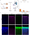
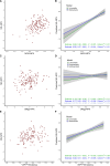
Neuroaxonal injury is a major driver of irreversible disability in demyelinating conditions. Accurate assessment of the association between demyelination and axonal pathology is critical for evaluating and developing effective therapeutic approaches. Measuring neurofilament light chain (NfL) in the blood could putatively allow longitudinal monitoring of neuroaxonal injury at 'single protein resolution' with high pathological specificity. Here, we demonstrate a robust association between blood and tissue NfL-based assessment of neuroaxonal injury and severity of inflammatory demyelination in experimental autoimmune encephalitis (EAE). In EAE, high levels of NfL were evident at the peak of demyelination and correlated with tissue evidence of NfL loss when using antibodies that target the same NfL epitopes. In addition, we validate the longitudinal NfL dynamics in relation to de- and remyelination in an inducible genetic model of inflammatory-independent myelin loss. Through inducible knockout of myelin regulatory factor (Myrf) in proteolipid protein (PLP) expressing cells in Myrffl/fl PLP1-CreERT (MyrfΔiPLP) mice, serum NfL peaked at the time of demyelination and reduced following effective remyelination. In people with multiple sclerosis, the most common demyelinating condition, we confirmed the association between NfL and myelin breakdown proteins in two independent cohorts using Olink proximity extension assays, the ReBUILD clinical trial and the multiple sclerosis participants in the UK-Biobank. Our study provides a translational framework to understand the biology behind NfL changes in the context of de- and remyelination and reveals novel aspects related to monitoring potentially reversible neuroaxonal pathology in humans and rodents.
Keywords: demyelination; multiple sclerosis; neurofilament light chain; neuroprotection; remyelination.
© The Author(s) 2025. Published by Oxford University Press on behalf of the Guarantors of Brain.
Conflict of interest statement
The authors report no competing interests in relation to this work.
Figures


Comment in
-
Extending the role of neurofilament light in multiple sclerosis beyond measuring irreversible neurodegeneration.Brain. 2025 Aug 1;148(8):2598-2600. doi: 10.1093/brain/awaf224. Brain. 2025. PMID: 40489576 No abstract available.
References
-
- Khalil M, Teunissen CE, Lehmann S, et al. Neurofilaments as biomarkers in neurological disorders—Towards clinical application. Nat Rev Neurol. 2024;20:269–287. - PubMed
-
- Abdelhak A, Kuhle J, Green AJ. Challenges and opportunities for the promising biomarker blood neurofilament light chain. JAMA Neurol. 2023;80:542. - PubMed
-
- Benkert P, Meier S, Schaedelin S, et al. Serum neurofilament light chain for individual prognostication of disease activity in people with multiple sclerosis: A retrospective modelling and validation study. Lancet Neurol. 2022;21:246–257. - PubMed
-
- Quanterix granted breakthrough device designation from U.S. FDA for NfL test for multiple sclerosis. 2022. Accessed 14 July 2022. https://www.quanterix.com/press-releases/quanterix-granted-breakthrough-...
MeSH terms
Substances
Grants and funding
LinkOut - more resources
Full Text Sources
Molecular Biology Databases
Research Materials
Miscellaneous

