. 2025 Jul;32(1):177.
doi: 10.3892/mmr.2025.13542. Epub 2025 Apr 25.

Understanding the role of iron/heme metabolism in the anti‑inflammatory effects of natural sulfur molecules against lipopolysaccharide‑induced inflammation

Affiliations

Understanding the role of iron/heme metabolism in the anti‑inflammatory effects of natural sulfur molecules against lipopolysaccharide‑induced inflammation

Dong Young Kang et al. Mol Med Rep. 2025 Jul.

Abstract

Iron transport and heme synthesis are essential processes in human metabolism, and any dysregulation in these mechanisms, such as inflammation, can have deleterious effects. Lipopolysaccharide (LPS)‑induced inflammatory responses can result in a number of adverse effects, including cancer. Natural mineral sulfur, methylsulfonylmethane (MSM) and nontoxic sulfur (NTS) suppress inflammatory responses. The present study hypothesized that MSM and NTS may inhibit LPS‑induced inflammatory responses in THP‑1 human monocytes. Reverse transcription‑quantitative PCR and western blotting assays were performed to analyze the molecular signaling pathways associated with sulfur‑treated and untreated cells. A comet assay was used to evaluate DNA damage, flow cytometry was performed to analyze cell surface receptors and chromatin immunoprecipitation was used to examine molecular interactions. Notably, LPS‑induced inflammation increased iron/heme metabolism, whereas MSM and NTS inhibited this effect. Furthermore, LPS treatment activated the Toll‑like receptor 4/NF‑κB signaling axis, which was downregulated by NTS and MSM. These sulfur compounds also suppressed the nuclear accumulation of LPS‑induced NF‑κB, which could induce the production of proinflammatory cytokines, such as TNF‑α, IL‑1β and IL‑6. Finally, MSM and NTS inhibited LPS‑induced reactive oxygen species generation and DNA damage in THP‑1 monocytic leukemia cells. These results suggested that natural sulfur molecules may be considered promising candidates for anti‑inflammation studies.

Keywords: anti‑inflammation; iron/heme metabolism; lipopolysaccharide; natural sulfur; oxidative stress.


Conflict of interest statement

The authors declare that they have no competing interests.

Figures

Figure 1.
Figure 1.
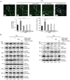
Sulfur compounds inhibit LPS-induced inflammation and iron/heme metabolism. (A) Flow cytometry showing Fe2+ levels in THP-1 cells following treatment with LPS (10 ng/ml) + NTS (3 µg/ml), MSM (200 mM) or TLR-C34 (40 µM) for 48 h. Western blot analysis of (B) transferrin receptor, ferroportin, DMT1 and STEAP3; and (C) MFRN, ABCB6, ABCB10, ALAS1, HO-1, FECH and FLVCR proteins in THP-1 cells following treatment with LPS (10 ng/ml) + NTS (3 µg/ml), MSM (200 mM) or TLR-C34 (40 µM) for 48 h. ALAS1, 5′-aminolevulinate synthase 1; DMT1, divalent metal transporter 1; Fe2+, ferrous ion; FECH, ferrochelatase; FPN, ferroportin; HO-1, heme oxygenase-1; LPS, lipopolysaccharide; MFRN, mitoferrin; MSM, methylsulfonylmethane; NTS, nontoxic sulfur; TfR, transferrin receptor; TLR, Toll-like receptor.
Figure 2.
Figure 2.
Sulfur compounds inhibit LPS-induced TLR expression. (A) Western blot analysis of TLR2 and TLR4 expression in THP-1 cells treated with LPS (10 ng/ml) + NTS (3 µg/ml), MSM (200 mM) or TLR-C34 (40 µM) for 48 h. (B) Reverse transcription-quantitative PCR analysis of the relative expression levels of TLR2 and TLR4 normalized to GAPDH following treatment with LPS (10 ng/ml) + NTS (3 µg/ml), MSM (200 mM) or TLR-C34 (40 µM) for 48 h. ***P<0.001; #P<0.001 vs. non-treated control (one-way ANOVA and Tukey's test). (C) Flow cytometry showing the inhibition of LPS-induced TLR4 expression by 48-h treatment with NTS (3 µg/ml), MSM (200 mM) and TLR-C34 (40 µM). LPS, lipopolysaccharide; MSM, methylsulfonylmethane; NTS, nontoxic sulfur; TLR, Toll-like receptor.
Figure 3.
Figure 3.
Sulfur compounds inhibit LPS-induced ROS. (A) Western blot analysis of iNOS expression in THP-1 cells treated with LPS (10 ng/ml) + NTS (3 µg/ml), MSM (200 mM) or TLR-C34 (40 µM) for 48 h. (B) Reverse transcription-quantitative PCR analysis of iNOS mRNA expression in cells treated with LPS (10 ng/ml) + NTS (3 µg/ml), MSM (200 mM) or TLR-C34 (40 µM) for 48 h. Data are normalized to GAPDH. ***P<0.001; #P<0.001 vs. non-treated control (one-way ANOVA and Tukey's test). (C) Flow cytometric analysis of cellular and mitochondrial ROS in THP-1 cells following treatment with LPS (10 ng/ml) + NTS (3 µg/ml), MSM (200 mM) or TLR-C34 (40 µM) for 48 h. iNOS, inducible nitric oxide synthase; LPS, lipopolysaccharide; MSM, methylsulfonylmethane; NTS, nontoxic sulfur; TLR, Toll-like receptor; ROS, reactive oxygen species.
Figure 4.
Figure 4.
Sulfur compounds inhibit the LPS-induced PKC-dependent and canonical NF-κB pathways. Western blot analysis of THP-1 cells treated with LPS (10 ng/ml) + NTS (3 µg/ml), MSM (200 mM) or TLR-C34 (40 µM) for 48 h showing the inhibition of LPS-induced (A) p-PKC-α, p-ERK and p-p38 expression; and (B) p-IKKα/β and p-IκBα expression. LPS, lipopolysaccharide; MSM, methylsulfonylmethane; NTS, nontoxic sulfur; p-, phosphorylated; TLR, Toll-like receptor.
Figure 5.
Figure 5.
Sulfur compounds induce DNA damage response following LPS-induced DNA damage. (A) Images of the comet assay were captured by fluorescence microscopy at ×10 and ×40 magnification levels, showing the fragmented DNA migrating out of the nucleoid body, which formed a comet tail following treatment with LPS (10 ng/ml) + NTS (3 µg/ml), MSM (200 mM) or TLR-C34 (40 µM) for 48 h. *P<0.05 and ***P<0.001; $P<0.05 vs. non-treated control; and $$$P<0.001 vs. non-treated control (one-way ANOVA and Tukey's test). (B) Western blot analysis of THP-1 cells; NTS (3 µg/ml) and MSM (200 mM) inhibited the LPS-induced expression of p-ATM, p-ATR, p-Chk2, p-BRCA1, and p-p53. However, the expression levels of p-MDM2 were suppressed by LPS treatment, and were increased by NTS (3 µg/ml), MSM (200 mM) or TLR4-C34 (40 µM). LPS, lipopolysaccharide; MSM, methylsulfonylmethane; NTS, nontoxic sulfur; p-, phosphorylated; TLR, Toll-like receptor.
Figure 6.
Figure 6.
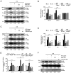
Sulfur compounds inhibit LPS-induced NF-κB, COX-2 and proinflammatory cytokine expression. (A) Western blot analysis of the expression levels of NF-κB, COX-1 and COX-2 in THP-1 cells treated with LPS (10 ng/ml) + NTS (3 µg/ml), MSM (200 mM) or TLR-C34 (40 µM) for 48 h. (B) RT-qPCR analysis of the relative expression levels of NF-κB, COX-1 and COX-2 normalized to GAPDH following treatment with LPS (10 ng/ml) + NTS (3 µg/ml), MSM (200 mM) or TLR-C34 (40 µM) for 48 h. ***P<0.001; #P<0.001 vs. non-treated control (one-way ANOVA and Tukey's test). (C) Western blot analysis of the expression levels of IL-1β, IL-6 and TNF-α in THP-1 cells treated with LPS (10 ng/ml) + NTS (3 µg/ml), MSM (200 mM) or TLR-C34 (40 µM) for 48 h. (D) RT-qPCR analysis of the relative expression levels of IL-1β, IL-6 and TNF-α normalized to GAPDH in cells following treatment with LPS (10 ng/ml) + NTS (3 µg/ml), MSM (200 mM) or TLR-C34 (40 µM) for 48 h. ***P<0.001; #P<0.001 vs. non-treated control (one-way ANOVA and Tukey's test). (E) Chromatin immunoprecipitation assay of THP-1 cells treated with LPS (10 ng/ml) + NTS (3 µg/ml), MSM (200 mM) or TLR-C34 (40 µM) for 48 h showing the relative binding of NF-κB to the promoters of IL-1β, IL-6 and TNF-α. ***P<0.001; #P<0.001 vs. non-treated control (one-way ANOVA and Tukey's test). (F) Nuclear protein extract analysis of THP-1 cells treated with LPS (10 ng/ml) + NTS (3 µg/ml), MSM (200 mM) or TLR-C34 (40 µM) for 48 h showing the protein expression levels of nuclear NF-κB. TBP was used as the housekeeping protein for the nuclear extract and β-actin was used to show the efficacy of nuclear protein extraction. LPS, lipopolysaccharide; MSM, methylsulfonylmethane; NTS, nontoxic sulfur; RT-qPCR, reverse transcription-quantitative PCR; TBP, TATA-binding protein; TLR, Toll-like receptor.
Figure 7.
Figure 7.
Molecular mechanism of LPS-induced regulation of the inflammatory response through iron/heme metabolism and TLR4/NF-κB expression through the canonical NF-κB and PKC-mediated inflammatory pathways. The anti-inflammatory activities of NTS and MSM were achieved by inhibiting iron/heme metabolism and suppressing the expression of TLR4/NF-κB signaling molecules, thus blocking the binding of NF-κB to the gene promoters of proinflammatory cytokines. DMT1, divalent metal transporter 1; Fe2+, ferrous ion; FPN, ferroportin; TfR, transferrin receptor; TLR4, Toll-like receptor 4.

Similar articles

References

    1. Wessling-Resnick M. Iron homeostasis and the inflammatory response. Annu Rev Nutr. 2010;30:105–122. doi: 10.1146/annurev.nutr.012809.104804. - DOI - PMC - PubMed
    1. Martins AC, Almeida JI, Lima IS, Kapitao AS, Gozzelino R. Iron metabolism and the inflammatory response. IUBMB Life. 2017;69:442–450. doi: 10.1002/iub.1635. - DOI - PubMed
    1. Alam Z, Devalaraja S, Li M, To TKJ, Folkert IW, Mitchell-Velasquez E, Dang MT, Young P, Wilbur CJ, Silverman MA, et al. Counter regulation of spic by NF-kappaB and STAT signaling controls inflammation and iron metabolism in macrophages. Cell Rep. 2020;31:107825. doi: 10.1016/j.celrep.2020.107825. - DOI - PMC - PubMed
    1. Wang L, Harrington L, Trebicka E, Shi HN, Kagan JC, Hong CC, Lin HY, Babitt JL, Cherayil BJ. Selective modulation of TLR4-activated inflammatory responses by altered iron homeostasis in mice. J Clin Invest. 2009;119:3322–3328. - PMC - PubMed
    1. Kang DY, Sp N, Jo ES, Lee JM, Jang KJ. New insights into the pivotal role of iron/Heme metabolism in TLR4/NF-κB Signaling-mediated inflammatory responses in human monocytes. Cells. 2021;10:2549. doi: 10.3390/cells10102549. - DOI - PMC - PubMed

MeSH terms