. 2025 Jun;301(6):108531.
doi: 10.1016/j.jbc.2025.108531. Epub 2025 Apr 23.

The mechanism of peptidoglycan O-acetylation in Gram-negative bacteria typifies bacterial MBOAT-SGNH acyltransferases

Affiliations

The mechanism of peptidoglycan O-acetylation in Gram-negative bacteria typifies bacterial MBOAT-SGNH acyltransferases

Alexander C Anderson et al. J Biol Chem. 2025 Jun.

Abstract

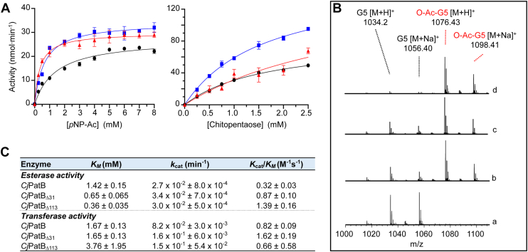
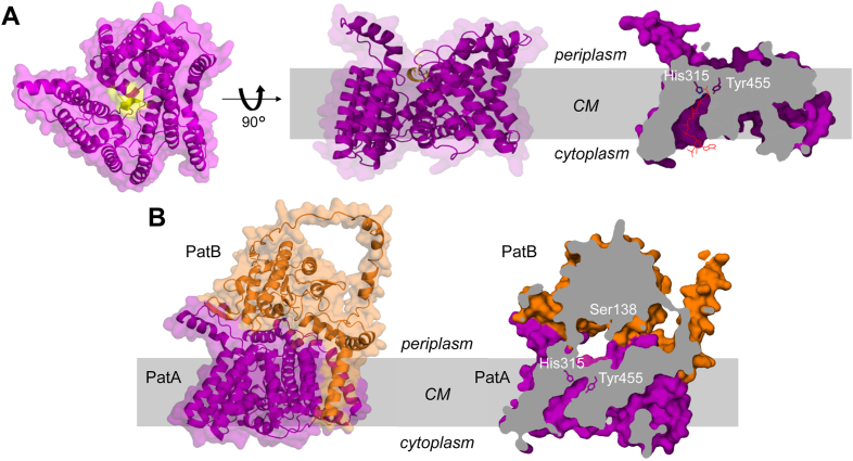
Bacterial cell envelope polymers are commonly modified with acyl groups that provide fitness advantages. Many polymer acylation pathways involve pairs of membrane-bound O-acyltransferase (MBOAT) and SGNH family proteins. As an example, the MBOAT protein PatA and the SGNH protein PatB are required in Gram-negative bacteria for peptidoglycan O-acetylation. The mechanism for how MBOAT-SGNH transferases move acyl groups from acyl-CoA donors made in the cytoplasm to extracellular polymers is unclear. Using the peptidoglycan O-acetyltransferase proteins PatAB, we explore the mechanism of MBOAT-SGNH pairs. We find that the MBOAT protein PatA catalyzes auto-acetylation of an invariant Tyr residue in its conserved C-terminal hexapeptide motif. We also show that PatB can use a synthetic hexapeptide containing an acetylated tyrosine to donate an acetyl group to a peptidoglycan mimetic. Finally, we report the structure of PatB, finding that it has structural features that shape its activity as an O-acetyltransferase and distinguish it from other SGNH esterases and hydrolases. Taken together, our results support a model for peptidoglycan acylation in which a tyrosine-containing peptide at the MBOAT's C-terminus shuttles an acyl group from the MBOAT active site to the SGNH active site, where it is transferred to peptidoglycan. This model likely applies to other systems containing MBOAT-SGNH pairs, such as those that O-acetylate alginate, cellulose, and secondary cell wall polysaccharides.

Keywords: O-acetyltransferase; X-ray crystallography; bacterial cell wall; o-acetylation; peptidoglycan.


Conflict of interest statement

Conflict of interest The authors declare they have no conflicts of interest with the contents of this article.

Figures

Figure 1
Figure 1
PatA forms a covalent acetyl-Tyr intermediate.A, previous model for PatAB catalyzed O-acetylation of PG. As an MBOAT acyltransferase, PatA was proposed to translocate acetyl groups from a cytoplasmic donor (1) involving a catalytic His residues to an unknown cytoplasmic acceptor (2) for their subsequent transfer to PG by PatB (3) via an acetyl-Ser intermediate. B, partial multiple sequence alignment of representative bacterial MBOAT acyltransferases encoded with SGNH proteins. The microprotein SaDltX is not an MBOAT but rather it binds to an MBOAT and an SGNH protein to provide the C-terminal portion of other bacterial MBOATs. The His and Tyr residues essential for PatA activity are in red, and other invariant residues are in bold type. C, SDS-PAGE autoradiography of identified PatA variants incubated in the presence of [14C]acetyl-CoA. D, MS/MS sequencing analysis of O-acetylated peptide from tryptic digest of PatA incubated with acetyl-CoA. Ba, Bacillus anthracis; Cj, C. jejuni; Ng, N. gonorrhoeae; Pa, Pseudomonas aeruginosa; Pf, Pseudomonas fluorescens; Sa, S. aureus.
Figure 2
Figure 2
Activity of recombinant PatB variants. A, acetylesterase (left) and transferase (right) activity of full-length CjPatB (black circle •), CjPatBΔ31 (blue square), and CjPatBΔ113 (red triangle) on pNP-Ac and chitopentaose as acetyl donor and acceptor substrates, respectively. The enzyme variants (1 μM) in 50 mM sodium phosphate pH 7.0 at 37 °C were incubated with the substrates at the concentrations indicated. Error bars denote SD (n = 3). B, LC-MS analysis of the reaction products of PatB variants acting as O-acetyltransferases (y-axis, relative abundance). Chitopentaose (G5) and pNP-Ac served as acceptor and donor substrates, respectively, for (a) no enzyme control, (b) full-length CjPatB, (c) CjPatBΔ113, and (d) CjPatBΔ31. C, Michaelis–Menten parameters determined for each PatB variant acting as esterases and transferases.
Figure 3
Figure 3
The crystal structure of PatB. The native structure of NgPatBΔ100 (7TJB) is presented as a representative of all the structural models of the PatB SGNH domain. A, ribbon presentation of PatB depicting a central α/β fold typical of the SGNH hydrolase family. Two extended β-hairpin motifs at the C-terminal face of the central β-sheet are present, which is not a feature of any structurally resolved SGNH hydrolase member. B and C, surface representations, oriented as in (A), showing the (B) catalytic triad and oxyanion hole residues (as sticks) arranged on the surface of NgPatB and (C) the surface electrostatic potential of PatB. D, B-factor putty model of PatB. The width and coloring of residues is based upon B-factor. E, the active site of NgPatBΔ100 depicting the positions of the block I, II, III and V residues. An unusual block II motif results in a family-atypical type I β-turn that composes the oxyanion hole, together with the block III Asn residue. A hydrophobic “wall” is formed behind the active site. A sulfate ion is coordinated by Ser133, Ser161, Asn196, and His305.
Figure 4
Figure 4
O-acetylated C-terminal peptide of PatA as acetyl donor for PatB.A, representative tracings of activity curves of NgPatBΔ100 acting as an esterase on the (a) C-terminal peptide FIYANF of NgPatA and (b) peptide IFYFAN as substrates. The arrow denotes the addition of the enzyme. B, LC-MS analysis of the reaction products of the transferase reaction involving incubation of NgPatBΔ100 with chitopentaose (G5) as acceptor in the (a) absence (negative control) and (b) presence of the O-acetylated FIYANF as donor substrate. y-axis, relative abundance.
Figure 5
Figure 5
Model structures of the PatA and PatAB complex.A, AlphaFold predicted model of CjPatA. From left to right, cartoon presentation of top (from periplasm) and side views of PatA, and cross-section of space-filling model. The latter reveals the transmembrane tunnel from the cytoplasm to the catalytic Tyr455 and His315 residues, which could accommodate the acetylated 4-phosphopantetheine arm of acetyl-CoA (red) as depicted with the superimposition of the X-ray structure of acetyl-CoA (extracted from its complex with galactoside acetyltransferase, PDB 1KRR). The C-terminal peptide comprising Tyr455 is depicted in yellow. B, cartoon (left) and cross-section (right) views of space filling model of the CjPatAB complex predicted by Colabfold. The catalytic residues of PatA (His315 and Tyr455) and PatB (Ser138) are positioned opposing each other within the active-site tunnel. CM, cytoplasmic membrane.
Figure 6
Figure 6
Proposed mechanism for PG O-acetylation in Gram-negative bacteria. A His residue in PatA catalyzes the transfer of acetyl groups from acetyl-CoA in the cytoplasm (1) to an invariant periplasmic Tyr residue in its C-terminal motif (2). The acetyl-Tyr intermediate serves as the donor substrate for PatB (3), which transfers the acetyl to PG via a covalent Ser intermediate (4). CM, cytoplasmic membrane.

Update of

References

    1. Silhavy T.J., Kahne D., Walker S. The bacterial cell envelope. Cold Spring Harb. Perspect. Biol. 2010;2:a000414. - PMC - PubMed
    1. Vollmer W., Blanot D., De Pedro M.A. Peptidoglycan structure and architecture. FEMS Microbiol. Rev. 2008;32:149–167. - PubMed
    1. Whitfield C., Trent M.S. Biosynthesis and export of bacterial lipopolysaccharides. Annu. Rev. Biochem. 2014;83:99–128. - PubMed
    1. Bera A., Herbert S., Jakob A., Vollmer W., Götz F. Why are pathogenic staphylococci so lysozyme resistant? The peptidoglycan O-acetyltransferase OatA is the major determinant for lysozyme resistance of Staphylococcus aureus. Mol. Microbiol. 2005;55:778–787. - PubMed
    1. Kristian S.A., Datta V., Weidenmaier C., Kansal R., Fedtke I., Peschel A., et al. d-Alanylation of teichoic acids promotes group A Streptococcus antimicrobial peptide resistance, neutrophil survival, and epithelial cell invasion. J. Bacteriol. 2005;187:6719–6725. - PMC - PubMed

MeSH terms

LinkOut - more resources